HYPERTROPHYC GINGIVATIS
Gingival enlargement, (also termed gingival overgrowth, hypertrophic gingivitis, gingival hyperplasia or gingival hypertrophy, and sometimes abbreviated to GO), is an increase in the size of the gingiva (gums). It is a common feature of gingival disease. Gingival enlargement can be caused by a number of factors, including inflammatory conditions and the side effects of certain medications. The treatment is based on the cause. A closely related term is epulis, denoting a localized tumor (i.e. lump) on the gingiva.

Classification
The terms gingival hyperplasia and gingival hypertrophy have been used to describe this topic in the past.These are not precise descriptions of gingival enlargement because these terms are strictlyhistologic diagnoses, and such diagnoses require microscopic analysis of a tissue sample. Hyperplasia refers to an increased number of cells, and hypertrophy refers to an increase in the size of individual cells.As these identifications obviously cannot be performed with a clinical examination and evaluation of the tissue, the term gingival enlargement is more properly applied. Gingival enlargment has been classified according to etiology into 5 general groups:
· Inflammatory enlargement
· Drug induced enlargement
· Enlargement associated with systemic diseases or conditions
· Neoplastic enlargement
· False enlargement.
Inflammatory enlargement
Gingival enlargement may be caused by a multitude of causes. The most common is chronic inflammatory gingival enlargement, when the gingivae are soft and discolored. This is caused by tissue edema andinfective cellular infiltration caused by prolonged exposure to bacterial plaque, and is treated with conventional periodontal treatment, such as scaling and root planing.
Gingivitis and gingival enlargement are often seen in mouth breathers, as a result of irritation brought on by surface dehydration, but the manner in which it is caused has not been demonstrated.
The accumulation and retention of plaque is the chief cause of inflammatory gingival enlargement. Risk factors include poor oral hygiene,as well as physical irritation of the gingiva by improper restorativeand orthodontic appliances.
Drug-induced enlargement
This type of gingival enlargement is sometimes termed “drug induced gingival enlargement”, “drug influenced gingival enlargement”,”drug induce gingival overgrowth” abbreviated to “DIGO”.[7] Gingival enlargement may also be associated with the administration of three different classes of drugs, all producing a similar response: Gingival overgrowth is a common side effect of phenytoin, termed “Phenytoin-induced gingival overgrowth” (PIGO).[
· anticonvulsants (such as phenytoin, phenobarbital, lamotrigine, valproate, vigabatrin, ethosuximide, topiramate and primidone)[10]
· calcium channel blockers, such as nifedipine, amlodipine, and verapamil. The dihydropyridine derivative isradipidine can replace nifedipine and does not induce gingival overgrowth.
· cyclosporine, an immunosuppresant.
Of all cases of DIGO, about 50% are attributed to phenytoin, 30% to cyclosporins and the remaining 10-20% to calcium channel blockers.
Drug-induced enlargement has been associated with a patient’s genetic predisposition, and its association with inflammation is debated. Some investigators assert that underlying inflammation is necessary for the development of drug-induced enlargement,while others purport that the existing enlargement induced by the drug effect compounds plaque retention, thus furthering the tissue response.
Enlargement associated with systemic factors
Many systemic diseases can develop oral manifestations that may include gingival enlargement, some that are related to conditions and others that are related to disease:
· Conditioned enlargement
· puberty
· vitamin C deficiency
· nonspecific, such as a pyogenic granuloma
· Systemic disease causing enlargement
· leukemia
· granulolomatous diseases, such as Wegener’s granulomatosis, sarcoidosis or orofacial granulomatosis
· neoplasm
· benign neoplasms, such as fibromas, papillomas and giant cell granulomas
· malignant neoplasms, such as a carcinoma or malignant melanoma
· false gingival enlargements, such as when there is an underlying bony or dental tissue lesion
Management
The first line management of gingival overgrowth is improved oral hygiene, ensuring that the irritative plaque is removed from around the necks of the teeth and gums. Situations in which the chronic inflammatory gingival enlargement include significant fibrotic components that do not respond to and undergo shrinkage when exposed to scaling and root planing are treated with surgical removal of the excess tissue, most often with a procedure known as gingivectomy.
In DIGO, improved oral hygiene and plaque control is still important to help reduce any inflammatory component that may be contributing to the overgrowth. Reversing and preventing gingival enlargement caused by drugs is as easy as ceasing drug therapy or substituting to another drug. However, this is not always an option; in such a situation, alternative drug therapy may be employed, if possible, to avoid this deleterious side effect. In the case of immunosuppression, tacrolimus is an available alternative which results in much less severe gingival overgrowth than ciclosporin, but is similarly as nephrotoxic. The dihydropyridine derivative isradipidine can replace nifedipine for some uses of calcium channel blocking and does not induce gingival overgrowth.
GINGIVAL OVERGROWTH
Gingival overgrowth occurs mainly as a result of certain anti-seizure, immunosuppressive, or antihypertensive drug therapies. Excess gingival tissues impede oral function and are disfiguring. Effective oral hygiene is compromised in the presence of gingival overgrowth, and it is now recognized that this may have negative implications for the systemic health of affected patients. Recent studies indicate that cytokine balances are abnormal in drug-induced forms of gingival overgrowth. Data supporting molecular and cellular characteristics that distinguish different forms of gingival overgrowth are summarized, and aspects of gingival fibroblast extracellular matrix metabolism that are unique to gingival tissues and cells are reviewed. Abnormal cytokine balances derived principally from lymphocytes and macrophages, and unique aspects of gingival extracellular matrix metabolism, are elements of a working model presented to facilitate our gaining a better understanding of mechanisms and of the tissue specificity of gingival overgrowth.
INTRODUCTION
Clinically detectable fibrotic overgrowth of gingiva is caused by a variety of etiological factors and is exacerbated by local bacterial plaque accumulation. Gingival overgrowth can be inherited (hereditary gingival fibromatosis), or is of idiopathic origin, or is sometimes associated with other systemic diseases. The majority of cases, however, occur as a side-effect of systemic medications. These medications include the anti-seizure drug phenytoin, the immune suppressor cyclosporin A, and certain anti-hypertensive dihydropyridine calcium-channel-blockers, most notably nifedipine.
There is now general agreement that gingival overgrowth lesions all contain fibrotic or expanded connective tissues with various levels of inflammation and an enlarged gingival epithelium. The degrees of inflammation, fibrosis, and cellularity depend on the duration, dose, and identity of the drug, on the quality of oral hygiene, and on individual susceptibility that stems from genetic factors and environmental influences. Many professors regard the importance of gingival overgrowth as a clinical problem, and highlights recent advances in the identification of molecular and cellular processes that are leading to an enhanced understanding of the biological mechanisms underlying the overgrowth. Many recent genetic studies on hereditary gingival fibromatosis are beginning to identify the unique cell biology and extracellular matrix metabolism that occur in human gingiva.
DRUGS AND GINGIVAL OVERGROWTH
Drug-induced gingival overgrowth is a side-effect and unwanted outcome of systemic medication and is limited to gingival tissues. Drug-induced gingival overgrowth does not originate in the periodontium, but it occurs exclusively in periodontal tissues and is generally not of the same magnitude in other tissues. Patients who receive anti-epileptic therapy through the systemic use of phenytoin, or immunosuppressive therapy with systemic cyclosporin A ,or anti-hypertensive treatment with systemic nifedipine develop gingival overgrowth with various degrees of prevalence. While the incidence of this side-effect can be as high as 65% in epileptics, 70% in transplant patients, and 30% in hypertension subjects, variation exists in the reported prevalence and severity of the clinical problem . Clinical and histological characteristics of drug-induced gingival overgrowth include hyperplasia in junctional epithelium and hypertrophy in keratinized epithelium and excessive connective tissue accumulation. In addition to being disfiguring and uncomfortable for affected individuals, moderate to severe forms of gingival overgrowth impair oral hygiene and may lead to increased accumulation of micro-organisms. Oral infections can potentially impair systemic health ,and elevated oral infections stemming from gingival overgrowth could possibly compromise the general health of patients. This is most obvious in the case of organ transplant recipients who require continuous therapy with cyclosporin A, thereby being rendered critically susceptible to life-threatening systemic infections, and who concomitantly develop gingival overgrowth. The effective management of these patients clearly requires the active involvement of both dental and medical professionals to minimize the possibility of complications.(Thomason JM, Seymour RA, Ellis JS,Kelly PJ, Parry G, Dark J, et al. Determinants of gingival overgrowth severity in organ transplant patients. J Clin Periodontol 1996;23:628-34)
Dose-dependent correlations with the severity of gingival overgrowth are weak, but decreased drug use in general results in reduced severity of gingival pathology. For example, phenytoin was reported to effuse into crevicular fluid without any correlation to the incidence of overgrowth ,while no direct link was shown between overgrowth and the concentrations of phenytoin and metabolites . A more recent study supports a correlation between diminished metabolism of phenytoin in affected individuals and overgrowth, but this has not been confirmed. Age, gender, concomitant medication with multiple drugs, local factors such as plaque accumulation, and genetic disposition are additional complicating risk factors in drug-induced gingival overgrowth .
Therapy of drug-induced gingival overgrowth would seem to be most simply accomplished by the use of alternate medications that do not induce gingival overgrowth, and new medications are under development. At this time, however, older medications are still in active use. Cyclosporin A, for example, increases the rates of survival of most organ transplant patients, although the more recently developed immunosuppressant FK506(tacrolimus) can be substituted in some patients . Tacrolimus has not yet been associated with gingival overgrowth, but does cause other side-effects that are important for some patients. The literature on the effective substitution of cyclosporin A with tacrolimus is relatively new. Cyclosporin A continues to be the most commonly prescribed drug for the prevention of graft rejection. Similarly, although several new anti-seizure drugs have been introduced and its prescription is markedly reduced, phenytoin remains the drug of choice for certain types of grand mal epileptic seizures . Finally, although hypertension cases are now being treated by alternative calcium-channel-blockers that either do not predictably cause gingival overgrowth or are linked to isolated cases of gingival pathologies, nifedipine remains a highly effective agent in the management of patients who do not respond sufficiently well to other anti-hypertensive medications .In summary, it should be anticipated that drug-induced gingival overgrowth will continue to be a problem until safe and equally reliable medications to control these systemic conditions are developed and fully utilized. In addition, because the administration of these medications in general is beyond the control of the dental professional, clinical management of the gingival overgrowth presents a continuous challenge.
Treatment of the gingival overgrowth lesion itself can be complicated due to the superimposed inflammation on the fibrotic tissue enlargement. Traditionally, periodontal therapy offers removal of the inflammatory component of the overgrowth through scaling and gingival curettage, followed by excision of the overgrown gingiva . For patients with severe gingival overgrowth and who require continuous drug therapy for medical reasons, gingivectomy must be repeated periodically due to the recurrent nature of drug-induced gingival overgrowth .
Studies on the direct effects of phenytoin , nifedipine , and cyclosporin A on collagenous and non-collagenous extracellular matrix metabolism by gingival fibroblasts in culture have provided variable results. The differences in these investigations depend in part on culture conditions . While cyclosporin A was found to increase glycosaminoglycan secretion by fibroblasts , and nifedipine and phenytoin increased heparan levels ,other studies failed to report any differences in vivo . Several studies demonstrate that these drugs inhibit gingival fibroblast extracellular matrix production and/or cell proliferation in vitro. These findings are inconsistent with the in vivo characteristics of drug-induced gingival overgrowth. Taken together, these studies support the conclusion that direct regulation of extracellular matrix metabolism or proliferation of gingival fibroblasts by these drugs is probably not the primary mechanism responsible for gingival overgrowth. As noted below, deregulated cytokine balances may contribute more significantly to the development and maintenance of gingival overgrowth. Several studies more consistently show that cultured human gingival fibroblasts treated with phenytoin, cyclosporin A, and nifedipine increase production of fibroblast cytokines and prostaglandin E2 ,although lymphocytes and macrophages may be important sources of these factors in vivo (Modéer et al., 1989; Zebroski EJ, Singer DL, Brunka JR. Cyclosporin A, nifedipine and phenytoin: comparative effects on gingival fibroblast metabolism. J Dent Res 1986).
Gingival tissues are generally in a state of injury and repair that involves repetitive cycles of production of chemotactic factors, inflammatory cell recruitment, and tissue resorption, replacement, and remodeling . Collagen turnover is unusually high in periodontal tissues .Wound healing and connective tissue turnover are largely controlled by chemokines and cytokines secreted by inflammatory cells such as macrophages and lymphocytes and, to a lesser degree, by fibroblasts. Proliferation and differentiation of connective tissue cells and production of extracellular matrix are controlled by cytokines that initiate signaling cascades mediated by specific receptors. In addition, extracellular matrix elements interact with cell-surface receptors, including integrins, that initiate or modulate signaling cascades .Recent studies demonstrate abnormally high levels of specific cytokines in gingival overgrowth tissues. These findings are of great interest, and suggest that substances that cause gingival overgrowth may do so by altering the normal balance of cytokines in gingival tissues. Cytokines and growth factors found at elevated levels in human drug-induced gingival overgrowth include interleukin-6 (IL-6), IL-1β, platelet-derived growth factor-B (PDGF-B), fibroblast growth factor-2 (FGF-2), transforming growth factor-β (TGF-β), and connective tissue growth factor CTGF.
There is a limited understanding of the mechanisms by which altered cytokine balances occur in drug-induced gingival overgrowth. A contributing factor may stem from immunomodulatory effects of the drugs. For example, the increased expression of the macrophage phenotype marker RM3/1 in phenytoin-induced gingival overgrowth is consistent with fibroproliferative disease . In contrast, macrophages in highly inflamed tissues express predominantly the 27E10 marker . Even though IL-1β levels have not been shown to be increased in cyclosporin-A-treated monocytes/macrophages, there was a significant up-regulation in PDGF-B in response to cyclosporin A and phenytoin. Similarly, phenytoin, cyclosporin A, and nifedipine gingival overgrowth tissues contain subpopulations of macrophages and other inflammatory cells that differ from those in healthy control gingival tissues. Studies on T- and B-lymphocytes showed that T-cells are increased in the peripheral blood of organ transplant patients with no apparent shift of subpopulations and nifedipine increased lymphocyte counts in blood , although these findings were not found in gingival tissues . A reduction in the number of Langerhans cells iifedipine and cyclosporin A gingival overgrowth occurs and suggests a modification of an inflammatory reaction that influences the level of helper T-lymphocytes and cytokine profiles. Inflammatory cell populations that are altered as a result of drug therapy are likely to modify the gingival tissue response. At this time, however, there is no consensus regarding functional relationships between and among drug therapies, the distribution of specific immune system cell subpopulations, and altered cytokine balances.
FUNCTIONAL STUDIES
The occurrence of abnormal cytokine levels does not alone prove a functional relationship to gingival overgrowth. Studies have begun to investigate functional relationships between cytokines and gingival extracellular matrix metabolism. These studies seem likely to result in a greater understanding of the biological mechanisms that may be unique to human gingival tissues and may be relevant to the development of therapeutic strategies for either the prevention or treatment of gingival overgrowth. TGF-β1 is a cytokine secreted by many cell types, including macrophages, and it has an important regulatory function in collagen metabolism in connective tissues. TGF-β1 slowly stimulates collagen and lysyl oxidase biosynthesis in early-passage human gingival fibroblast cell cultures , whereas IL-1β, IL-6, and PDGF-BB have little or no effect, andFGF-2 is inhibitory . In contrast, PDGF-BB and FGF-2 are potent mitogenic factors and contribute to the proliferation of gingival connective tissue and epithelial cells. The effects of TGF-β1 on gingival collagen and lysyl oxidase regulation are notable because the magnitude and kinetics of regulation are unexpectedly smaller and slower compared with studies on other connective tissue cells performed under the same conditions . To understand the unexpectedly slow kinetics of TGF-β1 on extracellular matrix synthesis in gingival fibroblasts, investigators in recent studies have focused on the presence and role of connective tissue growth factor (CTGF) as a possible matrix-stimulatory factor downstream of TGF-β1 in gingival overgrowth tissues. CTGF has been proposed to mediate the effects of TGF-β on extracellular matrix metabolism .Before studies performed in gingival cells and tissues are summarized, information on CTGF and the emerging related CCN family of factors is first offered as background information.
CTGF AND FIBROSIS
CTGF is found to occur at elevated levels in a variety of fibrotic pathologies, including the fibrous stroma of mammary tumors , chronic pancreatitis , cataract formation , nephropathy, systemic sclerosis , pulmonary fibrosis , inflammatory bowel disease , bladder fibrosis due to outlet obstruction , brain fibrosis following injury , atherosclerosis , and fibrotic skin disorders . CTGF alone does not promote fibrosis. This is illustrated by experiments in which the simultaneous application of CTGF and TGF-β1 is required for sustained skin fibrosis; neither factor alone was effective . CTGF is rapidly and potently induced by TGF-β1 in fibroblastic cells from a variety of different tissues, and contributes to the regulation of extracellular matrix genes .
CTGF is a member of the CCN family of factors .The name of this family is derived from the first three family members identified: Cyr61, CTGF, and NOV. Additional members of this family include WISP-1 to -3. These factors have a highly conserved structure that consists of four domains containing 38 conserved cysteine residues. The four conserved domains (or modules) are related to other extracellular matrix proteins: Module 1 is similar to insulin-like growth factor (IGF) binding proteins; module 2 is similar to von Willebrand type C domain; module 3 is related to thrombospondin-1; and module 4 contains a putative cysteine knot that could promote dimerization of these factors. WISP-3 lacks the fourth module. The biological functions of this family of proteins include positive and negative regulation of proliferation and differentiation of connective tissue cells, and regulation of extracellular matrix accumulation. Structure/function studies are still in an early stage of development .CTGF and cyr61 are closely related in structure, but promotion of extracellular matrix production or accumulation appears to be unique to CTGF . CTGF is expressed by endothelial cells, granulosa cells , fibroblasts , mesangial cells , chondrocytes , and osteoblast, and occurs in biological fluids and tissues in low-molecular-weight forms that contain domains 3 and 4 that retain mitogenic activity . It seems possible that the pleiotropic nature of the CCN family of factors may be related to proteolytic processing events that unmask latent activities encoded by different functionally independent domains. Moreover, recent studies indicate that CTGF binds to other growth factors, resulting in either inhibition or stimulation of their activity. Thus, CTGF binds to vascular endothelial growth factor (VEGF) and bone morphogenetic protein-4 (BMP-4), resulting in inhibition of VEGF and BMP-4 activity, respectively; whereas CTGF binding to TGF-β1 is reported to be stimulatory . Matrix metalloproteinase (MMP) hydrolysis of CTGF/VEGF complexes results in release of activeVEGF . CTGF binds to Vβ3, 6β1, and other β3 integrins on different cell types and activates signaling cascades . Taken together, these studies support the idea that CTGF is a ‘matricellular’ factor that works in concert with growth factors, growth factor receptors, extracellular matrix, and extracellular matrix receptors . This view is consistent with the relatively weak direct stimulatory effects of CTGF on extracellular matrix production compared with the effects of pro-fibrogenic cytokines such as TGF-β1, and may account for the requirement for the simultaneous presence of both CTGF and TGF-β for sustained fibrosis . Integrin receptor-mediated signals in combination with growth factor receptor-mediated signals seem likely to work together to result in tissue fibrosis (Cley C, Javisk K, Hassell T. Effects of cyclosporin A on human gingival fibroblasts in vitro. J Dent Res 1986 . )
CTGF is developmentally regulated , and interesting studies regarding the presence and function of CTGF in developing tooth germs have been reported . Inhibition of CTGF with a blocking antibody in murine tooth germ organ cultures inhibited tooth germ development and differentiation. Epithelial/mesenchymal interactions were found to be important for the expression of CTGF in tooth germs in tissue recombination experiments involving dental epithelium and mesenchyme, and TGF-β1 and BMP-2 were implicated as mesenchymal factors contributing to the maintenance of CTGF in tooth germs. (Stephen E. Wolverton et al. Comprehensive Dermatologic Drug Therapy. Saunderes Company 2000; 205-225)
CTGF contributes to fibrosis in many different tissues. The hypothesis was developed that CTGF could play a role in gingival overgrowth and fibrosis. Studies of CTGF regulation were initiated in vitro in human gingival fibroblast cultures, because increased collagen and lysyl oxidase biosynthesis induced by TGF-β1 occurred only at modest levels and with slow kinetics compared with effects seen in other cell types. The working hypothesis was that TGF-β1 might induce CTGF, which in turn would stimulate extracellular matrix production in gingival fibroblasts. Findings indicate that CTGF is rapidly and potently up-regulated by TGF-β1, and that CTGFstimulates insoluble collagen accumulation in human gingival fibroblast cultures . Moreover, a clinical study indicates that CTGF is present at elevated levels in phenytoin- and nifedipine-induced gingival overgrowth tissues, but not in cyclosporin-A-stimulated gingival overgrowth . The finding of elevated CTGF in phenytoin-induced gingival overgrowth was obvious and clear, even after adjustment for the level of inflammation determined by histomorphometric analyses . This study furthermore indicates that the more fibrotic tissues appear to contain the highest levels of CTGF. Analysis of the data obtained supports the notion that cyclosporin-A-induced gingival overgrowth tissues are significantly more inflamed and less fibrotic than phenytoin- or nifedipine-induced gingival overgrowth. Taken together, these findings identify clear and consistent molecular and cellular distinctions between and among phenytoin-, nifedipine-, and cyclosporin-A-induced gingival overgrowth tissues. It is apparent that gingival overgrowth is a clinical phenomenon that is heterogeneous with respect to the underlying biological mechanisms. Future studies will be necessary to identify additional molecular markers unique to specific forms of gingival overgrowth that will likely prove to be of functional significance in the etiology of different forms of this condition.
One of the earliest signs of gingivitis is bleeding on probing.
MOUTH BREATHING
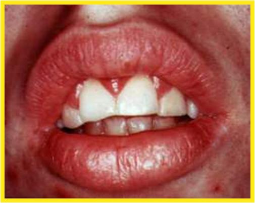
This type of gingivitis affects the anterior gingiva of chronic mouth breathers or individuals with incomplete lip closure. Note the erythematous, hypertrophic maxillary anterior gingiva.
PREGNANCY GINGIVITIS
Note the intense burgundy color and the marked gingival hypertrophy. These lesions bleed profusely.
PYOGENIC GRANULOMA
Pyogenic granuloma is considered to be a exuberant response to a chronic mild irritant. Its clinical appearance is similar to that seen in pregnancy gingivitis but generally confined to a single area. Pyogenic granulomas also bleed easily because they contain multiple capillaries.
DIABETES MELLITUS ASSOCIATED GINGIVITIS
Note the marked inflammatory reaction and hypertrophy of the free gingiva in this patient with diabetes mellitus. This reflects an increased gingival reaction to plaque with consequent increased risk of periodontal disease.
DIABETES MELLITUS PERIODONTAL ABSCESS
There is a greater increase risk for diabetic patients to develop periodontal abscesses due to increased gingival reaction to plaque and increased risk of periodontal disease. The arrow points to the abscess.
PHENYTOIN GINGIVAL HYPERTHROPHY
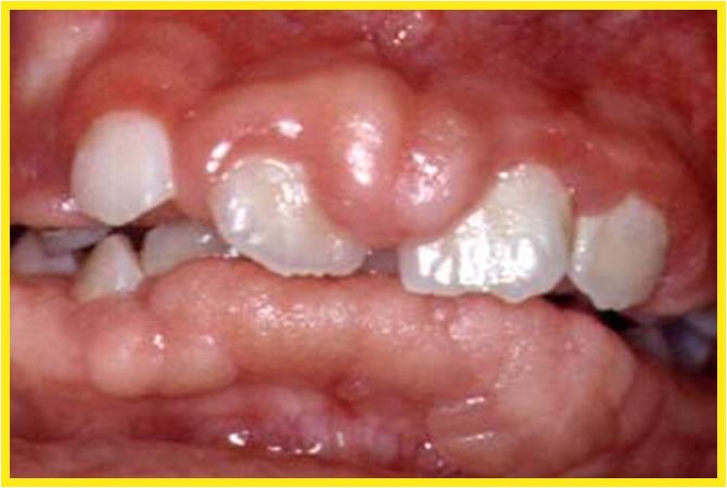
Phenytoin gingival hypertrophy has an incidence of 3 to 84.5%. This enlargement is produced by hyperplasia of the connective and epithelial tissues with secondary inflammation. It may have increased expression of platelet derived growth factor.
CALCIUM CHANNEL BLOCKERS – NIFEDIPINE
Nifedipine is used for coronary artery disease and hypertension to dilate blood vessel and is also used with immunosuppressant drugs in organ transplant. This medication induces gingival hypertrophy, as seen here, in 25% to 50% of patients.
Periodontal disease
The periodontal diseases are a family of chronic inflammatory diseases that involve the periodontium – the bone and soft tissues that support the teeth in the jaws. They are the most common infection in humans. These diseases are caused by bacterial plaque that grows on the teeth and the immune response to that chronic infection.
Gingivitis and periodontitis are the two main periodontal diseases and may be present concurrently. Gingivitis is inflammation of the gingival margin. It affects approximately 45% of the adult population in Australia and is characterised by redness and swelling or oedema of the gingival margin around the neck of the teeth. Periodontitis is a more severe condition. It afflicts 15-20% of the adult dentate population and causes loss of the bone supporting the teeth. Signs of periodontitis include, in addition to signs of gingivitis, recession of the gums, tooth looseness, changes in tooth alignment, and halitosis. The commonest sign of either gingivitis or periodontitis is bleeding gums which can be provoked by tooth brushing, flossing, and eating hard foods, but can also be spontaneous. The absence of bleeding does not necessarily indicate the absence of periodontal disease, particularly in smokers.
Periodontal practice today is largely evidence-based. It aims to reduce inflammation by cleaning the teeth to remove plaque, and by using various treatment modalities to prevent or limit further plaque accumulation. A patient’s management of their plaque control is central to successful periodontal treatment, but the response to treatment will also be influenced by factors such as genetic susceptibility to periodontal disease and background systemic disease.
Drug-induced gingival enlargement
Drug-induced gingival enlargement was first observed in patients who were taking phenytoin for epilepsy, with approximately 50% having gingival overgrowth. Cyclosporin is an immunosuppressant which has been reported to cause gingival enlargement in 25-80% of patients.1,2The calcium channel antagonists can also cause gingival enlargement. The dihydropyridines (e.g. nifedipine, felodipine, amlodipine) tend to be more commonly associated with gingival enlargement than the other sub-groups of calcium channel antagonists. Prescription of calcium channel blockers is relatively common, making it difficult to determine the true incidence of drug-induced gingival enlargement. Some of the variation in incidence of gingival enlargement can be attributed to differences between study populations and methods of classifying its severity.
Clinical presentation
Gingival enlargement usually develops in a susceptible individual within a few months of starting the medication. Drug-induced gingival enlargement consists of soft tissue growth that begins between the teeth and increases in all directions. As the tissue enlarges it develops a characteristically thickened and lobulated appearance. It may partially or completely cover the tooth surfaces, including the occlusal (chewing) surfaces, as well as extending the other way, into the sulcus. The epithelial surface is usually smooth and fibrotic, but can be nodular in cyclosporin-induced enlargement. If there is underlying periodontal disease then the tissues may be inflamed, red or purplish in colour, and highly vascularised, with a tendency to bleed profusely (Fig. 1).
Gingival enlargement tends to be more severe in areas where plaque accumulates, such as at the edges of fillings and around orthodontic appliances. It is rarely seen in edentulous areas. Gingival enlargement impedes effective plaque control and regularly traps plaque or food, producing halitosis or suppuration. There is a tendency for gingival enlargement to be distributed symmetrically and for the anterior teeth to be more severely affected than the posterior teeth.
Clinical parameters such as the standard of oral hygiene, drug dosage, and serum and salivary levels have some relationship to the incidence of gingival enlargement. Generally, a higher dose and poorer plaque control – or more plaque retentive sites – are more likely to be associated with gingival enlargement. However, there is no direct relationship and these factors do not fully explain the incidence or the characteristics of the lesion.
|
Management of gingival enlargement
In addition to plaque control and medical management, periodontal surgical treatment and multidisciplinary dental care are key strategies in managing gingival enlargement.
Mild gingival enlargement may only require local management as improvement in oral hygiene, together with professional cleaning of the teeth, can lead to resolution of inflammation and reduction in gingival enlargement. Treatment planning becomes more complex where there is periodontitis plus gingival enlargement that is a cosmetic or functional problem. Periodontitis can be treated using conventional clinical care, but the gingival enlargement may require changes to the medication regimen, periodontal surgery to remove excess tissue, or a combination of the two.
Plaque control
Effective plaque control can reduce and prevent gingival enlargement. Most people clean their teeth, but not particularly effectively. It is important that the dental professional encourages improved tooth cleaning in a supportive and positive manner, as well as providing information about the role of dental plaque in promoting gingival overgrowth. Mild gingival enlargement will often diminish with removal of plaque and calculus deposits. Even moderate gingival enlargement may reduce enough to avoid surgical intervention.
Attempts at improving oral hygiene are of limited benefit in severe gingival enlargement – surgical gingival resection is indicated. Chlorhexidine 0.1% should be rinsed two or three times daily for the first few postoperative days, with careful mechanical cleaning introduced gradually as it becomes more comfortable. Areas that are not included in the surgery can be cleaned as usual. The efficacy of chlorhexidine may be reduced by toothpaste because of a chemical interaction. The interval between tooth brushing and rinsing should therefore be at least 30 minutes.3
Many patients who take drugs that induce gingival enlargement will have some gingival growth between the teeth and thickening of the gums. It follows then that when they brush their teeth they risk either traumatising the soft tissue or inadequate cleaning of the crown of the tooth close to the gingival margin. Flossing is difficult and using interproximal brushes and wood sticks is often out of the question when gingival enlargement is present.
Plaque can be removed by cleaning each tooth separately, holding the brush in line with the long axis of the tooth. The narrower dimension of the head of the brush then fits in between the papillae (the part of the gum between the teeth). Another option is to use an electric toothbrush with a round head to clean the teeth in the same longitudinal fashion. Cleaning plaque from between the teeth can also be carried out if the patient is shown how to gently slide dental floss or tape along the tooth surfaces and under the edge of the gum. Thorough cleaning by brushing and flossing should be carried out at least once daily.
Medical management
Many cases of gingival enlargement will respond to local treatment, but consideration should be given to altering the medication if gingival enlargement covers more than about a third of the tooth surface. When possible, reducing the dose or changing to another drug may bring about partial or complete regression of the lesion. Most patients will observe an alteration in the soft tissues within a few days. If a person continues taking the same gingival enlargement-inducing medication then they should be warned of the possibility of gingival enlargement recurring despite periodontal treatment.
Several alternatives to phenytoin are available, but they may not be as well tolerated or they may not control seizures as well. Some patients can switch to a lower dose of phenytoin combined with another anticonvulsant.
If a patient develops gingival enlargement as a result of taking a particular calcium antagonist, they will usually also develop it in response to other calcium antagonists. Alternative classes of antihypertensive medication may be suitable for patients who are being treated for hypertension.
The dose of cyclosporin may be reduced in the course of medical treatment, and can also be reduced in some cases where patients are on a maintenance dose, with no adverse effects. Once the gingival enlargement is drawn to the treating physician’s attention, it may be possible to maintain a patient on a lower dose.
Changing from cyclosporin to tacrolimus can be considered if significant gingival enlargement recurs after excision. Tacrolimus has a different toxicity profile and is not associated with gingival enlargement. It has the same interactions with diltiazem, which could still be used, producing a residual but diminished gingival enlargement.
Gingival hypertrophy is the abnormal enlargement of the gums. The condition can be caused by medications, a systemic disease, poor oral hygiene, or a congenital disorder called occulodental syndrome. Many people with gingival hypertrophy experience bleeding from the gums and pain when eating. Gum enlargement needs to be evaluated by a dentist or physician to treat any underlying pathological causes.
The long-term build up of plaque around the teeth and gums is the leading cause of swollen gums. Poor oral hygiene allows bacteria to inflame the gum tissue. Dentures or partial dentures may irritate the delicate gum tissue, causing enlargement. If a dental appliance causes the swelling, it may need to be adjusted professionally.
Prescription medicines may have gingival hypertrophy as a side effect. Some of the medicines for the treatment of seizures as well as anti-convulsant drugs such as phenytoin, primidone, and topiramate are known to cause gingival enlargement. An immunosuppressant medication called ciclosporin can also cause gingival hypertrophy after taking it. Certain calcium channel blockers, namely nifidepine and verapamil, are also known to cause gum swelling. These oral side effects may subside after the medication is discontinued.
Natural changes in the body may cause gingival hypertrophy for a short time. Some women develop gingival hypertrophy during pregnancy. The swollen gums usually go back to the normal size after the woman gives birth. Adolescents may have enlarged gums during puberty, and it usually passes once the hormones circulating in their bodies even out.
There are systemic diseases that may cause gingival hypertrophy. People with leukemia may notice that the gum tissue has swollen and become painful. Growths called neoplasms may be responsible for the enlargement of gum tissue. The neoplasms may be benign or indicative of a malignant cancer.
Treatment of gingival hypertrophy depends on the cause of the enlargement. In the case of poor hygiene and plaque buildup, a dental procedure called scaling will be performed to reduce the plaque on the teeth. The plaque is scraped away from the teeth with dental tools. If the inflammation has reached the roots of the teeth, a process called root planing is necessary. It is a delicate process that removes the plaque and tarter from below the gumline.
A surgical procedure called a gingivectomy may be needed when the enlarged gum tissue does not reduce in size after the dental scaling or root planing. The swollen gum is snipped to a normal size, or electrosurgery may be used to remove the tissue and cauterize the wound simultaneously. A putty will be placed over the treated gums to protect them while they heal. The healing process may take up to three weeks.
Gingivectomy
Gingivectomy means excision of the gingiva. By removing the pocket wall, gingivectomy provides visibility and accessibility for complete calculus removal and thorough smoothing of the roots,creating a favorable environment for gingival healing and restoration of a physiologic gingival contour.
Indications
1. Elimination of suprabony pockets
2. Elimination of gingival enlargements
3. Elimination of suprabony periodontal abscesses.
Contraindications
2. When bottom of the pocket is apical to the Mucogingival junction
3. Esthetic considerations, particularly in anterior region of Maxilla
Gingivectomy can be performed by various techniques.
· Surgical gingivectomy
· Gingivectomy by electrosurgery
· Laser gingivectomy
· Gingivectomy by chemosurgery
Gingivectomy is periodontal surgery that removes and reforms diseased gum tissue or other gingival buildup related to serious underlying conditions. For more chronic gingival conditions, gingivectomy is utilized after other non-surgical methods have been tried, and before gum disease has advanced enough to jeopardize the ligaments and bone supporting the teeth. Performed in a dentist’s office, the surgery is primarily done one quadrant of the mouth at a time under local anesthetic. Clinical attachment levels of the gum to teeth and supporting structures determine the success of the surgery. Surgery required beyond gingivectomy involves the regeneration of attachment structures through tissue and bone grafts.
Purpose
Periodontal surgery is primarily performed to alter or eliminate the microbial factors that create periodontitis, and thereby stop the progression of the disease. Periodontal diseases comprise a number of conditions that affect the health of periodontium. The factors include a variety of microorganisms and host conditions, such as the immune system, that combine to affect the gums and, ultimately, the support of the teeth. The primary invasive factor creating disease is plaque-producing bacteria. Once the gingiva are infected by plaque-making bacteria unabated due to immuno-suppression or by oral hygiene, the bacterial conditions for periodontitis or gum infections are present. Unless the microorganisms and the pathological changes they produce on the gum are removed, the disease progresses. In the most severe cases, graft surgery may be necessary to restore tissue ligament and bone tissue destroyed by pathogens.
In healthy gums, there is very little space between the gum and tooth, usually less than
Gingivitis
Gingivitis occurs when gum tissue is invaded by bacteria that change into plaque in the mouth due to diseasefighting secretions. This plaque resides on the gums and hardens, becoming tartar, or crystallized plaque, known also as calculus. Brushing and flossing cannot remove calculus. The gum harboring calculus becomes irritated, causing inflammation and a loss of a snug fit to the teeth. As the pockets between the gum and the teeth become more pronounced, more residue is developed and the calculus becomes resistant to the cleaning ability of brushing and flossing. Gums become swollen and begin to bleed. A dentist or periodontist can reverse this form of gum disease through the mechanical removal of calculus and plaque. This cleaning procedure is called curettage, which is a deep cleaning process that includes scraping the tartar off the teeth above and below the gum line and planing or smoothing the tooth at the root. Also known as dental débridement , this procedure is often accompanied by antibiotic treatment to stave off further microbe proliferation.
Periodontitis
Periodontitis is the generalized condition of the periodontium in which gums are so inflamed by bacteria-produced calculus that they separate from the teeth, creating large pockets (more than
Demographics
According to a report by the U.S. Surgeon General in 2000, half of adults living in the United States have gingivitis, and about one in five have periodontitis. According to the same report, smokers are four times more likely thaonsmokers to have periodontitis, and three to four times more likely to lose some or all of their teeth. By region, individuals living in the Southern states have a higher rate of periodontal disease and tooth loss than other regions of the country. Severe gum disease affects about 14% of adults aged 45–54 years. One of the main risk factors for gum disease is lack of dental care. Initiatives by the Centers for Disease Control and Prevention have begun to study the relation between periodontal disease and general health. There is growing acknowledgment of the public health issues related to chronic periodontal disease.
The delivery of oral surgery, or even dental care, to individuals in the United States is difficult to determine. Race, ethnicity, and poverty level stratified individuals making dental visits in a year. While 70% of white individuals made visits, only 56% of non-Hispanic black individuals and only 50% of Mexican-American individuals made visits. Seventy-two percent of individuals at or above the federal poverty level made visits, while only 50% of those below the poverty level made visits. Since it is also estimated that more than 100 million Americans lack dental insurance, it is likely that periodontal surgery among the people most likely to have periodontal disease (low-income individuals with nutritional issues, with little or no preventive dental care, and who smoke) are the least likely to have periodontal surgery.
Description
Periodontal procedures for gingivitis involve gingival curettage, in which the surgeon cuts away some of the most hygienically unhealthy tissue, reducing the depth of the pocket. This surgery is usually done under a local anesthetic and is done on one quadrant of the mouth at a time.
Gingival or periodontal flap surgery (gingivectomy) is indicated in advanced periodontal disease, in which the stability of the teeth are compromised by infection, which displaces ligament and bone. In gingivectomy, the gingival flap is resected or separated from the bone, exposing the root. The calculus buildup on the tooth, down to the root, is removed. The surgery is performed under local anesthetic.
Small incisions are made in the gum to allow the dentist to see both tooth and bone. The surrounding alveolar, or exposed bone, may require reforming to ensure proper healing. Gum tissue is returned to the tooth and sutured. A putty-like coating spread over the teeth and gums protects the sutures. This coating serves as a kind of bandage and allows the eating of soft foods and drinking of liquids after surgery. The typical procedure takes between one and two hours and usually involves only one or two quadrants per visit. The sutures remain in place for approximately one week. Pain medication is prescribed and antibiotic treatment is begun.
Diagnosis/Preparation
Many factors contribute to periodontal disease, and the process that leads to the need for surgery may occur early or take many months or years to develop. Early primary tooth mobility or early primary tooth loss in children may be due to very serious underlying diseases, including hereditary gingival fibromatosis, a fibrous enlargement of the gingiva; conditions induced by drugs for liver disease; or gum conditions related to leukemia. Patient-related factors for chronic periodontal disease include systemic health, age, oral hygiene, various presurgical therapeutic options, and the patient’s ability to control plaque formation and smoking. Another factor includes the extent and frequency of periodontal procedures to remove subgingival deposits. Gum inflammation can be secondary to many conditions, including diabetes, genetic predisposition, stress, immuno-suppression, pregnancy, medications, and nutrition.
The most telling signs of early gum disease are swollen gums and bleeding. If gingivectomy is considered, consultation with the patient’s physician is important, as are instruction and reinforcement with the patient to control plaque. Gingiva scaling and root planing should be performed to remove plaque and calculus to see if gum health improves.
The protective responses of the body and the use of dental practices to overcome the pathology of periodontal disease may be thwarted and the concentration of pathogens may be such that plaque below the gum line leads to tissue destruction. Refractory periodontitis, or the form of periodontal disease characterized by its resistance to repeated gingival treatments, and often also associated with diabetes milletis and other systematic diseases, may require surgery to remove deep pockets and to offer regenerative procedures like tissue and bone grafts.
The level of damage is determined by signs of inflammation and by measuring the pocket depth. Healthy pockets around the teeth are usually between 0.04–0.11 in (1–3 mm). The dentist measures each tooth and notes the findings. If the pockets are more than 0.19–0.23 in (5–6 mm), x rays may be taken to look at bone loss. After conferring with the patient, a decision will be made to have periodontal surgery or to try medications and/or more gingival scaling.
Risks for infection must be assessed prior to surgery. Certain conditions, including damaged heart valves, congenital heart defects, immunosuppression, liver disease, and such artificial joints as hip or knee replacements , put the oral surgery patient at higher risk for infection. Ultimately, the decision for surgery should be based upon the health of the patient, the quality of life with or without surgery, their willingness to change such lifestyle factors as smoking and bad nutrition, and the ability to incorporate oral hygiene into a daily regimen. Expense is also a factor since periodontal surgery is relatively expensive. Long-term studies are still needed to determine if such medications as antibiotic treatments are superior to surgery for severe chronic periodontal disease.
Aftercare
Surgery will take place in the periodontist’s office and usually takes a few hours from the time of surgery until the anesthetic wears off. After that, normal activities are encouraged. It takes a few days or weeks for the gums to completely heal. Ibuprofen (Advil) or acetaminophen (Tylenol) is very effective for pain. Dental management after surgery that includes deep cleaning by a dental hygienist will be put in force to maintain the health of the gums. Visits to the dentist for the first year are scheduled every three months to remove plaque and tartar buildup. After a year, periodontal cleaning is required every six months.
Risks
Periodontal surgery has few risks. There is, however, the risk of introducing infection into the bloodstream. Some surgeons require antibiotic treatment before and after surgery.
Normal results
The gold standard of periodontal treatment is the decrease of attachment loss, which is the decrease in tooth loss due to gingival conditions. Normal immediate results of surgery are short-term pain; some gum shrinkage due to the surgery, which over time takes on a more normal shape; and easier success with oral hygiene. Long-term results are equivocal. One study followed 600 patients in a private periodontal practice for more than 15 years. The study found tooth retention was more closely related to the individual case of disease than to the type of surgery performed. In another study, a retrospective chart review of 335 patients who had received non-surgical treatment was conducted. All patients were active cases for 10 years, and 44.8% also had periodontal surgery. The results of the study showed that those who received surgical therapy lost more teeth than those who received nonsurgical treatment. The factor that predicted tooth loss was neither procedure: it was earlier or initial attachment loss.
Morbidity and mortality rates
The most common complications of oral surgery include bleeding, pain, and swelling. Less common complications are infections of the gums from the surgery. Rarer still is a bloodstream infection from the surgery, which can have serious consequences.
Alternatives
Alternatives to periodontal surgery include other dental procedures concomitant with medication treatment as well as changes in lifestyle. Lifestyle changes include quitting smoking, nutritional changes, exercise , and better oral hygiene. There have been some medication advances for the gum infections that lead to inflammation and disease. Medication, combined with scaling and root planing, can be very effective. New treatments include antimicrobial mouthwashes to control bacteria; a gelatin-filled antibiotic “chip” inserted into periodontal pockets; and low doses of an antibiotic medication to keep destructive enzymes from combining with the bacteria to create plaque.
Gingivitis Definition
Gingivitis is an inflammation of the gums whose range is variable. In most cases gingivitis is related to an imperfect dental hygiene : a bad tooth brushing can indeed cause a buildup of plaque and tartar at the gum collar.
But gum disease can appear in other circumstances. Temporary hormonal changes that occur during pregnancy can also cause inflammation of the gums which disappears after delivery. Some medications such as antidepressants, can also cause gingivitis.
Gingivitis Causes

Gingivitis (img thanks to jeffrey-cohen.blogspot.com)
They can appear in an infectious context, or as part of a metabolic disease as a diabetes example, where there is an imbalance of oral flora, saliva components and lead to the development of gingivitis. They can also be due to a vitamin deficiency. Sometimes some are severe gingivitis with ulcerations, it takes time to find a biopsy diagnosis. Here gingivitis is only one sign among many of the underlying disease, it will be supported with this disease. The pregnancy andmenopause favor the development of gingivitis, hormonal and physiological changes in the cause.
Gingivitis Symptoms
It is important to detect signs of inflammation that often because there is no pain. Thus, the person with no one realizes that at an advanced stage. Bleeding is a symptom that can be permanent or ad hoc. This phenomenon is likely to occur only at night. In the absence of bleeding, a single abnormal redness of the gums is a sign of inflammation.
Moreover, the gums may swell due to hypertrophic gingivitis. Depending on the severity, there may be a potentially disturbing detachment and mobility of the fabric. Sometimes the gum swells and obstructs so the teeth or it retracts so as to reveal the snares. Excessive swelling with redness and bleeding usually announces a hypertrophic variant located (epulis).
Other symptoms include edema of the palate and gums that shine.
The gums become painful especially wheecrotic ulcerative gingivitis. Among the extreme cases, there is a destruction of tissue connecting the teeth and the jaw portion. This is the beginning of receding gums that eventually tooth loss.
Gingivitis Sign
During outbreaks of gingivitis, the gums are red, swollen, it becomes very sensitive and bleed easily with brushing.
Without treatment, the underlying tissues can be achieved, it is periodontitis, an inflammation of the tissues supporting the tooth.
The bone can in turn be achieved thereafter causing a loosening of the tooth that is implanted.
Gingivitis Risk Factor
Some factors in your daily life influence the onset and development of periodontal disease and may be risk factors or aggravating factors.
Tobacco:
If you smoke, you have 2.5 to 6 times more likely to suffer from severe periodontitis thaon-smokers. You have more scale and space between the gum and teeth thaon-smokers.The rate of change of bone loss is faster in smokers and stabilization more difficult to obtain.Smoking cessation produces almost immediate effects on the response to periodontal treatment.
Diabetes:
Uncontrolled diabetes or poorly controlled increases the risk of periodontitis, irrespective of the biofilm or scale. The risk of periodontitis is 60% in diabetics as against 36% among non-diabetics.
The oral hygiene:
The presence of deep pockets and attachment loss is related to the presence of biofilm and calculus. better your oral hygiene , the better your periodontal status : less bleeding, less deep pockets and less care needs complex. A regular consultation with your dentist is required for proper evaluation of your oral health.
Brushing too aggressive:
A misuse of the toothbrush and other cleaning means can lead to injuries conduciveto the development of periodontal disease.
It is not necessary to brush hard, but very oftenbrush longer .
Periodontal disease increases with age (increasing the number of teeth affected, the number of deep pockets and bone loss). Moreover, the presence of gingivitis in childhood predisposes to the development of periodontal disease.
Sex :
On average, men have more plaque, gingivitis and periodontal pockets than women. In children and adolescents, boys are generally more plaque, bleeding and pockets than girls.This better periodontal status in girls is related to better hygiene.
Gingivitis Treatment
A scaling and full recovery of a meticulous brushing several times a day are usually signs disappear within days.
If gingivitis is related to drug or hormonal causes, treatment may be more complex and longer.
Hypertrophic gingivitis
Irritating symptoms may have bleeding gums, swollen, bad breath and so on. Late lesions due to fibrosis rather tough texture, leaving the gums, inflammation is also reduced, known as hypertrophic gingivitis.
Hypertrophy during pregnancy gingivitis, pregnancy gingivitis, said (Pregnancy gingivitis). Sometimes individual papillae swollen into a ball and often pedunculated, said gingival tumor during pregnancy. After delivery, hypertrophy of the gums are generally self-limiting.
Some systemic diseases also can occur gingival hypertrophy or hyperplasia of change. Should be noted that identification, such as:
Because white blood cells of leukemia increased deposition in the peripheral blood vessels, causing swelling and gingival hypertrophy, gingival color pale, such as inflammation of the gingival margin associated with congestion and a bleeding phenomenon. Severe cases, nipple or gingival margin gingival necrosis, and bad breath.
Vitamin C deficiency gingivitis. The gum was purple, and easy bleeding. In severe cases, necrosis of the gingival margin, bad breath, obviously.
Long-term use of phenytoin sodium in the patient, there may be gingival hyperplasia, characterized by the buccal, lingual gingival hyperplasia with is, nodular, solid mass, color pale, easy bleeding.
Treatment
1. Removal of local factors such as stimulus scaling, elimination of food impaction, poor removal of such restorations, mouth breathing, should for reasons to be addressed. With abnormal occlusion may be grinding the teeth or orthodontic treatment change.
2. Drug treatment is available 1 to 3% hydrogen peroxide wash the gingival pocket, bag or metronidazole on the iodine mixture, spiramycin membrane, and a mouthwash rinse and so on. If abscess occurs, can be treated with antibiotics.
3. Surgical treatment even after treatment by the gums can return to normal physical appearance, could be gum surgery.
Systemic disease caused by the gum hypertrophy, should be the treatment of systemic disease, together with appropriate local treatment. Such as leukemia caused, shall be first medical treatment, the plaque should not be lightly scrape or surgery.
Fourth, simple periodontitis (Simple Periodontitis)
Simple periodontitis is caused primarily by local factors of chronic inflammatory periodontal support. Age of onset is more common after the age of 35, also known as adult periodontitis. Often come from the further development of gingivitis, such as the failure to timely treatment of gingivitis, inflammation of the gums may spread to the deep periodontal ligament, cementum and alveolar bone and periodontitis. Since no obvious symptoms of early and more easily overlooked, until there are symptoms often have more serious treatment, or eveo longer retained teeth. Thus the need to strengthen missionary, so early treatment and timely treatment of patients.
The etiology of periodontitis and gingivitis simple basically the same, including plaque, calculus, food impaction, and poor restorations and other factors. In addition, if accompanied by an obvious bite wound, can increase the destruction of periodontal tissue, periodontitis complex, also known at this time.
Clinical manifestations
Obvious early symptoms, patients often inspired by nature only the performance of bleeding gums or bad breath, and symptoms similar to gingivitis. Examination shows gingival margin, gingival swelling of the nipple and attached gingiva, the quality soft, dark red or dark red, easy bleeding on probing. With the further spread of inflammation, the following symptoms:
Periodontal pocket formation: the expansion of the inflammation, periodontal ligament is damaged, the gradual absorption of alveolar bone, gums and root separation, so that the formation of gingival sulcus deepened periodontal pockets. Periodontal pocket depth measurements can probe. X-ray examination can be found at different levels of absorption of alveolar bone.
Overflow periodontal abscess: periodontal pocket wall ulceration and inflammatory granulation tissue formation, purulent discharge bag retained, so touch the gums, we can see pus overflow. And often bad breath:
Loose teeth: the destruction of periodontal tissue, especially alveolar bone resorption increased, the lack of strength to support the teeth, loose teeth appear, shift and so on.
At this point patients often feel bite, weakness, dull pain, bleeding gums and bad breath worse. When the body is lowered immunity, poor drainage when the periodontal pocket exudate, swollen to form periodontal expansion. At this point oval protruding gums, redness, swelling, tooth mobility increased with percussion pain. Patients with a sense of local severe rebound tenderness, and sometimes simultaneous multiple parts of the abscess, said multiple periodontal abscesses. At this point the patient may have fever, general malaise, submandibular lymph node enlargement, tenderness and other symptoms.
Simple early periodontitis should be noted that the identification with marginal gingivitis. When periodontal abscess alveolar abscess and should be identified.
In short, the treatment of periodontitis by a series of comprehensive measures to complete the treatment. In order to consolidate the efficacy, relapse prevention, oral health in the church should be regularly reviewed, if necessary, and then use appropriate treatments, such as scraping plaque, calculus or drug therapy.
High incidence of periodontal disease, regular dental cleaning is the most effective prevention method. So far, people’s awareness is still very weak scaling, you need to step up publicity efforts, to promote scaling on a regular basis. If you have never heard of scaling ‘Please hurry to the dentist take a look. One day, overheard a radio show is, is related to dental care, the speaker is the second Medical University Affiliated Ninth People’s Hospital, SHU Rong doctor. Content of the program is very rich, on dental caries, the periodontal disease, but also about scaling. Mildly entertaining, thought it was a new thing, then they would understand, scaling that is attached to the tooth surface to remove clumps of bacteria (plaque), and calculus. Ashamed to say, this reporter is the king of dental caries, often make complete tooth, the doctor will remove the plaque by the way helped. To be honest, the taste can be painful, especially after a week, but sour and astringent teeth. Later, after filling insisted omitted the “with.” Now, listen to the people the doctor said beam to promote scaling, the reporter wondered carefully, so face to face consult a doctor about the beams. Coincidentally enough, the day the interview is just the National Love Teeth Day, we talk on the potential “country look.” According to beam doctors, a high incidence of periodontal disease, and people place more emphasis on dental caries, teeth that appear black spots went to dental work, but very vague on the concept of periodontal disease. In fact, periodontal disease is from the extended to the entire periodontal gum disease, the harm is more serious. Dental caries is a bad thing, periodontal disease is a harmful one, and some teeth properly, because periodontal disease caot chew. Currently, the treatment of periodontal disease is also an international problem, only to start from prevention. Speaking of prevention, the topic into the conversation. Beam doctors told us that Yin is plaque and tartar cause gum disease bane, regular periodontal scaling is the most effective way to deal with. We eat every day, though also brush their teeth every day, but the gap is still part of the tooth brush caot. Small bacteria and food debris on the tooth surface over time, and gradually mineralized layers, forming tartar. Plaque and tartar cause gingivitis first, and then extended to the entire periodontal disease, resulting in alveolar bone destruction, gingival recession, root exposure, leading to periodontal disease. Therefore, only the periodic removal of plaque and calculus, that people commonly known as the “scaling” in order to prevent the teeth with the disease. For periodontal disease, the treatment is the first step in scaling. In addition, scaling can prevent dental caries, to bad breath. “I washed teeth, why is there after washing teeth feel strange or allergies? Scaling, the teeth broad, the teeth will not loose?” A reporter his doubts. In this regard, beam doctors explained: the calculus seems like jacket, once off ‘teeth exposed in the “long lost” environment, it will have all kinds of strange feeling. If you are healthy teeth, over time, strange feeling will disappear. If the big teeth, indicating that periodontal disease itself, and gingival recession, and after the removal of calculus gives the illusion of teeth widened. This is not therapy, but from periodontal disease itself causes. If not remove the tartar, the gums will shrink further, it will lead to loose teeth. “Why do teeth cleaning on a regular basis?” When a reporter asked this question, the beam appears to be some excitement doctors: “In the U.S., it will be every three months to your dentist to scaling, which has become a habit. I have received a 6-year-old foreign children, accompanied by the mother to scaling, the teeth look very clean.’re with a friend’s daughter to Canada to study. living in a local households. landlord first proposed to her on a regular basis scaling, shows the importance attached to this. You think, face to face, none other than the most pleasant to the eye teeth. Regular scaling, to the treatment and prevention are important. Therefore, foreign dental caries and periodontal disease incidence is very low. In contrast, the domestic situation is completely different. very few people go regularly scaling, scaling and sometimes doctors recommend that patients will also be misinterpreted as get money, and some units even as the scaling refused reimbursement beauty items medical expenses, this is too funny! scaling is a treatment program approved by the Ministry of Health. attributable to the medical practice, how to relate with the cosmetic side of it? “” I’m afraid to open my teeth cleaned with the beauty salon business of it. “Speaking of beauty, reporters want to know a beauty parlor the difference between scaling and the hospital. Beam doctors immediately understand the meaning of the press, but also said: “There are many beauty salons opened the scaling operations, the lack of professional knowledge and technology, not only disinfection is a problem, but the
specific process and danger. Because there is inflammation of the mouth cause cross-infection, and some infectious diseases such as AIDS, hepatitis B and other infections through blood, scaling may be routes of transmission of these diseases. in the regular hospital before scaling, the doctor must first detailed history, according to different case. such as blood diseases, infectious diseases, hepatitis and patients with cardiac pacemakers caot scaling, when the first oral anti-inflammatory acute inflammation. There are now more than ultrasonic scaling, if handled badly, can damage the gums, teeth instead of an surface is not smooth, more likely to lead to calculus, the formal scaling must also be polished later. All this beauty is generally difficult to achieve, therefore, scaling, or should go to the regular hospital for good. For people, it should be every regular hospital every six months to wash teeth. “beam when the doctor says and we shake hands. Scaling of course better.
Chronic hypertrophic gingivitis
Chronic hypertrophic (hyperplastic) gingivitis – a chronic inflammation of gum tissue, accompanied by her razrostaniem. The basis of chronic hypertrophic gingivitis are changes in hormonal status with endocrine disorders, puberty, pregnancy and menopause).
Chronic hypertrophic gingivitis promote common diseases (leukemic reticuloendotheliosis), chronic intoxication, taking certain drugs (nifedipine, carbamazepine, cyclosporin).
Chronic hypertrophic gingivitis: an increase in the volume of gingival papillae, formation of false periodontal pockets.
Chronic hypertrophic gingivitis seen an increase in the volume of gingival papillae with the formation of false periodontal pockets. Gingival epithelial attachment in chronic hypertrophic gingivitis is not broken. Pathological changes in bone alveoli in chronic hypertrophic gingivitis because it is not.
Forms of chronic hypertrophic gingivitis
According to clinical and morphological changes in isolated edematous and fibrous form of chronic gingivitis. Morphologically edematous form of hypertrophic gingivitis manifests edema of the connective elements papilla, vasodilatation, swelling of collagen fibers, lymphoplasmacytic infiltration of tissues.
The clinical picture of edematous forms of hypertrophic gingivitis proyalvetsya complaints patient’s aesthetic defect because of the unusual form of gum, on pain of brushing teeth, and during the meal. When viewed from the mouth papilla enlarged, edematous, hyperemic or cyanotic, bleed when probed. Papillae have a glossy finish, after pressure on the surface of the papilla blunt instrument is part of the groove. Can be detected of tooth deposits.
Morphologically fibrous form of hypertrophic gingivitaproyavlyaetsya proliferation of connective tissue elements papilla, coarsening of collagen fibers, parakeratosis phenomena. Edema and inflammatory infiltration of fibrous tissue in the case of hypertrophic form of gingivitis is not expressed.
The clinical picture is the fibrous form of hypertrophic gingivitis manifests patients’ complaints to the unusual form of gum disease and the associated aesthetic defect. When viewed from a patient with a fibrous form of hypertrophic gingivitis determine the increase papilla, they are pale pink, dense to the touch, pain and bleeding are absent. Can detect the hard and soft of tooth subgingival deposits.
Diagnosis of hypertrophic gingivitis
Diagnosis of hypertrophic gingivitis difficulties, usually does not cause. To assess the dental status of the patient rather questioning, inspection, palpation gums, clinical probing of pockets, Schiller-Pisarev (in the form of edema). In doubtful cases showed radiological investigation.
To exclude blood disease, all patients should be an overall analysis of the blood. Patients with hypertrophic gingivitis should consult medical specialists appropriate profile (gynecologist, endocrinologist, hematologist, etc.), in some cases require in-depth study of the hormonal status of the patient.
Treatment of chronic hypertrophic gingivitis
Treatment of chronic hypertrophic gingivitis conducted taking into account the etiologic factors, the morphological patterns and clinical forms of the disease.
Treatment of edematous forms of hypertrophic gingivitis
When the edematous form of hypertrophic gingivitis treatment begins with anti-inflammatory therapy:
· removal of tooth deposits
· applications of anti-inflammatory and antimicrobial agents
· appointment of an anti-inflammatory physiotherapy (galvanization, electrophoresis, darsonvalization)
With the ineffectiveness of the activities listed in the edematous form of hypertrophic gingivitapokazana sclerotherapy. It is performed by placing the edge of the gums and an introduction to clinical pockets turundas moistened with various sclerosing compounds: 20-30% solution of resorcinol, 10-25% solution of zinc chloride, 5-10% alcohol solution of propolis. At home in the edematous form of hypertrophic gingivitis and mouth rinses appointed bath with a decoction of herbs.
With the ineffectiveness of applicative sclerotherapy in the edematous form of hypertrophic gingivitis resorted to injecting the introduction of papilla of hypertonic solutions such drugs as 10% solution of calcium chloride, 40-60% glucose, 10% solution of calcium gluconate 90% solution of ethanol (deep sclerosing therapy). The introduction of sclerosing funds in the edematous form of hypertrophic gingivitis is performed under anesthesia.
As decongestants in the edematous form of hypertrophic gingivitis are also used steroids, for example, injections in papillae of 0,1-0,2 ml emulsion of hydrocortisone. Effectively as a daily rubbing in the papilla of ointments, which contain glucocorticoid hormones (“Ftorokort”, “Lorinden”, “Deperzolon”, “Gioksizon”). Glucocorticoids can also be used as part of periodontal dressings.
With the ineffectiveness of conservative treatment of the edematous form of hypertrophic gingivitis spend excision of hypertrophied gingival margin – Gingivectomy operation.
Gingivectomy with edematous form of hypertrophic gingivitis is done under anesthesia in 6-8 teeth simultaneously. Excision of hypertrophic gums perform a cut that starts closer to the transition to the crease and obliquely to the sea bottom “false” pocket. In this case, only the outer part of the resected hypertrophied gingival margin.
Treatment of fibrous forms of hypertrophic gingivitis
When the fibrous form of hypertrophic gingivitis demonstrates the use of cytotoxic drugs, such as novembihina: 10 mg dissolved in 10 ml of isotonic sodium chloride solution and injected into the hypertrophic papillae of 0,1-0,2 ml weekly, 3-5 injections per course.
When the fibrous form of hypertrophic gingivitis effective point diathermocoagulation hypertrophied papilla. Surgery for a fibrous form of hypertrophic gingivitis is done under anesthesia. Electrode (the root of the needle) is introduced into the fabric of the papilla at a depth of 3-
However, most often with a fibrous form of chronic hypertrophic gingivitis resort to surgical excision of enlarged gums – gingivectomy operations.
In pregnant patients with fibrous form of hypertrophic gingivitis of tooth removed deposits, conduct anti-inflammatory therapy. If postpartum condition of your gums is not normal, use sclerotherapy and surgical methods.
In juvenile (youth), hypertrophic gingivitis sit on the fence, all efforts are focusing on maintaining good hygiene and oral health. Treatment of chronic hypertrophic gingivitis conducted if the pathological changes in the gums do not disappear after puberty.
Removing the source of the infection is primarily how simple gingivitis is treated.
· By brushing teeth regularly with a toothbrush and fluoride toothpaste approved by dentists, plaque buildup can be kept to a minimum.
· Flossing is another means of removing plaque in between teeth and other areas hard to reach.
· Regular checkups with a dentist are also important. A dentist is able to remove plaque that is too dense to be removed by a toothbrush or dental floss.
· Severe gingivitis may require antibiotics and consultation with a physician. Antibiotics are medications used to help the body’s immune system fight bacterial infection and have been shown to reduce plaque. By reducing plaque, bacteria can be kept to a level manageable by the human immune system. Taking antibiotics is not without risks and should only be done after consultation with a dentist or doctor.
Scaling? Root planing ? These are everyday dental terms which sort of sound like something used in the field of engineering or construction. To a certain extent, there seems to be a muddle up of understanding about these two terms, what they are, and why they are even needed. This article aims to clear up the air of confusion.
Like nearly all dental problems it all originates from plaque. Plaque is a soft sticky bio-film formed by bacteria which is rather easily cleaned through the use thorough and propertooth brushing habits. However, should plaque be allowed to build up (due to improper tooth brushing technique or total neglect of oral hygiene.) it may take up trace minerals in ordinary salivary and harden to form what is known as dental calculus, a tenacious solid mass which is nearly impossible to remove through tooth brushing. Without removal of these substances, you are opening the door to gum infection, tooth loss and even serious internal diseases.
Scaling is the removal of plaque, calculus along with any other superficial stains. These two deposits adhere on the surface of the tooth; at and above the level of the gums (supragingival scaling), and below that of the level of the gums (subgingival scaling). The armamentarium used in scaling can range from manually driven hand scalers to ultrasonic scalers. Ultrasonic scalers use the kinetics of high frequency vibration to loosen and break up large chunks of calculus from the surface of the tooth. Hand scalers on the other hand, are finely crafted instruments designed to conform to the shape of the tooth. They are used in a sort of scraping motion in order to remove the fine deposits of calculus left behind by the ultrasonic scaler. In some places however, the usage of ultrasonic scalers is completely shunned and hand scalers are preferred from start to finish. Root planing is the removal of any infected tooth structure and the planing the root of the tooth in order to smoothen it. As subgingivalscaling tends to leave behind some remnants of infected cementum at the root structure and the sharp instruments leaving rough surfaces on the root, it leaves a window open for delayed healing and even a repeated build up of plaque and calculus due to a rough surface that increases the adherence of these deposits. This procedure is usually indicated immediately after subgingival scaling especially in cases with deep periodontal pockets (deep pockets in the gums) which provide low-oxygen conditions allowing for dangerous bacteria to grow.
The problem with scaling and root planing is at times they have both become an item together. However, there is a fine line to limit the extent of treatment planning by your dentist. Scaling is indicated when the calculus deposits are lined up at or above the level of the the gum. The teeth in question do not show signs of severe gum disease like tooth mobility and deep pocketing. This procedure is usually done as prophylaxis; that is to prevent further progression to a more severe form of gum disease.
Scaling AND Root Planing is done once signs of severe gum disease are detected. Gingivitis if left unchecked can and will progress to periodontitis, a more severe form of gum disease. The hallmarks of periodontitis are bone loss, deep pocketing and tooth mobility. Scaling is done below the gumline, in order to remove bacterial deposits and toxins. This is the succeeded by root planing which attempts to scrape of infected cementum on the root of the tooth, and smoothen the roughened surfaces of the root. This is to prevent those rough surfaces from becoming a plaque stagnating area. As such, a procedure like this is expected to cost significantly more as well as require a longer appointment duration.
Your dentist or periodontist would know what would be the best treatment to be done and design it accordingly based on their findings. When in doubt however, do not hesitate to question them regarding their diagnosis as you are on the paying side of it. Refrain from making personal attacks and seek a secondary opinion if you are unsatisfied.
1. To have your teeth cleaned (or scaling)…. to remove all the tartar that stick all around your teeth. (Blushing alone without scaling is not enough. You require professional help!!)
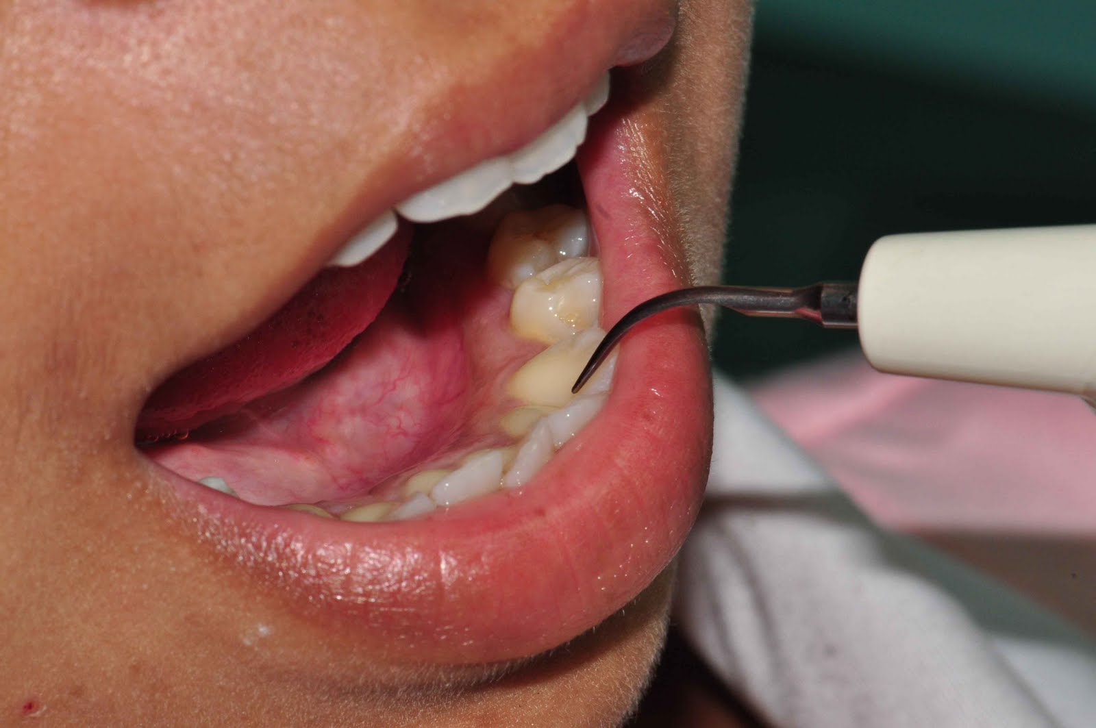
if not, you teeth is going to end up like these
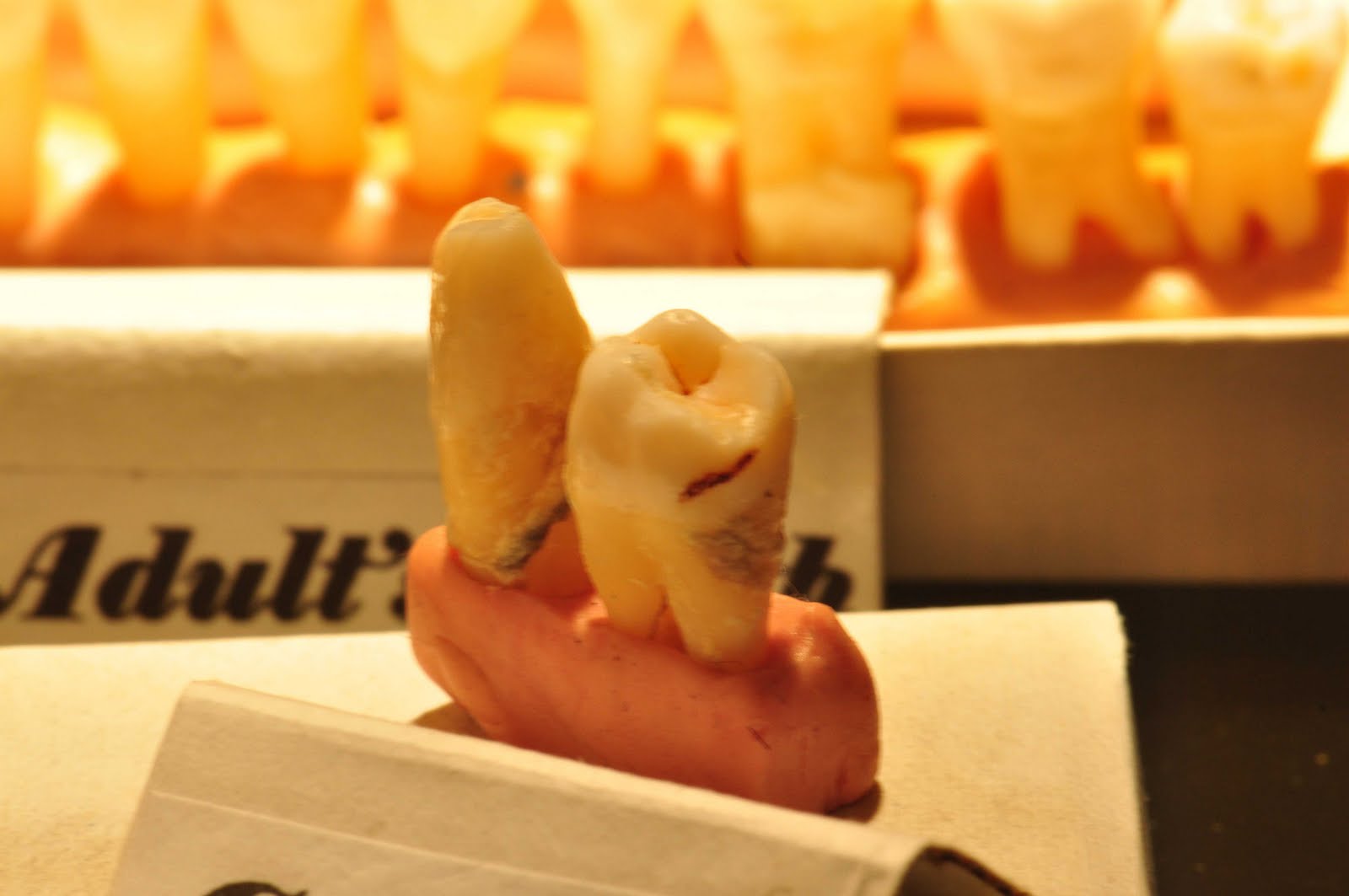
The tartar that attach to your teeth surface will lead to gum inflammation (gingivitis) and later; gum recession and bone resorption (periodontitis). Then finally… teeth become shaky (mobile) and drop out by themselves!! -too late!!
2. To have your teeth checked. Almost all tooth decay started without any symptom (eg. pain, sensitive). It takes about a year to grow significantly big. And treatment for SMALL DECAY is very simple — SMALL FILLING which is cheaper, painless and lasting.
However, when the tooth decay is too big (usually it gives you sensitive, pain or disturb your sleep), then the approach is different. The tooth might required large filling — uhh!! (expensive, painful, required injection) or maybe root canal treatment followed by a crown (because the pulp is infected) or maybe extraction!!
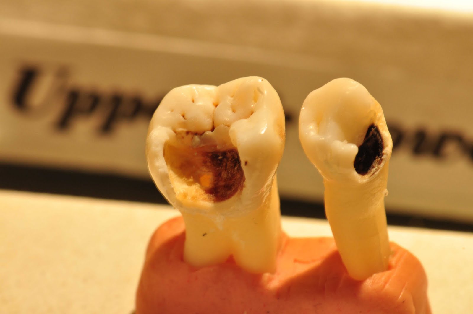
Example of a very large tooth decay
Scaling & Polishing
The simplest method of stain removal, eliminates many food stains, the majority of which can be attributed to coffee or tea. However, excessive staining from beverages or tobacco products may require more aggressive treatment.
It utilizes a strong oxidizing agent to lighten the teeth, is a relatively conservative and often highly effective way to brighten the smile. Bleaching is frequently used on mild-to-moderate tetracycline and fluoride stains, as well as on those stains that result from damage to the tooth. Your dentist may recommend either in-office or home bleaching or a combination of both, depending on the nature and the severity of the discoloration.
Bonding
Involves the application of a plastic material to the existing tooth. The composite resin bonding technique is used frequently to cover stains in a conservative and esthetically pleasing manner. This process can be particularly effective in masking tetracycline stains, white or brown spots, stains due to excessive wear and in lightening teeth that have become discolored by amalgam, the blend of metals found in silver fillings.
Treatment methods in periodontics depend upon the type of disease and how far the condition has progressed. The emphasis in our practice is conservative periodontal therapy. Many times, the early stages of periodontal disease are best treated with non-surgical periodontal therapy. This usually consists of good oral hygiene training, scaling and root planing and irrigation with medication, and, if needed, antibiotic therapy.Scaling and root planing is done under local anesthesia and involves the use of special instruments which go beneath the gum line to effectively remove calculus, debris and necrotic root surface. These procedures are often performed by our hygienist. 4 to 6 weeks or later, the areas are reevaluated for changes such as decreased bleeding and probing depth reduction.

