Spine

The following information is intended as a resource and should not be used to self-diagnose or treat. Use of non-steroidal anti-inflammatory drugs (NSAID’s) may be used to reduce inflammation and pain associated with that inflammation. Dancers should be aware that dancing while taking NSAID’s can mask pain, which can lead to further tissue injury.
Structure
The spine is made up of 24 moveable segments and 9 fused segments (at the bottom). The upper segments are called cervical vertebrae, the segments around the rib cage are called thoracic vertebrae, and the lower back segments are called the lumbar vertebrae. The two lower, fused segments of the spine are the sacrum and the coccyx. Due to the extreme ranges of motion and artistic demands placed on dancers, the lumbar spine vertebrae typically are the most often injured segments. Dancers can also sustain injuries to the sacrum, particularly with the joint articulation between the sacrum and the lowest lumbar spine segment, or with the sacrum and the two pelvic bones.
Common Injuries
Low Back Strain
Kissing Spines
Schuermann’s Disease
Spondylolysis
Spondylolithesis
Herniated Disc
Sacroiliac Joint Sprain
Back Injury Prevention/Tips
Low back muscle strain and spasm
” I pulled something in my back.”
Causes
Muscle strains and lumbar sprains are the most common causes of low back pain. A low back muscle strain occurs when the muscle fibers are abnormally stretched or torn. Causes can include an acute injury such as lifting a heavy object or a sudden movement or fall. Other causes include repetitive injuries such as improper technique or working on the same lift over and over. Muscle tearing such as this will lead to guarding and spasm of the back musculature to protect the area from further harm. Dancers will typically experience pain exclusively in the low back area.
Treatment
Dancers will do well with conservative treatment of low back strains and spasm. Initial treatment will include rest, ice, and anti-inflammatory medicines. A consult with a physical therapist can help identify areas of weakness, tightness, or postural faults that may have predisposed the dancer to injury. The dancer may also want to critically look at any technical faults including lifting technique to help prevent future injury.
Kissing spines – interspinous sprain
” I have pain when I overarch my back.”
Kissing spines is a term for a condition in which the spinous processes of adjacent vertebra are touching. It is also known as Baastrup’s disease or syndrome.
Causes
Kissing spines can either be caused by trauma or degenerative factors. Injuries that involve sudden, forceful flexion of the spine, such as driving accidents, falls, sudden torsions, or severe direct blows can be causative factors. It can also be caused by degenerative changes in the interspinous ligaments along the tips of the spinous processes of the vertebrae. It can affect the cervical vertebrae, but in dancers it commonly affects the lower lumbar vertebrae. Dancers will typically notice pain and limitation with both extension and flexion motions.
Treatment
Initially, ice and rest are indicated to reduce local tissue inflammation and swelling around the injured tissue. A physician may recommend anti-inflammatory medication to assist with pain and edema. A physical therapist consult is also valuable to help the dancer regain strength and mobility deficits. The dancer should also be instructed in proper body mechanics with everyday tasks (e.g., getting in/out of bed) to ensure no further unnecessary stress is applied to the injured area. Symptoms usually decrease after 3 days and should subside between 1-6 weeks. A safe return to full class or performance is ideally only possible when the dancer feels neither paior discomfort, so that muscles can react and perform appropriately. Any pain-avoiding behavior caused by remaining symptoms could place the patient at risk for re-injury.
Schuermann’s disease
” My back hurts at the end of the day.”
Scheuermann disease (also known as juvenile kyphosis) is a deformity in the thoracic or thoracolumbar spine in children. It involves a degeneration of bony segments of the spine, gradually increasing to the point where the natural curvature of the spine begins to change.
Causes
The exact cause of the disease is not known. Some attribute the disease to trauma to the growing spine or hormonal and nutritional deficiencies. Parents of dancers will typically notice a change in their child’s posture, usually a flattening out or rounding of the spine. In later stages, there will be tenderness over the spinous process segments on the back of the spine. Dancers will typically complain of backache at the end of physically strenuous days.
Treatment
A physician will typically confirm the diagnosis of Schuermann’s disease with an X-Ray. The major goal of management is to prevent progression of the disease and further curving of the spine. In the early stages of the disease, extension exercises and postural education are beneficial. A consult with a physical therapist can help identify areas of muscle weakness or tightness that the dancer may need to improve. Bracing, rest, and anti-inflammatory medication may also be helpful to decrease pain. In most cases, the dancer may continue with class and rehearsals, but should avoid painful movements. Swimming may allow the dancer to maintain a strength training and conditioning regimen without putting excessive stress on the back. Surgery is seldom needed except in the most severe cases. In these cases, the spinal column is fused, or joined together where necessary.
Technical Tip:
Unfortunately there is no way to prevent this disease occurring in the young dancer. However kyphosis or curvature of the spine can occur later in life as a result of osteoporosis, so maintaining good bone health by eating well, and taking in enough calcium can be helpful in preventing osteoporosis.
Spondylolysis
” My back hurts when I arch.”
Spondylolysis is the occurrence of a stress fracture in one or more of the vertebrae of the lumbar spine. (See diagram below) It commonly begins on one side of the vertebrae, and then may extend to the other side.
Causes
Spondylolysis can have a hereditary component, but also is attributed to repeated stress to the lumbar spine. Activities such as dance and gymnastics put a great deal of stress on the lower back and require over-stretching or hyperextension of the spine. Dancers may notice no symptoms until there is sudden trauma, such as a hyperextension injury. Pain will typically occur with port de bras or cambré backwards. The dancer may notice pain initially only with dancing. Pain may then occur with normal activities of daily living, and further progress to pain which interferes with sleep.
Treatment
Physicians can diagnose spondylosis with an x-ray to the lumbar spine. Dancers will likely be required to reduce their activity level and/or modify their technique in class. For severe cases, a short period of bed rest can be beneficial. Tissue healing can take as long as 2-3 months. During this time, participation in activities such as swimming, biking and limited weight lifting is usually permissible as long as it is pain-free. Physicians may prescribe a brace such as the modified
Spondylolisthesis
” I have back and buttock pain when I arch back.”
Spondylolisthesis is the forward slippage of a vertebra on the one below. (See diagram below) It commonly will be present with spondylolysis and is typically seen in girls more than boys.
Causes
Causes of spondylolisthesis include stress fractures (caused by repetitive hyper-extension of the back), and traumatic fractures caused by a direct force or sudden twist. The dancer will typically complain of localized pain or a pain that radiates into both buttocks, stiffness in the lower back, and increased irritation after activity. Dancers with spondylolisthesis usually display a significant lumbar spine curvature (lordosis) with tightness in the hamstrings.
Treatment
Treatment varies depending on the severity of the spondylolisthesis. Most dancers require only strengthening and stretching exercises issued by a physical therapist, combined with activity modification (avoiding hyperextension of the back). Some physicians recommend the use of a rigid brace to assist with stabilization of the joint. Conservative therapy for mild spondylolisthesis is successful in about 80% of cases. For cases with severe paiot responding to therapy, if the slip is severe, or there are neurologic changes, the slipping vertebra might need to be surgically fused. This surgery will limit lumbar spine range of motion and has a higher incidence of nerve injury than most other spinal fusion surgeries. Therefore surgery is only considered after all conservative treatments have been exhausted.
Herniated Lumbar Disc
” I have low back pain and pain occasionally shoots down my leg.”
Between each vertebrae are discs, made up of a combination of strong connective tissues which hold one vertebra to the next. These discs act as a cushion between the vertebrae. As individuals age, the center portion of the disc (nucleus pulposus) may start to lose water content, making the disc less effective as a cushion. This may cause a displacement of the disc’s center through a crack in the outer layer (known as a herniated or ruptured disc). A herniated lumbar disc can ultimately press on the nerves in the spine and may cause pain, numbness, tingling or weakness of the leg called ” sciatica” .
Causes
A disc herniation may occur suddenly in an event such as a fall or an accident. Often, a twisting or torsional movement is involved. Disc problems may also occur gradually with repetitive straining of the lumbar spine.
Symptoms
Most commonly, dancers will experience low back pain, but also leg pain over the outside of the thigh, the lower leg, or foot. The pain is often described as an electric shock type of symptom.
Severe cases of herniated lumbar disc injury will appear as bowel or bladder problems. Individuals with bowel or bladder complaints or who are having numbness around the genitals require immediate medical attention.
Treatment
An evaluation by a physician and physical therapist is critical to resolution of the dancer’s symptoms. The physician may request an x-ray or MRI to identify the location and severity of the disc herniation. Anti-inflammatory medications may be prescribed to assist with acute pain and local edema. A physical therapist will determine where physical deficits exist and instruct the dancer on postural corrections and activity modifications that might need to be made.
Conservative management of a herniated disc can often be sufficient to allow a dancer to return to full activity. If conservative management fails, surgical treatment may be recommended if there is a significant neurological component (i.e. leg weakness or numbness). Surgery is performed to remove a portion or all of the disc, and free up space around the compressed nerve. Recovery times from disc surgery vary from person to person, but a dancer should expect to have activity restrictions for 6-8 weeks following surgery.
Sacroiliac Joint Sprain
” I have pain low in my back, especially when I lie on my side.”
The sacroiliac joint is a firm, small joint that lies at the junction of the spine and the pelvis. The joint does not have a lot of motion, but it is critical to transferring the load of your upper body to your lower body and can become quite painful when injured.
Causes
Certain situations increase the risk of straining the sacroiliac joints. During pregnancy, the ligaments in the sacroiliac area soften and lengthen. This may also occur with prolonged bending or lifting and with degenerative arthritis. In dancers, potential for sacroiliac injury is significant due to the extreme ranges of motion and artistic demands placed on dancers. Dancers with sacroiliac pain may or may not recall a method of injury. Symptoms may present over the sacroiliac joint, or it may be referred, usually to the groin and the posterior thigh, and less often to the leg. Pain may become worse when they lie on the affected side
Treatment
During the acute phase of injury, pain may be relieved by rest and anti-inflammatory medication. Physical therapy to assist with joint mobilization and stretching and strengthening exercises can be very helpful. As with any ligamentous injury, a period of decreased intensity of class or rehearsals may be required for healing. Dancers are nearly always able to return to their usual daily routine after a few days or, at most, a few weeks of therapy.
Prevention / Tips for dancers
- Muscular imbalances or weaknesses of abdominal and posterior spinal muscles may constitute a risk factor to sustain an injury. Keep the abdominal and back muscles strong and the hamstring muscles flexible to help avoid back injury.
- The stabilization of the spine depends on appropriate and fast muscle reactions to suddenly changing postures of the spine. Proprioceptive training of the trunk muscles is a vital component in rehabilitation of low back injuries.
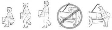
- A good upright posture while standing, sitting, and lifting during everyday life and implementing exercising routines takes unnecessary strain off the spinal structures and help avoid injury.
- Try to limit the amount of dancing each day, especially repetitive movements such as back bending – this will help prevent overuse injuries.
- Make sure you always fully warm up before class, rehearsal or performance.
- Try to maintain careful technique, and resist temptation to ‘cut corners’ to achieve movements such as forcing turnout, or tilting the pelvis.
- It is particulalry important to remember to maintain correct technique in positions which stress the spine, such as arabesque and attitude, and being aware of ‘lengthening’ the torso during any back-bending movements.
- Seek medical care for chronic back pain. Early vertebral stress fractures, particularly in adolescents and young adults, may heal with rest.
- Aerobic fitness can increase blood flow and oxygenation to all tissues, including the muscles, bones, and ligaments of the spine. Dancers should be encouraged to cross-train year round to maintain aerobic fitness.
Symptoms and Treatment for Common Hip, Pelvic & Back Injuries
Greater Trochanteric Bursitis
Greater trochanteric bursitis is an inflammation of the trochanteric bursa, a specific part of the hip. This bursa has the function of a shock absorber and a lubricant for the movement of the muscles adjacent to it. When the bursa becomes irritated or inflamed, it causes pain in the hip. Symptoms usually include pain on the outside of the hip, pain when lying on the affected side, pain when you press on the outside of the hip, or pain when walking up stairs. Treatment mainly includes rest, nonsteroidal anti-inflammatory medication, cortisone shots, physical therapy, or a bursectomy surgical procedure.
Groin Strain (Adductor Muscle Tear)
A groin strain is a tear or rupture of one of the adductor muscles. There are five adductors in which function to pull the legs back toward the midline. A rupture or tear in the muscle usually occurs when sprinting, changing direction or in rapid movements of the leg against resistance. There are 3 different grades of strain with different symptoms for each, but the main symptoms may include discomfort or pain in the groin, tightness of the groin muscles, bruising, swelling, or pain during movement. Treatment may include RICE (rest, ice, compression, and elevation), crutches, stretching, taping, sports massage, or a surgical operation if the strain is severe enough.
Hip Flexor Strain
The hip flexors are made up of three different muscles all working together to provide stability for the lower extremities. Hip flexor strain is a muscle strain felt in the front part of the hip. This injury is often associated with speed training or compensating for another injury, especially Achilles tendonitis or plantar fasciitis. Other causes could be from acute trauma, tight muscles, or poor flexibility. Symptoms may include pain along the front of your hip and thigh, difficulty running, jumping or walking, or a small amount of swelling. Treatment can include rest, ice, and gentle stretching.
Hip Fracture
A hip fracture is a break in the upper quarter of the femur (thigh) bone. If the actual hip socket is broken, the injury is considered to be completely different with different methods of symptoms and treatment. Hip fractures almost always occur from a fall or a direct blow to the side of the hip. A patient with a broken hip will experience pain over the outer upper thigh or in the groin. Treatment options usually include open reduction internal fixation surgery. Open reduction internal fixation (ORIF) is a method of surgically repairing a fractured bone. Generally, this involves either the use of screws and plates or an intramedullary (IM) rod to stabilize the bone. The type of surgery used to treat the fracture usually depends on which type of fracture a patient is experiencing.
Hip Pointer
A hip pointer is a contusion to the iliac crest of the pelvis, the soft-tissue structures surrounding the femur, or the greater trochanter of the femur. The term ‘hip pointer’ refers to the deep bruising of the muscle and bone. Typically, the injury is caused by a direct blow or fall. Treatment options may include RICE (rest, ice, compression and elevation), physical therapy, or nonsteroidal anti-inflammatory medication. Surgery is rarely required.
Lumbar Herniated Nucleus Pulposus (HNP)
Spinal stenosis is the term used when the spinal canal becomes narrowed due to normal wear-and-tear effects of aging. When the space around the spinal cord narrows pressure is put on the spinal cord and spinal nerve root causing pain, numbness, or weakness in the legs. Lumbar spinal stenosis occurs when the injury takes place in the lower back. Arthritis, the degeneration of any joint in the body, is the most common cause of spinal stenosis. Symptoms may include back pain, numbness or burning pain in buttocks or legs, weakness in the legs, or a sudden relief of pain while leaning forward or sitting. Nonsurgical treatment may include physical therapy, lumbar traction, anti-inflammatory medications, steroid injections, acupuncture, or chiropractic manipulation. Surgical options may include a laminectomy or a spinal fusion.
Lumbar Osteoarthritis (Degenerative Joint Disease)
Spinal vertebrae are separated by cartilage disks filled with a gelatinous substance, which provide cushioning to the spinal column. Lumbar HNP, or slipped disk, is a condition in which part, or all, of the soft, gelatinous central portion of an intervertebral disk is forced through a weakened part of the disk. This results in back pain and leg pain due to nerve root irritation. This nerve damage may cause neurological symptoms such as sensory or motor changes. Other symptoms may include sharp pain in one part of the leg, hip or buttocks, numbness, weakness, or tingling sensations. Treatment may include rest, anti-inflammatory medication, physical therapy, steroid injections, or surgery.
Lumbar Spinal Stenosis
Degenerative joint disease, also referred to as osteoarthritis, is a syndrome in which a painful disc causes chronic lower back pain. The condition generally starts with a twisting injury to the disc space. The injury weakens the disc, inflames the proteins inside the disk, and begins to expose and irritate the disk which produces the pain back. For most people, this syndrome can be successfully treated with conservative care. Symptoms may include stiffness and pain in the lower back, swelling, warmth, or tenderness. Treatment may include hamstring stretching, dynamic lumbar stabilization exercises, low-impact aerobic conditioning, medication, oral steroids, epidurals, or lumbar spinal fusion surgery for extreme cases.
Osteoarthritis (Degenerative Joint Disease) of the Hip
Osteoarthritis of the hip occurs when the smooth cartilage on the ends of the hip bones, which help your hip joint glide, begin to wear thin. Those who have a family history of the disease are more likely to experience it; also those that are elderly, obese, or have an injury that puts pressure on the hip cartilage are more likely to develop the condition. Symptoms may include discomfort or stiffness in the groin, buttock, or thigh. As the disease progresses, the more painful movement will become. Nonsurgical treatment can include rest, physical therapy, nonsteroidal anti-inflammatory medication, or weight loss. If the condition is in late stages, a total hip replacement (arthroplasty) may be recommended.
Piriformis Syndrome
The piriformis muscle is one of the external rotators of the hip and leg. This means that as the muscle works, it helps to turn the foot and leg outward. When the piriformis muscle squeezes and irritates the sciatic nerve, piriformis syndrome develops. Piriformis syndrome is a neuromuscular disorder that occurs when the sciatic nerve is compressed or irritated by the piriformis muscle. Symptoms can include pain that radiates down the back of the leg, difficulty sitting, or a tingling sensation down the leg. Treatment usually begins as nonsurgical: steroidal anti-inflammatory medication, physical therapy, or botulism injection therapy. Surgery may be used as a last resort.
Sacroiliac (SI) Joint Dysfunction
The sacroiliac joints connect the spine to the pelvis, formed by the connection of the sacrum and the right and left iliac bones. The SI joints have a cartilage layer covering the bone which acts as a shock absorber between the bones. When the cartilage is damaged or worn away the bones begin to rub on each other, causing SI joint dysfunction. Other causes of this condition include pregnancy and increased stress on the SI joints. Symptoms can include pain in the lower back, groin or thighs. Pain is typically worse with standing and walking, and improved when lying down. Treatments may include cortisone injections, anti-inflammatory medications, and physical therapy. If pain persists, surgery may become an option.
Neuromuscular scoliosis
![]()
Disease Information
Overview In-Depth Tests Treatment
& Care Research &
Innovation Your
Story Contact Us
In-Depth
At Children’s Hospital Boston, our Spinal Program team develops innovative treatments for scoliosis and other spine defects. We’re home to the world’s most extensive pediatric hospital research enterprise, and we partner with elite health care and biotech organizations around the globe. But as specialists in innovative, family-centered care, our physicians never forget that your child is precious, and not just a patient.
In dealing with your child’s neuromuscular scoliosis, you may want to know the basics about the spine and about the several forms of this spinal condition.
What is the spine?
Spine viewed from the front and side
Made up of many individual bones called vertebrae, the normal spine is joined together by muscles and ligaments. Flat, soft discs separate and cushion each vertebra from the next. Because the vertebrae are separate, the spine is flexible and can bend. Together the vertebrae, discs, muscles and ligaments make up the spine or vertebral column.
Various regions of the spine are named differently: The cervical spine refers to the neck, the thoracic spine to the chest, and the lumbar and sacral spines to the lower back.
What is scoliosis?
Although the spine has natural curves from front to back, it shouldn’t curve sideways very much. A side-to-side curve is called scoliosis, and may take the shape of an abnormal “S” (double curve) or a long “C” (single curve). The scoliotic spine is also rotated or twisted, pulling the ribs along with it to form a multi-dimensional curve. In serious cases, lung and heart function can be affected, as well as other organ systems.
What are the three main types of scoliosis?
Scoliosis occurs, and is treated, as three main types:
- neuromuscular scoliosis: the second-most common form of scoliosis, associated with disorders of the nerve or muscular systems like cerebral palsy, spina bifida, muscular dystrophy or spinal cord injury.
- congenital scoliosis: Congenital scoliosis results from a fetus’ abnormal spinal development in utero, such as a partial or missing formation or a lack of separation of the vertebrae.
- idiopathic scoliosis: the most common form of scoliosis, most commonly seen in adolescent and pre-adolescent girls. “Idiopathic” simply means that there is no definite cause. Most cases require no intervention.
What are some of the defining characteristics of neuromuscular scoliosis?
Some characteristics of neuromuscular scoliosis include:
- occurs in children with underlying neuromuscular disease
- usually a “C”-type curve
- onset much younger than many other forms of scoliosis
- more common and severe ion-ambulatory patients (those whose neuromuscular disease prevents them from walking)
- curve virtually always progressive; progression can continue into adulthood
- progresses (worsens) more during growth spurts
- progressing (worsening) curve can lead to collapsed torso and raised diaphragm, reducing lung space, impeding lung function and increasing risk of pneumonia; more severe effect if lung function is already compromised by weak muscles that control breathing (as in muscular dystrophy and high-level spinal cord paralysis)
- many organ systems involved due to underlying nerve or muscular disease, which can cause:
- contractures of hips and knees
- vision and hearing loss
- lung function impairment
- seizures
- bracing useful for functional support but rarely stops the curve
- commonly requires spinal fusion surgery ion-ambulatory, severe patients by adolescence, usually sooner than with idiopathic scoliosis
- fused portion of the spine is longer than with idiopathic scoliosis—often extends to sacrum and pelvis
- may require both anterior and posterior surgeries
- some posterior elements may be missing from spine (as in spina bifida)
- helps achieve a spine with torso in balance over pelvis (usually ion-ambulatory patients)
- spine surgery’s goals are to:
- stabilize the curve and stop its progression
- balance the spine and pelvis (usually in non-ambulatory patients)
- regain the ability to sit upright (in children who have lost this ability)
- improve/preserve lung function
What are the signs and symptoms of neuromuscular scoliosis?
You, your child’s doctor or his caregiver will most likely notice his spinal curve because of a deteriorating ability to sit or a change in his body’s overall position:
- he may begin leaning toward one side of his seat in an uneven seating posture
- he may increasingly need to use his arms for seating support
You may notice that his:
- shoulders are of uneven heights
- head isn’t centered with the rest of his body
- hips are of uneven heights or positions, or uneven buttocks
- shoulder blades are of uneven heights or positions
- arms hang beside his body unevenly
- left and right sides of his back appear different in height when he bends forward
How do you diagnose neuromuscular scoliosis?
Doctors will use medical and family histories, physical exams and diagnostic tests to determine the nature, extent and effects of your child’s scoliosis and underlying neuromuscular disease. Your Children’s multi-disciplinary care team will evaluate his nutritional status, lung and heart function, functional level, joints and extremities (for contractures), balance and ambulatory (walking) level. Testing can include:
- x-rays
- magnetic resonance imaging (MRI)
- computerized tomography scan (CT or CAT scan)
- blood tests
- ultrasound (sonogram)
- bone scans
- bone density scans (dual-energy x-ray absorptiometry, DEXA, DXA)
- muscle biopsy
- pulmonary function tests
- Electromyogram (EMG) and nerve conduction studies
How do you treat neuromuscular scoliosis?
Most neuromuscular conditions are diagnosed early in a child’s life, and neuromuscular scoliosis may also manifest early. When it appears, your child’s team will use non-surgical treatments such as bracing, wheelchair modification, physical therapy and environmental adaptation to help your child adapt to his everyday environments, and to help improve his mobility.
While most non-ambulatory pre-adolescent children are usually treated with bracing to control (not correct) spinal curves while they grow, stabilization by spinal fusion surgery is the most common treatment for neuromuscular scoliosis by the time a child reaches adolescence. But treatment will vary according to the type and severity of a child’s neuromuscular condition.
What if the curve is severe in my very young child before he’s ready for spinal fusion?
A young child with a severe early-onset curve can be treated with growth-friendly (growth preserving) procedures such as growing rods and/or VEPTR. The metal rods inserted in these procedures can help control the curve until he’s ready for spinal fusion. The rods are made longer as the child’s spine grows.
What are the benefits of spinal fusion surgery?
A spinal fusion aims to:
- stabilize the curve and stop its progression
- balance the spine and pelvis (usually in non-ambulatory patients)
- regain the ability to sit upright (in children who have lost this ability)
- improve/preserve lung function
If my child needs surgery, what will happen beforehand?
Before surgery, your child’s surgical team will assess his:
- lung and heart function
- nutritional and protein status
- likelihood of post-operative feeding difficulties
- neurologic status and seizure medications
- urologic status
- bone health
- swallowing and regurgitation evaluation (for cerebral palsy patients)
- clotting and bleeding evaluation (for children subject to seizures, since some seizure medications can affect blood clotting)
Will there be complications after the surgery?
Because neuromuscular scoliosis is associated with other underlying neuromuscular conditions, the complication rate after surgery is higher than with some other types of scoliosis. Complications are common, anticipated and treatable. They can include:
- respiratory problems: Post-operative pneumonia is a risk for a child with a poor cough reflex or some paralysis of the chest (intercostal) muscles.
- wound problems: Skin breakdown and infection can occur.
- nutritional/feeding problems: A feeding tube may be necessary until the child can feed orally again.
- hip problems: Many children who have spinal fusion also have stiff or abnormal hips. Fusing the spine may make existing hip contractures more troublesome.
- intestinal obstruction (ileus): This can result from intestinal sluggishness (hypomotility).
Who will be on my child’s scoliosis treatment team at Children’s?
Your child’s scoliosis team at Children’s may include his doctor, an orthotist (a specialist who makes braces), a physical therapist and a nurse, who will guide you through the treatment process. Specialists, such as neurologists and physiatrists as well as social workers, speech/language specialists and others—will be part of the team as needed to address your child’s underlying neuromuscular condition.
As part of our family-centered approach, your child’s nurse will help with all your questions and appointments. The nurse can also:
- help you understand how best to care for your child, his brace, wheelchair, orthotics or other equipment
- design a schedule for you and him to follow
- help you meet others whose children have neuromuscular disease with scoliosis—in person and/or online
Will my child be OK?
The outlook for your child mostly depends on the nature, severity and effects of his neuromuscular disease, and less on his scoliosis.
With successful surgery and attentive post-operative care, your child can:
- return to the functional level he had attained before surgery
- have his spine solidly fused and in balance, reducing the deformity
- have the potential for improved lung function and decreased susceptibility to pneumonia
- find it easier to sit up; requirements for seating adaptation will usually be improved
Children’s Hospital Boston’s research into spinal problems—including neuromuscular scoliosis—means that we will provide your child with the most innovative care available.
FAQ
Q: What is scoliosis?
A: Scoliosis is a condition in which the spine, in addition to the normal front to back curvatures, has an abnormal side-to-side “S-” or “C”-shaped curvature. The spine is also rotated or twisted, pulling the ribs along with it. Sometimes, a child’s lung function can be compromised when the curvature is severe or starts early in life.
Q: What is neuromuscular scoliosis?
A: Neuromuscular scoliosis is the form that’s associated with another, underlying condition involving the nerves and/or muscles, such as cerebral palsy, spina bifida, myopathy, muscular dystrophy and paralysis from spinal cord injury.
Q: If my child has neuromuscular scoliosis, will he be OK?
A: The outlook for your child greatly depends on the nature, severity and effects of his scoliosis and his underlying neuromuscular disease. With successful surgery and attentive post-operative care, your child can return to the functional level he had attained before surgery. His spine will be solidly fused and in balance—usually reducing the deformity and improving his breathing.
Q: How does Children’s treat neuromuscular scoliosis?
A: Most neuromuscular conditions are diagnosed early in a child’s life, and neuromuscular scoliosis may also present early. When it appears, your child’s team will use non-surgical treatments such as bracing, wheelchair modification, physical therapy and environmental adaptation to help your child adapt to his everyday environments, and to help improve his mobility.
While most non-ambulatory pre-adolescent children are usually treated with bracing to control (not correct) spinal curves while they grow, stabilization by spinal fusion surgery is the most common treatment for neuromuscular scoliosis by the time a child reaches adolescence. But treatment will vary according to the type and severity of a child’s neuromuscular condition.
Q: What are the signs and symptoms of neuromuscular scoliosis?
A: You, your child’s doctor or his caregiver will most likely notice his spinal curve because of a deteriorating ability to sit or a change in his body’s overall positioning.
Q: How is scoliosis usually diagnosed?
A: Doctors will use medical and family histories, physical exams and diagnostic tests to determine the nature and extent of your child’s scoliosis and underlying neuromuscular disease. Your Children’s multi-disciplinary care team will evaluate his nutritional status, lung and heart function, functional level, joints and extremities (for contractures), balance and ambulatory (walking) level. Testing can include:
- x-rays
- magnetic resonance imaging (MRI)
- computerized tomography scan (CT or CAT scan)
- blood tests
- ultrasound (sonogram)
- bone scans
- bone density scans (dual-energy x-ray absorptiometry, DEXA, DXA)
- muscle biopsy
- pulmonary function tests
- electromyogram (EMG) and nerve conduction studies
Q: What should I expect after my child has spinal fusion surgery for neuromuscular scoliosis?
A: After surgery, most patients go to the ICU (intensive care unit), where some may be put on a ventilator, and some may require a longer stay than others. Your child’s hospital stay will probably be between five and 10 days.
During your child’s hospital recuperation, his Children’s caregivers will closely monitor his lungs, fluids and nutrition. They will get him up and moving as soon as possible, and will extend his time upright a little more each day. His wheelchair will be modified for his improved sitting position.
Q: What’s the long-term outlook for my child with neuromuscular scoliosis?
A: With successful surgery and attentive post-operative care, your child can:
- return to the functional level he had attained before surgery
- have his spine solidly fused and in balance, reducing the deformity
- have the potential for improved lung function and decreased susceptibility to pneumonia
- find it easier to sit up; requirements for seating adaptation will usually be minimized
His longer-term outlook greatly depends on the extent and severity of—and the prognosis for—his underlying neuromuscular condition.
Q: Will a straighter spine after surgery improve the function of my child’s organ systems?
A: A straighter spine can improve your child’s lung function and decrease his vulnerability to pneumonia. It can reduce deformity and help him with his sitting posture. And sometimes, a straighter spine will improve the organ function of a child with neuromuscular disease—but not always. (Rarely, a straighter spine can actually be detrimental to function.) However, not correcting his curve surgically will almost always result in breathing or positioning problems later on.
Q: What is Children’s experience treating neuromuscular scoliosis?
A: At Children’s Spinal Program, we’re known for our clinical innovations, research and leadership. We offer the most advanced diagnostics and treatments—several of which were pioneered and developed by Children’s researchers and clinicians.
Q: What are Children’s spine research and innovations?
A: Children’s Orthopedic Clinical Effectiveness Research Center (CERC) helps coordinate research and clinical trials to improve the quality of life for children with musculoskeletal disorders, such as scoliosis. CERC physicians are pursuing several areas of basic and clinical research based at Children’s and the Harvard Orthopaedics Biomechanics Laboratory.
For details see Children’s spine Research & Innovations.
Causes
Neuromuscular scoliosis is associated with an underlying neuromuscular condition such as cerebral palsy, muscular dystrophy, spina bifida, myopathy or paralysis from spinal cord injury.
Signs and symptoms
You, your child’s doctor or his caregiver will most likely notice his spinal curve because of a deteriorating ability to sit or a change in his body’s overall position:
- he may begin leaning toward one side of his seat in an uneven seating posture
- he may increasingly need to use his arms for seating support
You may notice that his:
- shoulders are of uneven heights
- head isn’t centered with the rest of his body
- hips are of uneven heights or positions, or uneven buttocks
- shoulder blades are of uneven heights or positions
- arms hang beside his body unevenly
- left and right sides of his back appear different in height when he bends forward
When to seek medical advice
Because your child has an underlying neuromuscular disease, your doctor and caregivers will be evaluating him frequently. His scoliosis will usually become apparent as the result of changes in his sitting ability and/or body position. Consult your child’s doctor if you notice that he is:
- leaning toward one side of his seat in an uneven seating posture
- increasingly needing to use his arms for seating support
Also consult the doctor is you notice that his:
- shoulders are of uneven heights
- head isn’t centered with the rest of his body
- hips are of uneven heights or positions
- shoulder blades are of uneven heights or positions
- arms hang beside his body unevenly
- left and right sides of his back appear different in height when he bends forward
Questions to ask your doctor
If your child is diagnosed with neuromuscular scoliosis, you may feel overwhelmed with information. It can be easy to lose track of the questions that occur to you. Lots of parents find it helpful to jot down questions as they arise—that way, when you talk to your child’s doctors, you can be sure that all of your concerns are addressed.
Some of the questions you may want to ask include:
- What is happening to my child, and why?
- Are other tests needed to diagnose my child?
- What actions might you take after you reach a diagnosis?
- What will happen with growth over time?
- Will there be restrictions on my child’s activities?
- Will there be long-term effects?
- What can we do at home?
Who’s at risk
Children most at risk for developing neuromuscular scoliosis include:
- non-ambulatory children with quadriplegic cerebral palsy
- children who have spina bifida with a high level of paralysis
- children with spinal cord paralysis before age 10 years
Complications
Because neuromuscular scoliosis is associated with other underlying neuromuscular conditions, the complication rate after surgery is higher than with some other types of scoliosis. The complications are common, anticipated and treatable. They can include:
- respiratory problems
- wound problems
- nutritional/feeding problems
- hip problems
- intestinal obstruction (ileus)
Long-term outlook
With successful surgery and attentive post-operative care, your child can:
- return to the functional level he had attained before surgery
- have his spine solidly fused and in balance, reducing the deformity
- have the potential for improved lung function and decreased susceptibility to pneumonia
- find it easier to sit up; requirements for seating adaptation will usually be minimized
His long-term outlook greatly depends on the extent and severity of—and the prognosis for—his underlying neuromuscular condition.
For teens
Besides the typical issues any teenager faces—from social acceptance to body changes and more—if you have a neuromuscular disease that includes scoliosis, it’s true that you’ll also have to deal with medical appointments, feeling different and assuming a big personal responsibility for maintaining your health. You may also wonder why you need surgery.
It’s important for you to know that surgery for your scoliosis will fuse your spine to correct the curve and prevent further curvatures. You’ll be able to sit more upright in your wheelchair. You’ll also probably breathe better and be less susceptible to respiratory problems.
Even knowing the benefits of surgery and treatments, this can be a tough time for you. If you feel overwhelmed, depressed or anxious during this important time in your transition to adulthood, speak to your doctor or counselor to get help.
What to expect after your child’s surgery
After surgery, most patients go to the ICU (intensive care unit), where some may be put on a ventilator, and some may require a longer stay than others. Your child’s hospital stay will probably be between five and 10 days. Your child’s caregivers will closely monitor his lungs, fluids and nutrition. They’ll get him up and moving as soon as possible, and will extend his time upright a little more each day. His wheelchair will be modified for his improved sitting position.
Prevention
Neuromuscular scoliosis is associated with your child’s underlying neuromuscular disease. Nothing you or your child did caused it, and there’s nothing you could have done to prevent it.
Neuromuscular scoliosis glossary
Adams forward bending test: a screening measure for assessing scoliosis
- ambulatory: pertaining to walking, able to walk
- anterior fusion: spinal fusion surgery on the front of the spine approached from the side of the body; sometimes combined with posterior fusion, usually performed on the same day or in stages
- brace, bracing (spinal orthosis): A brace supports your child’s spine in a balanced position over the pelvis. A bracing program may help delay surgery.
- The Center for Families at Children’s: dedicated to helping families find the information, services and resources they need to understand their child’s medical condition and take part in their care
- Cobb angle: an angular measurement on x-ray to evaluate the severity and degree of scoliosis curves
- congenital scoliosis: a form of scoliosis resulting from abnormal in utero spinal development, such as a partial or missing formation or a lack of separation of the vertebrae
- contractures, joint contractures: joints with limited range of motion and that cannot be fully straightened or bent; frequent among patients who don’t walk; can be a complication in hips after surgery
- diagnosis: identifying disease or injury through examination, testing and observation
- ICU: intensive care unit
- idiopathic scoliosis: the most common form of scoliosis, mainly affecting adolescent girls. “Idiopathic” simply means that there is no definite cause. Nothing you or your child did caused the defect, and there’s nothing you could have done to prevent it.
- instrumentation: the metal rods, hooks, screws and wires implanted during spinal fusion surgery to correct the spinal curve and secure the spine in position while the fusion heals and becomes solid
- neuromuscular: affecting, or characteristic of, both neural (nerve) and muscular tissue
- neuromuscular scoliosis: scoliosis that is associated with disorders of the nerve or muscular systems like cerebral palsy, spina bifida, muscular dystrophy or spinal cord injury
- orthopedics: the medical specialty concerned with diagnosing, treating, rehabilitating and preventing disorders and injuries to the spine, skeletal system and associated muscles, joints and ligaments
- orthopedist, orthopedic surgeon: a physician specializing in surgical and non-surgical treatment of the spine, skeletal system and associated muscles, joins and ligaments
- orthotics: the science of designing and fitting of devices such as braces to treat orthopedic conditions
- physical therapy:a rehabilitative health specialty that uses therapeutic exercises and equipment to help patients improve or regain muscle strength, mobility and other physical capabilities
- posterior fusion: spinal fusion surgery approached from the back of the body; sometimes combined with anterior fusion, performed either simultaneously or in two stages
- progression, curve progression: worsening of a scoliosis curve
- scoliometer: a surface measurement device for evaluating the angle of torso rotation (ATR or scoliometer angle, which is not the ‘Cobb’ angle measured on x-ray)
- scoliosis: a spinal condition in which the spine, in addition to the normal front to back curvature, has an abnormal side-to-side “S-” or “C”-shaped curvature. The spine is also rotated or twisted, pulling the ribs along with it. Scoliosis occurs in three main types: neuromuscular (associated with neuromuscular diseases); congenital (present at birth); and idiopathic (no definite cause).
- seat supports, seating supports: supports for helping to sit upright (usually in a wheelchair) for children with insufficient balance for self-support. Seating supports help children sit upright, but they don’t correct curves.
- spinal cord: a nerve bundle within the vertebral column that extends down from the brain stem; it conducts signals in both directions between the brain and extremities, and allows for bodily motion and sensation
- spinal fusion: usually a solid fusion (solidification) of the curved part of the spine, achieved by operating on the spine, adding bone chips and allowing the vertebral bones and bone chips to slowly heal together to form a solid mass of bone called a fusion. A fusion partially corrects a scoliosis curve, stabilizes the curve and stops its progression, as well as balancing the spine and pelvis (usually ion-ambulatory patients).
- spine (spinal column, vertebral column): the series of moving vertebrae forming the axis of the skeleton and protecting the spinal cord
- VEPTR™ (titanium rib) procedure: an operation that expands the chest and allows continued growth of the chest and spine. A curved metal rod fits the back of the chest and spine, helping the spine to become straighter and allowing the lungs to grow and fill with enough air to breathe. The device can be made longer as your child grows.
- vertebra, vertebrae: the individual spine bones that form the spinal column
Traumatic Pelvic Fractures
Epidemiology
The mechanisms of injury of pelvic fractures include motor vehicle crashes, 57%; pedestrians hit by motor vehicles, 18%; motorcycle crushes, 9%: falls, 9%; and crush injuries, 5% (Failinger & McGanity, 1992; Heetveld, Harris, Schlaphoff, & Sugrue, 2004). Mortality of the patient increases with the severity of the pelvic injury. The patient with a closed pelvic fracture who is hemodynamically unstable has a mortality of approximately 27%, which increases to approximately 55% for open pelvic fractures (Heetveld et al., 2004).
The unstable pelvic fracture can cause the most damage and is life threatening. The “open book” pelvic fracture is a type of fracture, and it will be discussed further in this article. The complications that can occur are massive, such as exsanguination (blood loss), genitourinary trauma, sepsis, chronic pain, bowel damage, neurological damage, and laceration of the vagina (Walsh, 2002).
Anatomy of the Pelvis
The pelvis is a ring structure made up of three bones: the sacrum and two innominate bones. The fusion of three ossification centers, the ilium, the ischium, and the pubis, makes up the innominate bone. The pelvic ring is formed by the connection of the sacrum to the innominate bones at the sacroiliac (SI) joints and the symphysis pubis. The ligamentous structures help with the stability of the pelvis. The strongest and the most important ligamentous structures occur in the posterior aspect of the pelvis at the SI joints. The posterior ligaments have to hold up the weight-bearing forces that occur from the lower extremities to the spine and are transferred to the SI joints (Kellam & Mayo, 2003). The interosseous SI ligaments are considered the strongest in the body. They act to stabilize the SI complex. The symphysis acts more as a supporting “strut” because the SI joint is the stabilizing structure of the pelvic ring. If this “strut” is disturbed, this will not lead to instability of the pelvic ring and surgery (Young & Burgess, 1987).
The organs and structures that can be affected during a trauma within the pelvis are the genitourinary system (bladder and urethra), the vagina, prostate, and gastrointestinal system. The lumbar and sacral plexuses that run posterior can also be affected during pelvic trauma (See Figures 1-3).
|
|
FIGURE 1. Ligamentous anatomy of the pelvis. From |
|
|
FIGURE 2. Neuroanatomy of the pelvis. From |
|
|
FIGURE 3. Vascular anatomy of the pelvis. From |
History and Physical Examination
Diagnostic Tests
Radiology
Radiographic images are obtained for patients who have experienced blunt trauma. It helps to determine the extent of the pelvic fracture. Radiographic images are considered the gold standard in determining a pelvic fracture. An anterior-posterior (AP) view of the pelvis will show most of the pelvic fractures and dislocations. However, this view does not allow one to determine the degree of bone displacement; small fractures can be missed. Inlet and outlet views are also obtained for the pelvis to obtain better images of the fracture (Cwinn, 2002).
The outlet view is taken from the foot of the bed directed at 45[degrees] toward the head. This type of X ray helps in determining the displacement or leg-length discrepancy, sacral fractures, and disruptions of the SI joints. The inlet and outlet X-ray views can be taken by simply angling the beam, thus not having to move the patient (Kellam & Mayo, 2003).
Computed Tomography
A CTscan can also help diagnose associated visceral injuries and hemorrhage.
Classifications of Pelvic Injuries
A classification is essentially a description of the bone injury; it does not define the injury. In defining the injury you need to look at all of the following: degree of displacement; stability of the pelvic ring; force direction; state of the soft tissues, including whether the fracture is open or closed; anatomic injury in the pelvis and the associated injuries (Tile, Helfet, & Kellam, 2003).
At this time there is no universally accepted classification system of determining pelvic fractures. There are different descriptions or classifications of the pelvic ring fractures. Letournel classified the fractures of the pelvic ring anatomically. Bucholz, Tile, and the Orthopaedic Trauma Society classified the fractures by the stability of the fracture. Based on the Pennal classification, Young and Burgess classified pelvic fractures by the mechanism of injury, in other words, how the injury was obtained (Guyton & Crockarell, 2003). This article will focus on the Tile classification and the Young and Burgess classification.
Young and Burgess Classification
According to the Young and Burgess classification, injuries are grouped by mechanism of injury. They show it is important to think of three vectors of force and how this disrupts the pelvic ring. First, the most common vector of force is the lateral compression (LC). The vector force is applied from the lateral side of the pelvic ring from a T-bone (intersectional motor vehicle crash) or in a situation in which a pedestrian is struck from the side or a person lands from a great height on his or her side. Usually, in this type of injury the individual is injured mostly from associated injuries of the abdomen, thoracic contents, and the cervical spine. The force of the vector is not strong enough to open up the pelvis but can cause great damage to the internal organs from fracture fragments (Scalea & Burgess, 2004). An exception to this classification is when a LC-III injury occurs. If the lateral force that contacts one side of the pelvis creates an internal rotation, which continues to the opposite side of the pelvis creating an external rotation injury, an LC-III injury results. This is called a “windswept” pelvis. This type of injury is brought on by both initial impact and secondary crush injury (Burgess et al., 1990). It commonly occurs when a pedestrian is struck, then rolled over by a motor vehicle.
The last example of vector force is the vertical shear (VS) that causes vertical shear injury. This usually occurs when a person jumps from a great height and lands on an extended lower extremity or other associated events. The injury will demonstrate an ipsilateral disruption of all the restraining ligaments of the hemipelvis, including the symphyseal, sacrospinous, sacrotuberous, and SI ligaments. This type of injury usually does not involve major arterial injuries of the posterior pelvic vascular complexes (Scalea & Burgess, 2004).
The complex mixed (CM) pattern of injuries does not fit into the other categories neatly. These injuries result from a combination of the aforementioned types of injuries (See Figure 4 and Table 1).
|
|
FIGURE 4. Young-Burgess classification of injury. A, Lateral compression, grade I. B, lateral compression, grade II. C, Lateral compression, grade III. E, Anterior-posterior compression, grade II. F, Anterior-posterior compression, grade III. G, Vertical shear, From Tornetta, P. III. (1999). Pelvis and acetabulum: Trauma. In J. H. Beaty (Ed.). Orthopaedic knowledge update 6. |
|
|
TABLE 1. Burgess-Young Classification |
Tile’s Classification
Type C fractures are unstable. These fractures are always caused by severe trauma such as from a crushing injury, motor vehicle crash, or fall from a great height. This injury involves the complete disruption of the posterior SI complex, involving vertical shear forces. This fracture can involve one or both sides of the pelvis (Tile et al., 2003) (See Table 2). Table 3 shows the comparison of the difference between the Tile classification and the Young and Burgess classification of pelvic fractures. The table demonstrates that the Young and Burgess classification is more condensed than the Tile classification.
|
|
TABLE 2. Tile Classification |
|
|
TABLE 3. Classification of Pelvic Ring Disruption Comparison |
Surgical Management of Pelvic Fractures
Pelvic Girdle
The pelvic girdle may be used for an unstable pelvic fracture. The girdle acts as a temporary fix until surgical interventions can be initiated. A bedsheet may also be used temporarily to help stabilize the pelvic girdle. The bedsheet is placed underneath the patient and crossed over the anterior pelvis area to reduce the pelvic fracture. The bedsheet will stabilize the fracture and reduce the internal bleeding until fixation can be performed. Bottlang, Drieg, Mohr, Simpson, & Madey (2002) showed some means of circumferential pelvic compression for immediate temporary reduction and stabilization of open-book pelvic fractures, such as a pelvic sling or girdle is effective, noninvasive and safe.
External Fixation
In unstable patients, an emergent external fixation can be placed in the emergency room. An external fixation is usually applied to the “open book” or externally rotated pelvic fracture, an internally rotated pelvis, or other unstable fracture patterns. This intervention can reduce the amount of blood loss by causing a tamponade effect on the retroperitoneal hematoma. After the application of the external fixation, the fracture moves less. This assists in clot formation and allows the patient to become more mobile in transportation to the CT scan facility and other areas for evaluation. Early stabilization can help the nurse provide appropriate care to the patient, facilitate pulmonary toilet, decrease pain, and enable the patient to sit upright. Several types of external fixates can be used (See Figures 5 and 6). It is recommended to use a simple anterior frame with two 5-mm pins in each iliac wing. A C-clamp can also be used, which helps stabilize the posterior aspect of the pelvis.
|
|
FIGURE 5. Pelvic external fixation constuct, demonstrating double frame configuration. From Jones, A. L., & Burgess, A. R. (2001). Fractures of the pelvic ring. In R. W. Bucholz, & J. D. Heckman (Vol. Eds.), |
|
|
FIGURE 6. Clinical photograph of a two-pin external fixation construct. From Jones, A. L., & Burgess, A. R. (2001). Fractures of the pelvic ring. In R. W. Bucholz, & J. D. Heckman (Vol. Eds.), |
Internal Fixation
Internal fixation has become the treatment of choice for treating the patients after they become more hemodynamically stable. The benefits include the following: it helps in obtaining and maintaining anatomic reduction of the fracture; it is biomechanically a more stable fixation; it is a safer technique for stabilization; it assists in early mobilization of the patient; it shortens hospitalization; it eases nursing care of the patient; and it improves outcomes in the patient care management (Tile et al., 2003). There are different techniques and surgical procedures that can be performed in reducing a pelvic fracture by internal fixation. It depends on the fracture type, type of fixation used, and surgeon’s technique.
The following are the general guidelines in the fixation of pelvic fractures by Jones & Burgess (2001). In LC-I, and AP-I pelvic fractures, the injuries are minimally displaced, and ligamentous stability of the pelvic ring still exists with partial disruption of the bony structure. In this case, these patients are treated with protected weight bearing on the affected side and symptomatic treatment. Further surgical treatment might be needed if displacement of the fracture occurs after the patient starts to weight bear. Follow-up X rays can determine the displacement after 2-5 days of physical therapy.
In LC-II, LC-III, AP-III, and VS pelvic fractures, there is complete disruption of the posterior ring, and, in general, the patients require stabilization of both the anterior and posterior pelvic ring. According to Tile’s classification, Table 4 shows the type of pelvic ring fixation one would use.
|
|
TABLE 4. Pelvic Ring Fixation According to Tile’s Classification |
Nursing Management of Pelvic Fractures
Prefixation
Postfixation
Rehabilitation
Complications of Pelvic Fractures
Internal Injuries
Patients who sustain pelvic fractures usually have other complications associated with this injury. The most life-threatening injury is to blood vessels that can cause large amounts of blood loss. This large amount of blood loss can be associated with long bone fractures, and especially pelvic fractures. Aortic tears or lacerations usually occur in AP compression pelvic injuries. Closed-head injuries are also common in patients with pelvic fractures. Visceral injuries such as a bladder rupture (genitourinary injury), a diaphragmatic injury, or a small bowel injury can also occur. Urethral injuries are uncommon in women but they do exist. Urethral injuries are usually associated with bilateral pubic rami fractures, significant vertical displacement of the pelvis, and symphyseal disruption. Nerve injury occurs usually when there is a posterior pelvic injury, but it is difficult to determine because a pelvic fracture can also cause the same type of pain or mask the nerve pain in a particular area. It is important to complete a full physical examination of the patient at the time of admission to detect these other injuries (See Table 5).
|
|
TABLE 5. Associated Injuries (%) in Pelvic Fractures |
Delayed Complications
Conclusion
Acknowledgement
I would like to thank Dr. Martin Tile for his insight, knowledge, and suggestions for my article.
Definition of Pain
Pain is a sensory and emotional experience of the unpleasant consequences of tissue damage actual or potential. Nursing definition of pain is, anything that hurts the body that is said by the individual / person who experienced that there whenever the person is saying. The primary legislation in treating patients with pain is that all pain is real, although the cause is unknown. Therefore, the existence of pain is based only on patient report.
Definition of Low Back Pain
Low Back Pain (LBP) or lower back pain is a sensation of pain that is felt in the intervertebral discs, are generally lower lumbar, L4-L5 and L5-S1.
Etiology
Most lower back pain caused by one of a variety of musculoskeletal problems (eg, acute lumbosacral strain, lumbosacral ligament instability and muscle weakness, osteoarthritis of the spine, spinal stenosis, intervertebral disc problems, leg length inequality). Other causes include obesity, kidney disorders, pelvic problems, retroperitoneal tumors, abdominal aneurysm and psychosomatic problems. Most back pain due to musculoskeletal disorders will be aggravated by activity, whereas the pain due to other circumstances is not affected by the activity.
Clinical Manifestations
Patients usually complain of back pain is acute or chronic back pain and weakness. During the initial interview, review the location of pain, nature and propagation along nerve fibers (sciatica), also evaluated how the way patients, spinal mobility, reflexes, leg length, motor strength and sensory perception along with the degree of discomfort they experienced. Straight leg elevation in the state which resulted in pain suggests irritation of nerve fibers.
Diagnostic Evaluation
Necessary diagnostic procedures performed on patients suffering from lower back pain. Vertebral X-rays may reveal a fracture, dislocation, infection, osteoarthritis or scoliosis. Computed Tomography (CT) is useful to know the underlying disease, such as the existence of a hidden soft tissue lesions around the vertebral column and intervertebral disc problems. Ultrasound can help diagnose the narrowing of the spinal canal. MRI allows visualization of the nature and location of spinal pathology.
Nursing Assessment of Low Back Pain
Patients with back pain, led to describe the discomfort (eg, location, weight, duration, nature, propagation and associated leg weakness). A description of how the pain occur with certain actions or activities in which the weak muscles are used in excess and how patients handle it. Information on jobs and recreational activities can help identify areas for health education.
During this interview, the nurse can observe the patient’s posture, abnormal position and how the road. On physical examination, reviewed the curvature of the spine, the iliac crest and shoulder symmetry. Paraspinal muscle spasm palpated and recorded presence and tenderness. Patients assessed the existence of obesity because it can cause lower back pain.
Nursing Diagnosis of Low Back Pain
1. Acute pain associated with musculoskeletal problems.
2. Impaired physical mobility related to pain, muscle spasm, and reduced flexibility.
3. Deficient knowledge related to body mechanics techniques to protect the back.
4. Ineffective Role Performance related to impaired mobility and chronic pain.
5. Imbalanced Nutrition: more than body requirements related to obesity.
Intervention and Implementation of Low Back Pain
1. Relieves Pain
To reduce paiurses can encourage patients to bed rest and modification of the position is determined to improve lumbar flexion. Patients are taught to control and adjust the pains that go through the respiratory diaphragm and relaxation can help reduce muscle tension that contributes to lower back pain. Distract patients from pain with other activities such as reading books, watching TV and with imagination.
Massage of the soft tissue, gently is very useful for reducing muscle spasms, improve circulation and reduce the damming and reduce pain. When given the drug the nurse should assess the patient’s response to each drug.
2. Improving physical mobility
Physical mobility is monitored through continuous assessment. Nurses assess how patients move and stand. Once back pain is reduced, self-care activities may be performed with minimal strain on the injured structure. Change of position should be done slowly and assisted if necessary. Circular motion and sway should be avoided. Patients are encouraged to switch activities lying, sitting and walking around for a long time. Nurses need to encourage patients comply with exercise programs according to established, that one just does not exercise effective.
4. Health education
Patients must be taught how to sit, stand, lie down and lifting objects properly.
5. Improving the performance of the role
Responsibilities associated with the role may have changed since the occurrence of lower back pain. Once the pain healed, patients can return to his role of responsibility again. But when the activity is impacting on the bottom of back pain occurs again, it may be difficult to return to the original responsibility without bearing the risk of chronic low back pain with disability and depression caused.
6. Changing nutrition and weight loss
Weight loss through eating way of adjustment can prevent recurrence of back pain, by means of the rational nutrition plan that includes changes in eating habits to maintain a desired weight.
Evaluation
1. Experienced pain relief
– Rest in comfort
– Change the position comfortably
– Avoidance of drug dependence
2. Shows the return of physical mobility
– Return to activity gradually
– Avoid positions that cause discomfort which causes muscle
– Plan rest breaks throughout the day
3. Demonstrate body mechanics that maintain back
– Improved posture
– Changing the position of its own to minimize back stress
– Demonstrating the use of good body mechanics
– Participate in training programs
4. Back to the responsibilities associated with roles
– Use techniques facing problems to adjust to stressful situations
– Shows the reduction in dependence on others for self care
– Back to work when the back pain has been healed
– Return to full productive lifestyle
5. Achieving the desired body weight
– Identify the need for weight loss
– Participate in the development of weight loss plan
– Faithful to the weight-loss program
REFERENCES
Bottlang, M., Drieg, J. C., Mohr, M., Simpson, T. S., & Madey, S. M. (2002). Emergent management of pelvic ring fractures with use of circumferential compression. The Journal of Bone and Joint Surgery, 84-A(Suppl. 2), 43-47. [Context Link]
Burgess, A. R., Eastridge, B. J., Young, J. W. R., Ellison, T. S., Stribling Ellison, P., Jr, Poka, A., et al. (1990). Pelvic ring disruptions: Effective classification system and treatment protocols. The Journal of Trauma, 30(7), 848-856. [Context Link]
Cwinn, A. A. (2002). System injuries: Pelvis. In J. Marx, R. Jockberger, & R. Walls (Eds.), Rosen’s emergency medicine (pp. 625-642).
Failinger, M. S., & McGanity, P. L. J. (1992). Current concepts review unstable fractures of the pelvic ring. The Journal of Bone and Joint Surgery, 74-A(5), 781-791. [Context Link]
Guyton, J. L., & Crockarell, J. R. (2003). Fractures of acetabulum and pelvis. In S. T. Canale (Ed.),
Heetveld, M. J., Harris,
Jones, A. L., & Burgess, A. R. (2001). Fractures of the pelvic ring. In R. W. Bucholz, & J. D. Heckman (Vol. Eds.), Rockwood and Green’s fractures in adults: Vol. 2 (5th ed., pp. 1469-1512).
Kellam, J. F., & Mayo, K. (2003). Pelvic ring disruption. In B. D. Browner, J. B. Jupiter, A. M. Levine, & P. G. Trafton (Eds.), Skeletal trauma: Vol. 1 (3rd ed., pp. 1052-1108).
Reilly, M. C. (2005). Pelvis and acetabulum: Trauma. In A. R. Vaccaro (Ed.), Orthopaedic knowledge update: Home study syllabus 8 (pp. 387-398). Rosemont:
Scalea, T. M., & Burgess, A. R. (2004). Pelvic fractures. In E. E. Moore, D. V. Feliciano, & K. L. Mattox (Eds.), Trauma (5th ed., pp. 779-805).
Tile, M., Helfet, D. L., & Kellam, J. F. (2003). Fractures of the pelvis and acetabulum. (3rd ed.).
Walsh, C. R. (2002). Musculoskeletal injuries. In K. A. McQuillan, K. T. VonRuden, R. L. Hartsock, M. B. Flynn, & E. Whalen (Eds.), Trauma nursing, from resuscitation through rehabilitation (3rd ed., pp. 646-689).
Young, J. W. R., & Burgess, A. R. (1987). Radiologic management of pelvic ring fractures. Baltimore, MD: Urban & Schwarzenberg. [Context Link]

