Differential diagnosis of acute respiratory syndrome (flu, parainfluenza, respiratory syncytial infection, adenoviral infection, bacterial lesions). Differential diagnosis of typical and atypical pneumonia (ornithosis, mycoplasmosis, legionelosis, SARS), pulmonary tuberculosis. Respiratory distress syndrome of adults. Current approaches to pulmonary resuscitation in severe forms of respiratory infections
Influenza
http://www.medicinenet.com/influenza/article.htm
Influenza is acute infectious disease which occurs in epidemics and is caused by a virus, it is characterized by an abrupt onset and such manifestations as general intoxication and affection of the respiratory tract mucosa.
Together with the diseases of the cardiovascular system and tumors, influenza takes the leading position in the human pathology. Influenza and other acute respiratory diseases constitute about 75% of all infectious diseases, and 85 – 90% in epidemics, thus resulting in great social and economic damage. Thus, in Ukraine in the epidemic period 1968 – 1972 the economic damage equaled about $120 million. The main thing is that besides relatively mild cases of the disease, there are severe cases resulting in disability and sometimes death when children or old people contract a disease. According to the USA statistics influenza takes the tenth position concerning fatal outcomes.

Influenza virions
Pathogenesis
After penetrating the respiratory tracts, the virus sticks to the epithelial cells which have receptors – things of the lipid and carbohydratic nature. When the virus fixes on the cell surface receptors some complex enzymatic processes begin to occur, they ensure its penetration a cell in which it reproduces. This complex multistage process results in the cell death, and new virions born in the cells occupy new areas of the mucous membranes. The virus multiplication cycle lasts 7-10 hours. Every virion which penetrated a cell gives birth to 1000 virions and there will be 1027 of them in a day. That’s why the influenza incubation period is so short.

Host cell invasion and replication by the influenza virus. The steps in this process are discussed in the text.
If there were no obstacles for reproduction, the entire tissue of the respiratory tract would be affected in 1-2 days and it would result in a lethal outcome. It happens in rare cases – «quick influenza» develops and a patient dies in 2 days. But it doesn’t usually happen so, because a cell, in which virus reproduces, produces and secretes interferon. This interferon gets into the neighboring cells and after that they are not defenseless against the virus invasion. Interferon prevents virus protein from synthesis. The further development of virus infection depends on the struggle of these two forces -virus genome and cell interferon: either it stops at the very beginning or the disease lasts a short time and a patient gets well or the infection spreads in the lungs and fatal pneumonia develops.
The cells affected by a virus are rejected and the products of their decomposition are absorbed, causing a general feverish disease. At the same time in the submucous membrane there develop inflammatory processes with distinctive circulatory disorders, that clinically manifests by hemorrhage syndrome.
When the process spreads in the lung tissue, in severe cases with the development of influenza pneumonia, there are signs of general edema with scattered or confluent foci of hemorrhage.
Under these conditions the influenza virus easily penetrates the blood and virusemia develops. However, virusemia at influenza doesn’t last long, as the virus quickly dies under the influence of nonspecific immunity factors -interferon, complement, properdin, β-lysines, β-inhibitors, histones, leukins, etc.
It is quite possible that the affection of the internals at influenza is connected with virusemia. However, the great maiority of authors doubt the specificity of such affections, as there are no specific receptors in all the other organs, and they think that in the pathogenesis of affections the leading role doesn’t belong to the cytopathogenic phenomena, it belongs to the organism reaction to toxic products or other substances, which appear at the influenza virus reproduction process.
Besides, it is a fact that even in the mild cases of the disease there are signs of the organism hem poetic and immune system considerable depression. The number leukocytes in blood decreases and their functions are suppressed. Macrophages become less active. Due to it bacteria and viruses become more active and the accompanying diseases take an acute form. Influenza «opens» the gate for the enemy, that’s why it is called after Tarpeya, a legendary traitor, who opened the gate of Rome for the enemies. So influenza infection is mostly a combined virus-bacterial or virus-virus infection.
In conclusion it is necessary’ to note that interferon production is very important for the disease outcome in the struggle between viruses and the organism protective forces. Antibodies of class IgM appear only at the end of the first week of the disease when the organism wins the first main battle, and antibodies of class IgG in two weeks.
Pathologic Anatomy
There are three main groups of pathoanatomic changes at influenza: the first one – primary changes, caused by the virus itself; the second ones – secondary changes, caused by influenza virus in combination with cocci and bacterial flora; the third ones – late changes in patients who had influenza and died of complications or worsening of other diseases.
The most important morphological signs of the first group are dystrophic changes of the respiratory epithelium and lungs with distinctive disorders of microcirculation; sharp plethora, edema and pericellular infiltration of submucous membrane and thickening of basal membrane.
The interalveolar septum of lung tissue are considerably thickened due to plethora and edema with leukocytic-lymphoid infiltration. The walls of small vessels and capillaries are thickened, in some vessels there are fibrous and leukocyte thromboses. The cells of alveolar epithelium became partially hyperplastic, in some places – died, there is a small microphagic exudate in the alveoli lumens.
In the second group there remain signs of pure influenza infection, but more or less they are prevailed by the purulent affections of the respiratory system and serious blood circulation disorders in the lungs. Pyo-hemorrhagic and pyo-necrotic tracheitis with a destruction of epithelium is developed in trachea. The lung tissue is low-pneumatic, the surface of the incision is motley, with alternation of large dark-red and gray foci. During microscopy massive foci of pyo-hemorrhagic pneumonia are found.
In the third group there are different kinds of pneumonia with various inflammatory exudate: purulent, pyo-hemorrhagic and abscess, plethora, edema and in some places hemorrhages into parenchymal organs, and also changes, which are characteristic of the accompanying chronic diseases.

The different sites of infection (shown in red) of seasonal H1N1 versus avian H5N1. This influences their lethality and ability to spread.
Clinical manifestations
The incubation period at influenza is short – from several hours to 2 -3 days. Its duration depends on the dose and toxic characteristics of the virus. The incubation period is short if the dose is big and the virulence is considerable. Thus, its duration has a prognostic meaning for a doctor.
There have been different opinions about the preliminary symptoms of the disease. It should be admitted that there is a prodromal period, which is characterized by an elevated temperature for a short period of time (2-3 hours), slight malaise, chilliness, myalgias. These symptoms don’t last long and are usually ignored by both a patient and a doctor. The disease begins to develop on the next day. In some patients the disease develops so fast that a practically healthy person becomes seriously ill in several minutes or hours.
The first symptoms are chilliness (always more or less manifested), high temperature, headaches, dizziness, a syncope condition, fever, malaise, pains in different parts of the body i.e. the symptoms of general intoxication. The headache is located in the forehead, temples and over the brows, it can be of different intensity. There is an early distinctive symptom – pain in the eye pupils especially intense at the eye movement or pressing, hyperemia of conjunctivas and sometimes scleras. Dizziness and syncope conditions are characteristic of teenagers and old people. The fever which is one of the main symptoms of influenza does not last long – 1-4 days (in 86% patients). The ‘two-humped’ character of the temperature is connected with the condition when the chronic infection takes an acute form or a secondary flora joins. Such symptoms as unconsciousness, delirium, convulsions and meningeal manifestations are characteristic of children at intense toxicosis.
Such symptoms as malaise, pains in the limbs and muscles, bones or in the whole body appear during the first hours of the disease and disappear when fever and other signs of toxicosis decrease. Adynamia, malaise can be considerable and are manifested from the first day of the disease. The skin on the face is hyperemic during the first 2-3 days, in severe cases they become pale with cyanotic shade. It is often a bad prognostic sign. Sweating is a characteristic feature. Intoxication is a characteristic feature of influenza, its degree and frequency vary in case of different microbes. In different epidemics there is hemorrhage syndrome, in 10 -20% cases, its symptoms are nasal bleeding, sometimes reciprocal, hemorrhage in the fauces, metrorrhagia, short hemoptysis and gum bleeding sickness. Cough appears during the first days of the disease, dry, excruciating, heart-rending which is accompanied by the feeling of tickling, scratching behind the breast bone. Almost all the patients have a catarrhal syndrome which has such symptoms as rhinitis, pharyngitis, tracheitis. There are often such combined affections of the mucous membrane as rhinopharyngitis, laryngotracheitis, tracheobronchitis, etc. They usually appear in the first days of the disease. Such symptoms as herpetic rash is quite frequent, but appears; on the 3rd-4th day. Photophobia and lacrimation are finite rare.

Symptoms of influenza, with fever and cough the most common symptoms.
There are no specific changes on the skin. Different kinds of rash which were described result from other reasons (taking drugs, accompanying diseases). As it has been mentioned before, quite often there is herpetic rash, theoretically there is a possibility of petechiae, hemorrhages, if we take into consideration the affection of vessels and their hyperpermeability. There can be random rash.
A natural manifestation of the influenza infection is the affection of the respiratory organs, as different pathological processes take place in them, they are located on a certain level, but sometimes affect the entire area. The affection of the upper respiratory tracks is accompanied with hyperemia and swelling of mucous membrane, sometimes with slight hemorrhages. There is nasal obstruction, rough breathing, and discharge of different nature and consistence: mucous, mucopurulent and sttaguinolent – in severe cases. During rhinoscopy swelling and hyperemia of mucous membrane can be seen, especially at the middle turbinated bone. At the same time accessory nasal sinus can be affected (maxillary sinusitis, frontal sinusitis, eustachitis with the development of otitis) with different nature of affection – from catarrhal to purulent.
During fauces examination the hyperemia of tonsils, uvula palatina and posterior wall of the throat could be found. Sometimes there are granules with vascular injection and hemorrhages on the soft palate. The development of influenza laryngitis and false croup is extremely dangerous, especially in children. Patients become pale, cyanosis develops, they often breathe with the help of additional musculature, the voice remains. Lethal outcomes are not rare, because not only larynx is affected, but trachea and bronchi as well, they are full with croupous superposition. The swelling of the mucous membrane of trachea and bronchi results in their permeability and leads to the deterioration of lung ventilation. Depending on the severity of the disease the degree of manifestations is different – from the hidden forms, which can be found with the help of pharmacological tests (aerosolic injection of eusporinum) to the severe forms accompanied with dyspnea and cyanosis. The most common and dangerous complication of influenza is pneumonia. It is necessary to mention, that even during the first days of the disease there are roentgenologic strengthening of the vessel picture in the inferiomedial parts, that looks like indistinct infiltrate, and hurried breathing, shortening of the percussion sound and appearance of so called «conductive» rhonchi, resemble pneumonia. But they often disappear without any traces in 2 – 3 days. It may not be pneumonia, but some circulatory disorders. Not everything is clear in the problem of pneumonia origin. After the detection of pathogen it was considered that during the first three days pneumonia is of virus etiology, on the 3 -5 day – virus-bacterial, later – bacterial etiology. There is a picture of the so called «big motley lung» on the section. Hemorrhage pneumonia foci of different sizes can be seen along the whole length, they are small and large and separated by some parts of unaffected tissue. The foci of festering appear quite early. The rough beginning with severe toxicosis, catarrhal syndrome, significant and diverse changes in the lungs, are characteristic of influenza infection, which is complicated with pneumonia.
Diverse changes in the cardiovascular system have been described. The vascular system is usually affected, and sometimes considerably, it is probably connected with a toxic action of influenza virus on capillary vessels. Dilation of capillaries, turbid background, sometimes formation of the arterial aneurysms, are seen at the capillaroscopy. Arterial and venous pressure decreases, especially in case of pneumonia, the speed of blood flow slows down. The pulse is very often corresponds the fever, there is sometimes tachycardia, especially at the beginning of the disease, in some cases there is bradycardia. The heart sounds are muffled, heart borders are widened, slight systolic murmur and sometimes extrasystoles appear. All these manifestations disappear when the general condition of the patient becomes better. There is elongation of the PQ interval, decreasing and notching, and sometimes inversion of the wave T at different abductions on the ECG. These disorders are interpreted as toxic and dystrophic. They are unstable and disappear in 1 – 2 weeks. The myocarditis described at influenza is disputed by other authors. More severe and diverse disorders are found in patients with chronic affections of the cardiovascular system (coronary atherosclerosis, rheumatic heart diseases, etc.). These disorders are not pathognomonic for influenza, and arise because of the aggravation of the main disease under the influence of influenza infection.
There are various affections of the nervous system during the influenzal infection. The functional disorders of the vegetative nervous system are distinctively manifested. We have already got acquainted with such symptoms as sweating, changes of the pulse rate, dizziness, etc. However, all these changes quickly disappear. At the same time serious affections of the central and peripheral nervous systems are observed, they are manifested as meningitis, meningoencephalitis, radiculitis, neuritis, etc. The rate of these complications is different in different epidemic outbreak. The pathogenesis of these diseases is still a difficult question. Side by side with the theories of the toxic and parainfectious factors in their development, it is possible, that the virus invasion plays a significant role.
The complications in the digestive system are less often, and there are evidently no specific disorders, although fur, dryness in the mouth, decreased appetite, and heaviness in the epigastrium are observed. These symptoms are characteristic not only of influenza, but of any disease with fever. And now such forms of influenza as gastrointestinal, intestinal and abdominal which were the results of diagnostic mistakes are mentioned in conversations but not in literature.
The changes in the urinary tracts are manifested as pyelitis, pyelocystitis and sometimes nephritis, which result from metabolic-dystrophic manifestations of fever and bacterial superinfection.
The described affections of the endocrine system (adrenal gland, thyroid and pancreas glands) are very rare and it is not possible to completely exclude the influence of influenza virus in these cases.
The changes in the hemogram are manifested as leukopenia or normocytosis. If there are no complications and accompanying diseases, there is absence or decrease of the eosinophils, neutropenia and relative lymphocytosis in the hemogram at influenza (the percentage of lymphocytes increases whereas their absolute number is the same). ESR is normal or insignificantly increased. The connection of the bacterial complications is accompanied with leukocytosis and neutrophilia. It is important to take into account the absolute number of elements of white blood in the dynamic of the disease.
Differential diagnosis
http://www.ncbi.nlm.nih.gov/pmc/articles/PMC229118/
Besides careful clinical and epidemiological findings, modern methods of lab diagnostics are used for influenza diagnosis and differential diagnosis of other diseases.
Diagnosis does not seem to be difficult during epidemic outbreaks. However, at the same time besides influenza there 30-60 % patients with the respiratory tracts affection syndrome are registered, they are not of the influenza etiology, and clinical diagnosis is even more difficult during a non-epidemic period. As we see, influenza doesn’t have specific symptoms which are characteristic of it only, but there are 3 strongly pronounced symptoms: abrupt onset with chilliness, general intoxication and the affection of the upper respiratory tracts. But they also accompany other acute respiratory diseases, and that is why there are many cases when patients with the diagnosis «influenza» are taken to hospital, but they have different other infectious and non-infectious diseases. That is why it is always important first of all to take into account the epidemic situation in the region.
A short incubation period is characteristic of influenza that is why the contacts with sick people, especially in the foci 1-3 days before the disease should be taken into account. If it is possible it is advisable to make up a general conception of the disease clinical picture in the people the patient contacted.
A careful and detailed physical examination of the patients, analysis and a comparative evaluation of the reveled changes with the consideration of the time past from the disease onset is also of great importance. It is important to remember that the preceding therapy can have a considerable influence on the natural disease course, sometimes changing or allaying some symptoms, and in other cases, on the contrary, resulting in the development of the new symptoms with are not typical of influenza. These can be various manifestations of the medication disease: skin rash, lymphoadenopathy, the toxic affection of the liver, hemogenic system, development of asthmatic syndrome, etc. Only a careful analysis of all the clinical symptoms can reveal the main syndromes, the peculiar mosaic of which is characteristic of one or another nosological form.
There is not any typical temperature curve. A relatively short febrile temperature reaction (5-6) days with a quick rise and maximum values during the first 2-3 days and shortened lysis should be considered to be more or less typical if the fever lasts longer than this period, it is always necessary to think of a possibility of another disease or joining of a complication. The usage of antibiotics, analgetics, sulfanilamides and glucocorticoides can considerably change a natural course of the temperature curve.
An intoxication syndrome is the main in influenza, and various symptoms of the syndrome can be expressed in different ways and occur in different combinations. A headache and general malaise are the most frequent. But they are typical of many other diseases, mainly the infectious ones, and do not have a diagnostic value. In influenza there is no skin rash except a herpetic one. In acute meningitis of different etiology there is a complete or incomplete meningeal syndrome and typical changes of the spinal liquor. It is not meningitis but meningism that is typical of the severe hypertoxic form of influenza, meningism is characterized with incomplete meningeal syndrome, liquor hypertension without any inflammatory changes in the spinal liquor, the spinal puncture solves the diagnostic problem in these cases.
The development of edema swelling of brain – is accompanied by sopor, coma, convulsions, olygopnoe and bradycardia. These condition should be distinguished from coma and convulsion syndrome of another nature. A general malaise, dizziness, fainting, asthenisation, do not have a diagnostic value, but in combination with a headache and retroorbital pains as well as with catarrhal symptoms might help diagnose influenza. Nasal bleeding is the most frequent manifestation of the hemorrhagic syndrome in influenza, but they also occur in-other diseases and can help in diagnostics only in combination with other characteristic symptoms. The appearance of the blood admixture in sputum is almost always a bad symptom. Acute hemorrhagic toxic edema of the lungs is one of the variants of hypertoxic influenza, its clinical symptoms are asphyxia, cyanosis, bubbling breathing and liquid pink foamy sputum. It must be distinguished from poisoning connected with breathing in vapors of poisonous substances, acute left ventricular heart insufficiency. Taking into account the epidemic situation, high temperature, intoxication and tracheitis can help diagnose influenza.
The appearance of sputum containing blood (pus and blood) in influenza, which is complicated by pneumonia, often testifies about the latter’s staphylococcus nature. The pleura is often involved in the pathological process, severe respiratory and cardiovascular insufficiency develops. Hemorrhagic pneumonia and influenza should be distinguished from the croup pneumonia. In croup pneumonia there are no symptoms of the upper respiratory tract affection which are characteristic of influenza, the disease has a sudden onset with pains in the side and sudden temperature rise, there is “liver” dullness over the affected lobe and bronchial breathing with the following development of moist rale, the sputum is rusty though there can be admixture of crimson blood in it.
The fever and vomiting, which are observed in influenza may be diagnosed as alimentary toxic infection of salmonelesis or another etiology. But in influenza there are other symptoms of intoxication which are considerably expressed, they are combined with the nose stuffing, tickle in the throat, pain behind the sternum and dry cough. Sometimes even in influenza there are pains in the upper part of abdomen stipulated by mialgia. Diarrhea often occurs in this disease. That is why in case of moderate toxicosis, vomiting, pains in the abdomen and frequent watery stool with an admixture of slime and blood it is necessary to think of some acute alimentary disease, but not influenza.
The development of symptoms of the upper respiratory tract in influenza makes one distinguish this disease from other acute respiratory diseases caused by adenoviruses respiratory-syncytial viruses, paragrippal viruses, rhinoviruses, reoviruses, coronaviruses, ECHO-viruses, Koksaky viruses, mycoplasma pneumonia.
The catarrhal syndrome in influenza develops later and is less expressed than intoxication. Tracheitis, to be more exact laryngotracheitis is the main syndrome in influenza. Scattered dry rale together with hard breathing develop when the inflammatory process spreads along the bronchial tree. The symptoms of laryngotracheitis stay even in case of the development of pneumonia or other complications, this helps to suspect influenza as the main disease. Rhinitis and pharyngitis in influenza do not always occur and have peculiarities in the form of dryness and stagnant hyperemia of the nose and throat mucous membranes, absence of scanty discharge from the nose, spontaneous nasal bleeding.
Adenoviral infection is characterized by a more prolonged incubation period (7-14) days. The fact that there are simultaneous cases with various clinical picture in the foci of adenoviral infection is a characteristic feature; the clinical picture: acute rhinitis, rhinopharyngitis, pharyngoconjunctivatis, covering conjunctivitis, exhantema, hepatolienal syndrome, etc. A less acute than in influenza onset, moderate intoxication in spite of the high and sometimes prolonged temperature reaction is typical of the adenoviral infection. However the syndrome of intoxication is less important as compared with the expressed catarrhal changes on the part of upper respiratory tract and conjunctiva, which are of exudative character. The pathological process sort of “crawls over” from one zone to another, and the involvement of each new are of the respiratory tract is accompanied with a temperature rise which results in the two or three top character of the temperature curve. Together with this or some time later peculiar tonsillitis may develop together with exudative pharyngitis which manifests itself with edema and bright hyperemia of the back wall of the throat, on which one can see hypertrophic lymph follicles. If the disease starts with rhinitis (it can be limited by it), the discharge from the nose can be abundant, serouse. Laryngitis and tracheitis in contrast to influenza are not characteristic of adenoviral infection.
If the adenoviral infection is complicated by pneumonia, in adults it has approximately the same course as a moderate severe affection of the lungs in influenza and can be cured by usual antibacterial therapy. The adenoviral infection itself preserves its main clinical features, which allow to distinguish it from influenza. In case of the combination of influenza and adenoviral infection the disease has the symptoms characteristic of both nosological forms.
The respiratory-syncytial infection (RS) in adults is usually a sporadic disease, which equally effects all the age groups. In contrast to influenza the disease does not often have acute onset. The intoxication syndrome is expressed moderately or slightly. The temperature is subfebrile or moderately febrile. The changes of the upper parts of the respiratory tract are slightly expressed. The symptoms of acute bronchitis which are often accompanied with bronchial spastic component (continuous cough that is dry or has some scanty sputum, scattered dry rale and rare medium bubbling moist rale, prolonged inhaling, difficult exhalation, swelling of the lungs and others) dominate. The liver gets involved more often in the respiratory-syncytial viral infection in adults than in other acute respiratory diseases. At the high point of the disease it is enlarged and sensitive at pulpation, the Orthner symptom becomes positive (pain at beating on the costal arc).
Paragrippal diseases in adults like RS-infection have a more gradual onset the intoxication is slight or moderate as well as the temperature reaction, which in fact lasts longer than in influenza. Rhinitis and pharyngitis are moderately expressed, laryngitis is considered to be typical. There is no syndrome of false croup in adults as compared with children.
The rhinoviral infection occurs only in adults. The disease is characterized with subfebrile or normal temperature, slight intoxication symptoms or their complete absence and expressed exudative inflammation of the nose mucous membrane with abundant rhinorea, which is the main clinical symptom.
The coronaviral infection is also not severe disease, which is difficult to distinguish from a rhinoviral one and which affects not only adults but also children, and besides rhinitis the patients may have slight pharyngitis and even bronchitis.
ECHO- and Koksaky viruses can cause the diseases with affection of the upper respiratory tract. But the involvement of the brain meninx and spinal radices in the pathological process is more characteristic of the enteroviral infection.
Mycoplasma pneumonia can cause a respiratory disease in adults. It has a gradual onset and has a course with both low and febrile temperature, relatively slight symptoms of intoxication, slight affection of the upper parts of the respiratory tract, prolonged and persistent bronchitis.
The disease named “legionaries” disease is given to a new disease, which appeared in 1976 in the USA, its bacteriological nature was proved later. Now it is determined that this disease is widely spread in all the countries. The most cases are registered in the warm season. The elderly man suffering from different chronic diseases or alcoholics who use immunedepressors and smoke a lot fall ill more frequently. The disease takes a course of severe progressive abscedic pneumonia with parapneumonic pleuritis and affection of the parenchimal organs.
Ornitosis and Q-fever are diseases which must be no often differentiated from influenza complicated with pneumonia. Both diseases are not accompanied with affection of the upper respiratory tract but have an expressed intoxication and prolonged fever hepatolienal syndrome and atypical affection of the lungs. Well gathered epidemiological anamnesis (contact with birds or their discharge in ornitosis, contact with different animals usage of raw milk and other diary products, usage of cotton brought from endemic regions, etc. in Q-fever) helps to diagnose the disease.
It is necessary to say in conclusion that the differential diagnostics of influenza and its complications in spite of the seeming simplicity is actually quite difficult. The basis of diagnostics and differential diagnostics should be a careful analysis of clinical epidemiological data which can allow either to suspect influenza or doubt this diagnosis. The most simple clinical investigation of blood, urine and spinal liquor help in the diagnostics. The serological, bacteriological and immunefluorescent methods of investigation are of primary importance.
Diagnosis
The virusological methods of diagnostics are used to isolate and identify the influenza virus. As a rule these methods are used to find out the nature of the outbreaks but not the sporadic cases of the disease because they are very laborious and less sensitive as compared with the serologic methods.
The infection of the chicken embryos is universal method of the primary isolation and cultivation of influenza virus. This method is more accessible and sensitive than the infection of the laboratory animals. It is performed by insertion of the virus containing material in the amniotic or allantoic cavities and causes the infection of organs and tissues of the chicken embryo with the following accumulation of the influenza viruses in the embryos liquid. The presence of the influenza virus in the allantoic or amniotic liquid is stated by the hemagglutination reaction (GAR). The simultaneous erythrocytes of the chicken an guinea pig testifies in favor of the viruses A and B presence, various the agglutination of only chicken erythrocytes suggests the presence of virus type C. In case of the erythrocyte agglutination absence it is necessary to make 2-3 additional passages by the way of embryos infection with the mixture of allantoic and amniotic liquid from the previous passage. In case of the negative results of GAR after the passages the investigation of the material is finished.
The methods of the influenza virus isolation in the tissue culture are preliminary and demand the following pathogen cultivation on the chicken embryo. The trypsineted cultures from the kidneys of monkeys and human foetus are the most suitable for the influenza virus isolation.
The serological diagnostics of influenza ensures an accurate determination of etiology by the way of revealing the quantitative growth of specific antibodies in the disease dynamics in blood. The serological diagnostics is especially important in case of the atypical or symptomless course of the influenza infection. In such cases the discovery of antiinfluenza antibodies in the blood of the examined people in the dynamics of the increasing concentration independently of virusological investigation is the only truthful of the influenza virus participation in the development of the disease and its cooperation with the human organism. Among the methods of influenza serological diagnostic the reaction of hemagglutination inhibition (RHAI) and the reaction of complement banding (CB) is the most widely spread.
The immunofluorescent method is recommended by WHO as one of the reliable means of quick deciphering of the etiology of acute respiratory diseases. The sorting of patients with acute respiratory diseases is done on the bases of the immunefluorescent method data, it is especially important for the prevention of the cross infection of children of an early age. Being widely used this method is an important and reliable means of control of the etiological structure of the acute respiratory diseases in different periods according to the epidemic situation. The essence of the immunefluorescent method is in specific reaction of antigen-antibody which reveal the presence of viral antigens in the cells by the way of joining antibodies to them, the antibodies are connected with the fluorescent mark, which lights in the ultraviolet rays.
Treatment
http://www.cdc.gov/flu/pdf/freeresources/updated/treating_flu.pdf
Among antiviral agents which are indicated at influenza type A, Remantadin is recommended in such doses: at 1-st day 0,1 gr. 3 times per day, at 2-nd and 3-rd day 0,1 gr. 2 times, at 4-th 0,1 gr after meal.
The positive effect at influenza of type A and B is at using Adampromin. Synthetic preparation Ribamidil (Ribavirin) has positive influence on viruses of grippe of types A and B which is indicated at a daily dose 0,3 – 0,6 gr. during 5 days, however in clinical conditions rather inconsistent data are received. Perspective combined indication of Ribavirin with Remantadin or Adampromin as medical aerosols is represented. Adampromin influences on viruses of an influenza A and B. Similar antiviral property have Midantan, Deitiforin, Arbidole.
As agent of a choice can be a human leukocytic interferon: 3 – 5 drops in each nasal meatus through 1 – 2 hours not less than 5 times per day during 2 – 3 days or as aerosole with the same frequency. Treatment of virus rhinitis includes:– Unguent of Oxolini, grease a mucosa of nose 2 – 3 times per day 3 – 4 days. The preparation is indicated at herpetic superinfection, however its efficiency is low. The specified antiviral agents should be applied in the first days of disease, later they are not effective.
To decrease a body temperature, and to reduce a headache and muscular pain Analgin, Ascofen, Upsarin with vitamin C, Eferalgan, Paracetamol are indicated. As a preparation of a choice you can use noarcotic analgetic Amison, rendering analgetic, anti-inflammatory, antipyretic and inferonogenic action. The fever is the major adaptive and protective reaction of organism, induces synthesis of an endogenic interferon. Antipyretic preparations are indicated only at a hyperpyrexia and expressed cerebral and cardiovascular disorders in adegnote dose to lower a body temperature on 1 – 1.5° C.
As the stimulation of endogenic interferonotransformation apply Amixin 0,125 – 0,25 gm per day for 2 days, then on 0,125 gm in 48 hours for one week or in the first 2 – 3 days prescribe Mefanam acid 0,5 gm 2 times per day. For patients Polyvitamines, Ascorutin are indicated. At excruciating tussis indicate Codein phosphat, Codterpin, tablets against tussis, at labored nasal respiration – Halazolin, Farmasolin or Naphthyzin, Efedrin hydrochloride, Pinosol, at exaltation and disorders of sleeping – mixture of Behterev, Fenobarbitalum for the night.
In serious cases of influenza and the weakened patient, with indicated specified agents, infuse antiinfluenza donor immunoglobulin 3 ml. (I.M.) unitary, sometimes repeatedly in 6 – 12 hours.
In connection with the expressed toxicosis infuse Reopolyglucin, solution of Albumin, isotonic solution of Sodium chloride, 5 % a solution of glucose (I.V.). For prevention of hypertension in a small circle of a circulation and a fluid lungs, it is necessary to infuse no more than 500 – 800 ml of liquids slowly and simultaneously to use diuretic preparations – Furosemid, Diacarb, Etacrinic acid. Appoint Corglykon, Sulfocamphocain, Euphyllin, inhalations of Oxygen or Carbogen.
Patients with especially serious (hypertoxical) form of influenza should be treated in departament of intensive treatment. Antiinfluenza gamma-globulin or a serumal polyglobulin indicate 3 – 6 ml. in 4 – 6 hours (in muscle or even in vein). Infuse (I.V.) admixture of the following structure: blood plasma –150- 200ml; solution of glucose 40 % – 20 ml, Mesaton 1 % or Noradrenalin 0,2 % 1 ml.; strophanthin 0,05 % or Corglykon 0,06 % 0,5- 1 ml.; Furosemid (Lasix) 40 -80 mg; Hydrocortizon 250- 400 mg; Euphyllin 2,4 % 1 ml; ascorbic acid solution 5 % 5- 10 ml; calcy chlorid solution 10 % 10 ml; polyglobulin-3 ml. At disorders of cardiac activity use- Corglykon or Strophanthin. At increase of hypoxia and fluid of lungs prescribe to inhale Oxygen-alcohol mixture on extremity impose venous garrots, apply diuretic preparations.
In case of development of acute edema and brain swelling in a vein infuse Mannit (or. Mannitole, Furosemid (or. Lasix), preparations of a potassium, glucocorticoids.
Widely use tinctura of the herbs, with sudorific, anti-inflammatory, soothing, spasmolytic, expectorating and antimicrobial properties.
The collecting №1 consists of- root of Altea medicinal (2 parts), buds of a birch white (1 part), flowers of elder black (1 part), a rhizome with roots of Inula (1 part), a grass of St.-John’s wort (7 parts), berries and leaves of a raspberry ordinary (2 parts), a leaves of mint peppery (2 parts), buds of a pine ordinary (2 parts), a grass of a sage medicinal (2 parts), leaves of Eucaliptus (2 parts);
the collecting №2 consists of- root sweetflag (1 part), buds of a birch white (2 parts), herbs of Origana ordinary (3 parts), a root of Valeriana medicinal (1 part), a herb of St.-John’s wort (3 parts), leaves of Viburn ordinary (2 parts), a seed of flax sowing (2 parts), a herb of a yarrow ordinary (2 parts), fetuses of fennel garden (2 parts). It is necessary to fill 4 or 6 dining spoons of the collecting in a thermos (0.7 – 1.l) to fill up to top with abrupt boiled water, to sustain 3- 4 houres and to drink all within day in 3 – 4 receptions. Course of treatment by such shock doses lusts 3 – 5 days. The next days use usual doses- making 2 – 3 dining spoons of an admixture 0,5 l. of boiled water. Among other medicinal herbs for preparation tinctures it is possible to use leaves of Fragarias wood, Tussilagoes farfara, flowers of Camomily Calendulaes, an elder black, lindens. For inhalations use broths of leaves of sage, Eucalyptus, grasses of thyme, pine buds, buds and young branches currants, birches, raspberries, a root of willow-leaf inula, better acidified- then rinse a mouth, a throat and wash out a nose. Revaitl Garlick Pearls indicate to rise immunity . The heating of a thorax with the help of Sinapismuses, mustard wrappings or pepper Emplastr is prescribed. The same agents put to a plantar surface of the feet and shins.
Antibiotics at an influenza are indicated in following cases: 1) at serious current of disease (the hypertoxical form with encephalitis if disease begins with a pneumonia); 2) to children of the first 2 years of the life, the pregnant,to weaken patients, to persons of elderly and senile age; 3) at bacterial complications; 4) at accompanying chronic diseases of inflammatory character which may become aggravated at influenza. In other cases antibiotics contrindicative, as they strengthen allergization of organism and enlarge frequency of various complications.
Treatment of bacterial complications is necessary to start, before getting results of bacterial inoculation and definitions of sensitivity on antibiotics of the allocated microflora. At the pneumonia indicate benzylpenicillin or one of semisynthetic Penicillins. At a hypersensibility of organism to these preparations use Erythromicin, Oleandomycin or Doxycyclin. At ambulatory treatment also frequently indicate one of the combined preparations – Oletetrin, Tetraolen, and at more serious current of pneumonia – Vancomycin, Tienam and antiinfluenza a gamma-globulin or a polyglobulin. The expressed effect is spotted at a combination of preparations of Tetracyclines or Cefalosporines with semisynthetic Penicillins and Gentamicin, infused parenterally. At unsuccessful treatment after 5 – 7 days choose antibiotic in view of sensitivity of microflora of sputum. Alternative preparations may be a Fusidin – Natry, Bactrim, Nitroxolin.
At serious bacterial complications of influenza apply Macrolides of II – III generations: Sumamed, Claritromicin, Cefalosporines of III -IV generations – Cefotaxim, Cefoperason, Cedex, Cefpirom, combinations of Cefalosporines and Penicillins with inhibitors of β-lactamazes ( acid Clavulanic, Sulbactam, Tasobactam) and Aminoglicosides. Preparations of a choice may be Ftorhinolones – Ofloxacin, Ciprofloxacin, Pefloxacin and others, which have high antibacterial activity and wide spectrum of action, including influence on polyresistant of Gram-negative and Gram-positive bacteries.
Use antitussive (Glaucini hydrochloride, Libexin, Tusuprex), expectorating ( terpin hydrate, Natrii benzoic, broth of a herb of Termopsis, a root of althaea, mucolytic (Acetylcystein, Bronchoclar, Bromhexin, Ambroxole, Lasolvan, Fluditec) agents, physical methods of treatment.
Vaccination
The influenza vaccine is recommended by the World Health Organization and United States Centers for Disease Control and Prevention for high-risk groups, such as children, the elderly, health care workers, and people who have chronic illnesses such as asthma, diabetes, heart disease, or are immuno-compromised among others. In healthy adults it is modestly effective in decreasing the amount of influenza-like symptoms in a population. Evidence is supportive of a decreased rate of influenza in children over the age of two. In those with chronic obstructive pulmonary disease vaccination reduces exacerbations, it is not clear if it reduces asthma exacerbations. Evidence supports a lower rate of influenza-like illness in many groups who are immunocompromised such as those with: HIV/AIDS, cancer, and post organ transplant. In those at high risk immunization may reduce the risk of heart disease. Whether immunizing health care workers effects patient outcomes is controversial with some reviews finding insufficient evidence and others finding tentative evidence.

Giving an influenza vaccination
Due to the high mutation rate of the virus, a particular influenza vaccine usually confers protection for no more than a few years. Every year, the World Health Organization predicts which strains of the virus are most likely to be circulating in the next year (see Historical annual reformulations of the influenza vaccine), allowing pharmaceutical companies to develop vaccines that will provide the best immunity against these strains. The vaccine is reformulated each season for a few specific flu strains but does not include all the strains active in the world during that season. It takes about six months for the manufacturers to formulate and produce the millions of doses required to deal with the seasonal epidemics; occasionally, a new or overlooked strain becomes prominent during that time. It is also possible to get infected just before vaccination and get sick with the strain that the vaccine is supposed to prevent, as the vaccine takes about two weeks to become effective.
Vaccines can cause the immune system to react as if the body were actually being infected, and general infection symptoms (many cold and flu symptoms are just general infection symptoms) can appear, though these symptoms are usually not as severe or long-lasting as influenza. The most dangerous adverse effect is a severe allergic reaction to either the virus material itself or residues from the hen eggs used to grow the influenza; however, these reactions are extremely rare.
The cost-effectiveness of seasonal influenza vaccination has been widely evaluated for different groups and in different settings. It has generally been found to be a cost-effective intervention, especially in children and the elderly, however the results of economic evaluations of influenza vaccination have often been found to be dependent on key assumptions.
ADENOVIRAL INFECTION
http://www.medicinenet.com/killer_cold_virus_adenovirus_infection_ad14/article.htm
Definition
Adenoviral infection is a disease developing mainly in children and having the symptoms of the mucus affection of the respiratory tract, eyes. intestines as well as lymphoid tissue.
Adenovirus
Pathogenesis
The adenoviruses usually affect different organs and tissues: the respiratory tract – eyes, lymphoid tissues, intestines and urinary bladder.
The upper parts of the respiratory tract and conjunctivas are the most frequent entrance gates. The virus penetrates the lower parts from the upper part through the bronchial paths and causes atypical pneumonia in adults and children. The virus intensively reproduced in the parenchyma of the lungy and in the cells of the upper respiratory tract. Virusemia is one of the stages of the adenoviral infection. Because of virusemia the virus can penetrate not only the lower respiratory tract but also other organs and tissues by a hematogenic way. In the diseases connected with the adenoviruses of the academic type, virusemia is observed in the acute period from the 1st to the 8th day. In the latent type cases the period of virusemia lasts up to 2-3 weeks.
The viruses are supposed to affect the endothelium of the vessels and thus cause the exudative type of affection, inclination towards the prolapse of fibrin, necrotic changes in the mucus membrane (exudative pharyngitis, tonsillitis. film conjunctivitis).
Irrespective of the fact that an adenoviral disease has only respiratory or respiratory and intestines symptoms the reproduction of the adenoviruses is observed in the small intestine for longer periods of time (10 days and longer) than in the respiratory tract.
An association of the adenoviruses with the immune deficiency conditions has been described. They were isolated from the urine and excrements of the AIDS patients as well as from the urine of the patients who were ill with other immune deficiency illnesses.
Lymphadenopathy has such symptoms as the increase of the tonsils, periphery lymph nodes, liver, spleen, tracheobronchial. bronchopulmonal and mesenteric nodes.
Clinical manifestation
The disease caused by the adenoviruses is characterized by the polymorphism of the clinical manifestations, that do not develop simultaneously. There are symptoms of the affection of the respiratory tract, eyes, intestines mucous membrane, the disease is accompanied by a prolonged fever and a moderately expressed intoxication. The incubation period lasts 5-7 days with the fluctuations from 4 to 12 days. The adenoviral infection is mainly characterized by a gradual development of the disease with the accumulation of the clinical symptoms, the replacement of some symptoms by others and the prevalence of the local symptoms over the general ones. Besides, an acute onset of the disease is also possible. As a rule the expressed catarrhal symptoms with a labored nasal breathing come to the foreground. The intoxication is expressed by flabbiness, adynamia, the appetite worsening, moderate and inconstant headaches, sometimes vomiting. The rise of the temperature is usually gradual, in the beginning 37.2 °C. on the following days – 38 ° C and sometimes higher. The duration of the fever is 5-7 days less often – up to 12 days.
The acute respiratory disease is the most frequent clinical manifestation. There are usually no pathognomonic symptoms. In the beginning its diagnostics is considerably difficult, especially, in the first cases because they do not practically differ from catarrh caused by different other pathogens. The onset can be acute or gradual. Already on the first day there is a labored nasal breathing, and on the second-third day an abundant serous or serous-mucous discharge. There develops hyperemia of the nasopharynx mucous membrane, edema of the uvula, hyperplasia of the lymphoid tissue, especially, on the back wall of the throat. There is sometimes a vesicular rash on the mucous membrane of the mouth cavity. The submandibular lymph nodes and the ones on the back of the neck are enlarged. The cough is usually dry, it becomes rough, barking when laryngitis develops. sometimes the voice becomes hoarse, but there is no aphonia. In contrast to influenza croup develops in the first hours of the disease. The physical manifestations in the lungs are absent or they are poorly expressed.
Acute Pharyngitis. It is usually in the cold season of the year that the disease is observed, the general condition often remains satisfactory. The main complaint is a pain in the throat at swallowing. Moderate hyperemia of the airfoils, back wall of the throat with hyperplasia of the lyinphoid tissue can be noticed during the throat examination. The mucous membrane of the throat, airfoils, uvula, tonsils is loosened, edematic. On the surface of the tonsils there is a thin whitish patch in the form of dots which covers the tonsils. The exudate often spreads beyond the borders of the airfoils to the soft palate, back wall of the throat. The patches disappear during 5-6 days, but the edema of the mucous membranes of the throat and rhinitis usually remain longer. At the same time the peripheral lymph nodes are often enlarged. The cough is frequent, but not constant, it is moist, less often dry.
Pharyngoconjunctival fever is the most typical clinical variant of the adenoviral infection. The term “pharyngoconjunctival fever” (FCF) was proposed by J. A. Bell and his co-authors (1965) while describing an outburst in the children’s summer camp. They also most fully described the clinical form characterized by the triad: fever, pharyngitis with the enlargement of the lymph nodes and conjunctivitis. As a rule the disease starts with the increase of the temperature which often increases up to 39-40 °C and remains for 2-10 days (5-6 days on the average). There is a lytic temperature decrease. The main complaints of the patients are redness and uncomfortable sensation in the eyes, watery eyes. affection of the throat, headache. Somnolence and malaise often develop at the end of the feverish condition. Nausea, vomiting, diarrhea and nasal bleeding are observed very rarely. The bone-muscle aches and weakness are often observed in adults. During the throat examination the hyperemia of the back wall of the throat and lymphatic toll ides on it is observed. The submaxillary lymph nodes are often enlarged even it there is no pain in the throat. The disease is accompanied by the one-side nonpuruleiit follicular conjunctivitis, which remains from several days to three weeks and is manifested by the injection of the eye and eyelid vessels. The enlargement of the parotid lymph nodes is sometimes observed. There is no photophobia and pain in the eyes. The iris of the eye and cornea are usually not involved in the process. The exudale is almost always serous. The clinical symptoms (fever, pharyngitis and conjunctivitis) are manifested in different combinations. The pharyngoconjunctival fever in the form of sporadic cases or outbursts is registered in different countries.
Eye Affection. The intensely expressed inflammation of the conjunctiva with bright hyperemia and scarce discharge is a peculiarity of adenoviral conjunctivitis. The inflamed mucous membrane of the conjunctiva looks like a “conflagration without a fire”. Unlike in conjunctivitis of another etiology only the lower eyelid is usually affected. In the beginning the inflammatory process in the eye develops only on one side and only later the second eye gets involved in the process, but the changes in it are less expressed. There are such forms of the eye affection as catarrhal follicular. membranous conjunctivitis and keratoconjunctivitis. The last ones usually develop in adults; a long recurring course is typical of them.
In case of the catarrhal form hyperemia. tissue infiltration, edema of the eyelids and conjunctiva are observed. The edema and the infiltration of the tissues usually disappear in 2-5 days. but the hyperemia of the conjunctiva remains up to three weeks, sometimes – up to a month.
In case of the follicular form of conjunctivitis along with conjunctiva infiltration and edema of the eyelids there is abundant eruption of the large follicles on the conjunctiva. There is no discharge or it is scarce. One third of the patients have a hemorrhage into the sclera of the eyeball. The hemorrhage dissolves slowly, during 7-9 days. and then during 3-4 days a vessels’ netting “sclera injection” can be observed. Sometimes the hemorrhages are so large that the eye looks like a rabbit’s one.
In case of membranous conjunctivitis the tissue infiltration, eyelid edema are much more expressed (often a patient cannot even open the eye) than in catarrhal or follicular forms, and the edema of the eyelids is soft in contrast to the diphtheritic one. The hemorrhages into the conjunctiva and sclera of the eye are more massive. The gray dense films appear on the 4-6 day of the disease. The bleeding surface remains after their removal. The discharge is scanty, very often there is sanioserous discharge. Parents say that “the child cries with bloody tears”.
In case of keratoconjunctivitis the disease has an acute onset and is manifested by hyperemia and the conjunctiva edema. On the 2-3 rd day together with the eyelid edema, redness of the eyeball conjunctiva, lachrymal muscle and semilunar fold. Hemorrhages appear on the eyelid conjunctiva and the hypodermic fold. In some cases the films appear on the eyelid conjunctiva. The abundant eruption on the eyelid conjunctiva and transitional folds of the superficial follicles is very typical. The discharge is usually scanty. The enlargement and tenderness of the parotid and sometimes submandibular lymph nodes are important diagnostic symptoms.
Keratoconjunctivitis
The typical changes in the cornea appear on the 7-14th day of the disease. Their appearance often coincides with the disappearing of the inflammatory processes in the conjunctiva. On the cornea, usually in the center, in the pupil zone. there are delicate subepithelial round infiltrates, which do not tend to ulceration. The disease is sometimes accompanied by the temperature increase. Quite often the patients complain of a headache and general malaise. A patient considers the dimness of the cornea to be a foreign body. photophobia. and vision disorder. In half of the patients only one eye is affected, but in some time (7-10 days) the second eye can get involved in the process. The disease lasts from 8-10 days to 6-7 weeks. The foci of dimness on the cornea dissolve slowly, during 3 months. In some cases the dimness remains for a long time and causes the vision worsening.
Pneumonia. Among different forms of the adenoviral diseases pneumonia causes the greatest alarm, especially, in the children of the young age. Clinically the symptoms of the pneumonia in case of the adenoviral infection are expressed quite distinctly. The disease has an acute onset with the temperature increase up to 38-39°C; the temperature curve is usually irregular, with oscillations, quite often the fever period has a lingering character up to 20 days and longer. The temperature reaction is not expressed or absent in the children of the first months. Pneumonia is accompanied by the expressed catarrhal manifeslations in the upper respiratory tract. The fauces mucous membrane is hyperemi, edematic, the tonsils are enlarged and in some cases they are covered with whitish fur. The nasal discharge is abundant. The discharge is mucous or mucopurulent. The cough is painful, often excruciating, dry or with the discharge of the mucopurulent sputum. During the first days of the disease the physical changes in the lungs may not be found, they usually develop later. From the 3-4th day of the disease along with the shortening of the resonance with the tympanic inflection there is a big amount of dry and mixed moist rale. The rale can disappear, and then come back again. Sometimes an asthmatic component joins these manifestations. The massive affection of the lung tissues is revealed during the X-ray examination. The inflammatory foci flow together. they dissolve slowly. A tendency to relapses, exacerbation and a slow reparation of the inflammatory process in the lungs are characteristic of adenoviral pneumonia. A severe course of pneumonia with an unfavorable outcome is usually observed in the children of the early age, and in other patients who are weakened by previous diseases or accompanied diseases. Pleuritis and abscesses can complicate pneumonia, but it occurs comparatively rarely.
The changes of some inner organs and systems, which are typical of the adenoviral disease (lymphadenopathy, hepalosplenic syndrome, changes in the cardiovascular, nervous system, hematological changes, etc.). are more expressed in case of pneumonia and occur more often comparing with the uncomplicated course of the disease. Sometimes pneumonia is accompanied by conjunctivitis which is characteristic of the adenoviral disease, that helps in the etiologic diagnostics.
Diagnosis and Differential Diagnosis
The problems in the differential diagnostics of the diseases which form this group are due to the fact that different viruses can cause similar clinical syndromes and first of all the syndrome of the acute disease of the respirator, tract.
The differential diagnostics is possible only in case of the typical course of the disease during the clinical recognition of the nosologic forms taking into consideration the peculiarities of the location of the pathological process, the degree of the toxicosis, the presence and expressiveness of the catarrhal manifestations as well as changes in other organs and systems.
Times, in contrast to influenza in the adenoviral diseases the local limited outbreaks are registered, the incubation period in the infected patients is longer, the catarrhal manifestations with an abundant discharge are considerably expressed, there are typical changes in the fauces, the lymph nodes, liver and spleen are enlarged, relapses occur later.
In contrast to paragrippe the onset of the adenoviral diseases is often acute. the exudative component is more expressed, there is lymphoadenopathy. one-side conjunctivitis.
The clinical diagnostics of the RS infection is based on the primary affection of the lower parts of the respiratory tract, quite often with an asthmatic component and respiratory insufficiency. The changes in the upper parts are less expressed.
The confirmation of the diagnosis is based on the laboratory investigations. The collection of the material and the investigation methods are the same as in case of other viral infections of the respirators tract.
Treatment
http://emedicine.medscape.com/article/211738-treatment
Treatment will carry out in view of gravity of current and the clinical form of disease. Localy use the etiotropic preparation Desoxyribonucleasa which is instilated into conjunctival bag and nasal courses as the solution. Apply also a solution of Oxolin, Oxolin Unguent, or Florenal for pawning edges of blepharons and for greasing mucosa of nose. As agent of a choice use solution of human interferon as inhalations, dispersion or drops in a nose through every 1 – 2 houres during 2 – 3 days. Efficiency of treatment above, than earlier it is begun. Among inductores of interferon and imunomodulatores indicate amixin, amizin, cycloferon, proteflazid, erbisol. At serious forms of disease use human placental or serumal immunoglobulin (3 – 6 ml unitary), at absence of effect give repeatedly in 6 – 8 hours or oext day. According to the indications apply infusions of 5 % of solution of a glucose with ascorbic acid. Reopolyglucin, sault solutions, humidified Oxygen are used through a nasal catheter.
Recommend a hot drinks of tinctura of Raspberries, Lime color, flowers of a black Elder, tea with lemon, sudorific collectings. For inhalations use warm broths of leaves of eucalypt, sage, pine buds, grasses of thyme, buds of a birch (separately or in admixture), for a gargle of pharynx and an oral cavity give broths of flowers Chamomiles, Calendula, grasses of a Yarrow, Sage. Indicated also sinapismuses on thorax cell and soles of stop.
In case of rhinitis we instill into nose vasoconstrictive preparations,such as: naphthyzin , ephedrin hydrochloride, pharmasolin. For cupping of inflammatory process apply into nasopharynx Faringosept or Falimint. Among others agents recommend Pectusin or Terpin hydrate, Ascorutin,Calci of gluconate, Methyluracil, and also Diazolinum, Suprastin, Tavegil, Gismanal, Zestra, Loratidin, Alegra, Telfast.
In case of bacterial complications antibiotics and others chemotherapeutic agents are indicated in view of kind of the originator and its medicinal sensitivity. Use benzylpenicillin sodic salt, Ampicillin sodic salt, Carbapenicilin dinatri salt, Ampiox, Erythromycin, Oleandomycin phosphas or Doxycyclin hydrochloride, Cefalosporines (Cefazolin, Cefotaxim, Ceftriaxon).
PARAINFLUENZA
Human parainfluenza viruses (hPIVs) are the etiologic agents causing human parainfluenza. hPIVs are a group of four distinct serotypes of enveloped single-stranded RNA viruses belonging to the paramyxovirus family. These viruses are closely associated with both human and veterinary disease. They are approximately 150–250 nm in size and composed of negative sense RNA with a genome encompassing ~15,000 nucleotides.

Transmission electron micrograph of parainfluenza virus. Two intact particles and free filamentous nucleocapsid.
The viruses can be detected via cell culture, immunofluorescent microscopy, and PCR. hPIV remains the second main cause of hospitalisation in children under 5 years of age suffering from a respiratory illness (only respiratory syncytial virus causes more respiratory hospitalisations for this age group).
Classification
The first hPIV was discovered in the late 1950s. The taxonomic division is broadly based on antigenic and genetic characteristics, forming four major serotypes. These include:
- Human parainfluenza virus type 1 (hPIV-1) (most common cause of croup)
- Human parainfluenza virus type 2 (hPIV-2) (causes croup and other upper and lower respiratory tract illnesses)
- Human parainfluenza virus type 3 (hPIV-3) (associated with bronchiolitis and pneumonia)
- Human parainfluenza virus type 4 (hPIV-4) (includes subtypes 4a and 4b)
hPIV is closely related to a recently formed virus grouping, the “megamyxoviruses” (Hendra and Nipah) and closely linked to the metapneumovirus. hPIV is divided within two genera: Respirovirus (hPIV-1 & hPIV-3) and the Rubulavirus (hPIV-2 & hPIV-4).
Viral entry and replication
Viral replication is initiated only after successful entry into a cell by attachment and fusion between the virus and the host cell lipid membrane. Viral RNA (vRNA) is initially associated with nucleoprotein (NP), phosphoprotein (P) and the large protein (L). The hemagglutinin–neuraminidase (HN) is involved with viral attachment and thus hemadsorption and hemagglutination. Furthermore, the fusion (F) protein is important in aiding the fusion of the host and viral cellular membranes, eventually forming syncytia.
Initially the F protein is in an inactive form (F0) but can be cleaved by proteolysis to form its active form, F1 and F2, linked by di-sulphide bonds. Once complete, this is followed by the hPIV nucleocapsid entering the cytoplasm of the cell. Subsequently, genomic transcription occurs using the viruses own “viral RNA-dependant RNA polymerase” (L protein). The cells own ribosomes are then tasked with translation, forming the viral proteins from the viral mRNA.
Towards the end of the process, (after the formation of the viral proteins) the replication of the viral genome occurs. Initially this occurs with the formation of a positive-sense RNA (intermediate step necessary for producing progeny) and finally negative-sense RNA is formed which is then associated with the nucleoprotein. This may then be either packaged and released from the cell by budding or it can be used for subsequent rounds of transcription and replication.
The observable and morphological changes which can be seen in infected cells includes the enlargement of the cytoplasm, decreased mitotic activity and “focal rounding”, with the potential formation of multi-nucleate cells.
The pathogenicity of hPIV is mutually dependant on the virus having the correct accessory proteins which are able to elicit anti-interferon properties. This is a major factor in the clinical significance of disease.
Clinical significance
In the USA it is estimated that there are 5 million children with lower respiratory infections (LRI) each year. Estimates have shown that hPIV-1, hPIV-2 and hPIV-3 have been linked with up to a third of these infections. Upper respiratory infections (URI) are also important in the context of hPIV, however are caused to a lesser extent by the virus. The highest rates of serious HPIV illnesses occur among young children and surveys have shown that about 75% of children aged 5 or older have antibodies to HPIV-1.
For infants and young children it has been estimated that ~25% will develop “clinically significant disease”.
Repeated infection throughout the life of the host is not uncommon and symptoms of later breakouts include upper respiratory tract illness, such as cold and a sore throat. The incubation period for all four serotypes is 1 to 7 days. In immunosuppressed people, parainfluenza virus infections can cause severe pneumonia which can be fatal.
hPIV-1 and hPIV-2 have been demonstrated to be the principal causative agent behind croup (laryngotracheobronchitis) which is a viral disease of the upper airway and is mainly problematic in children aged 6–48 months of age. Biennial epidemics starting in Autumn are associated with both hPIV-1 and 2 however, hPIV-2 can also have yearly outbreaks. Additionally, HPIV-1 tends to cause biennial outbreaks of croup in the fall. In the United States, large peaks have presently been occurring during odd-numbered years.
hPIV-3 has been closely associated with bronchiolitis and pneumonia and principally targets those aged <1 year.
hPIV-4 remains infrequently detected. However, it is now believed to be more common than previously thought, but is less likely to cause severe disease. By the age of 10, the majority of children are sero-positive for hPIV-4 infection which may be indicative of a large proportion of asymptomatic or mild infections.
Important epidemiological factors that are associated with a higher risk of infection and mortality are those who are immuno-compromised and may be taken ill with more extreme forms of LRI. Associations between hPIV and neurologic disease are known, for example hospitalisation with certain types of hPIV has a strong association with febrile seizures. hPIV-4B has the strongest association (up to 62%) followed by hPIV-3 and 1.
hPIV has also been linked with rare cases of virally caused meningitis and Guillain-Barré syndrome.
HPIVs are spread person to person by contact with infected secretions through respiratory droplets or contaminated surfaces or objects. Infection can occur when infectious material contacts mucous membranes of the eyes, mouth, or nose, and possibly through the inhalation of droplets generated by a sneeze or cough. HPIVs can remain infectious in airborne droplets for over an hour.
Overall, hPIV remains best known for its effects on the respiratory system and this appears to be where the majority of the focus has been upon.
Airway inflammation
The inflammation of the airway is a common attribute of hPIV infection. It is believed to occur due to the large scale up-regulation of inflammatory cytokines. Common cytokines which would be expected to be up-regulated include IFN–α, various other interleukins (IL–2, IL-6) and TNF–α. Various other chemokines and inflammatory proteins are also believed to be associated with the common symptoms of hPIV infection.
Recent evidence seems to suggest that viral specific antibodies (IgE) may be responsible for mediating large scale releases of histamine in the trachea which are believed to cause croup. More detail on the pathways and interactions can be found here.
Diagnosis
Diagnosis can be made in several ways, encompassing a range of multi-faceted techniques:[3]
- Isolation and detection of the virus in cell culture.
- Detection of viral antigens directly within bodily respiratory tract secretions using immunofluorescence, enzyme immunoassays or fluroimmunoassays.
- Polymerase chain reaction (PCR).
- Analysis of specific IgG antibodies showing a subsequent rise in titre following infection (using paired serum specimens).
Because of the similarity in terms of the antigenic profile between the viruses, hemagglutination assay (HA) or hemadsorption inhibition (HAdI) processes are often used. Both complement fixation, neutralisation and enzyme linked immunosorbent assays – ELISA, can also be used to aid in the process of distinguishing between viral serotypes.
Morbidity and mortality
Mortality caused by hPIV in developed regions of the world remains rare. Where mortality has occurred, it is principally in the three core risk groups (very young, elderly and immuno-compromised). Long term changes can however be associated with airway remodelling and are believed to be a significant cause of morbidity. The exact associations between hPIV and diseases such as chronic obstructive pulmonary disease (COPD) are still being investigated.
In developing regions of the world, the highest risk group in terms of mortality remains pre-school children. Mortality may be as a consequence of primary viral infection or secondary problems such as bacterial infection. Predispositions, such as malnutrition and other deficiencies may further elevate the chances of mortality associated with infection.
Overall, LRI’ cause approximately 25–30% of total deaths in pre-school children in the developing world. hPIV is believed to be associated with 10% of all LRI cases, thus remaining a significant cause of mortality.
Medication
Ribavirin is one medication which has shown good potential for the treatment of hPIV-3 given recent in-vitro tests (in-vivo tests show mixed results). Ribavirin is a broadscale anti-viral and is currently being administered to those who are severely immuno-compromised, despite the lack of conclusive evidence for its use. Protein inhibitors and novel forms of medication have also been proposed to relieve the symptoms of infection.
Furthermore, antibiotics may be used if a secondary bacterial infection develops. Corticosteroid treatment and nebulizers and also a first line choice against croup if breathing difficulties ensue.
Preventions
Despite decades of research, no vaccines currently exist.
Recombinant technology has however been used to target the formation of vaccines for hPIV-1, 2 and 3 serotypes and has taken the form of several live-attenuated intranasal vaccines. Two vaccines in particular were found to be immunogenic and well tolerated against hPIV-3 in phase I trials. hPIV 1 and 2 vaccine candidates remain less advanced.
Vaccine techniques which have been used against hPIV are not limited to intranasal forms, but also viruses attenuated by cold passage, host range attenuation, chimeric construct vaccines and also introducing mutations with the help of reverse genetics to achieve attenuation.
Maternal antibodies may offer some degree of protection against hPIV during the early stages of life via the colostrum in breast milk.
Human respiratory syncytial virus
Human respiratory syncytial virus (RSV) is a virus that causes respiratory tract infections. It is a major cause of lower respiratory tract infections and hospital visits during infancy and childhood. A prophylactic medication (not a vaccine) exists for preterm (under 35 weeks gestation) infants, infants with certain congenital heart defects (CHD) or bronchopulmonary dysplasia (BPD), and infants with congenital malformations of the airway. Treatment is limited to supportive care (for example C-PAP), including oxygen therapy.
In temperate climates there is an annual epidemic during the winter months. In tropical climates, infection is most common during the rainy season.
In the United States, 60% of infants are infected during their first RSV season, and nearly all children will have been infected with the virus by 2–3 years of age. Of those infected with RSV, 2–3% will develop bronchiolitis, necessitating hospitalization. Natural infection with RSV induces protective immunity which wanes over time—possibly more so than other respiratory viral infections—and thus people can be infected multiple times. Sometimes an infant can become symptomatically infected more than once, even within a single RSV season. Severe RSV infections have increasingly been found among elderly patients. Young adults can be re-infected every five to seven years, with symptoms looking like a sinus infection or a cold (infections can also be asymptomatic).
RSV is a negative-sense, single-stranded RNA virus of the family Paramyxoviridae, which includes common respiratory viruses such as those causing measles and mumps. RSV is a member of the paramyxovirus subfamily Pneumovirinae. Its name comes from the fact that F proteins on the surface of the virus cause the cell membranes oearby cells to merge, forming syncytia.
|
|

Clinical manifestation
The incubation time (from infection until symptoms arrive) is 4–5 days. For adults, RSV produces only mild symptoms, often indistinguishable from common colds and minor illnesses. The Centers for Disease Control consider RSV to be the “most common cause of bronchiolitis (inflammation of the small airways in the lung) and pneumonia in children under 1 year of age in the United States”. For some children, RSV can cause bronchiolitis, leading to severe respiratory illness requiring hospitalization and, rarely, causing death. This is more likely to occur in patients that are immunocompromised or infants born prematurely. Other RSV symptoms common among infants include listlessness, poor or diminished appetite, and a possible fever.
Recurrent wheezing and asthma are more common among individuals who suffered severe RSV infection during the first few months of life than among controls;[5] whether RSV infection sets up a process that leads to recurrent wheezing or whether those already predisposed to asthma are more likely to become severely ill with RSV has yet to be determined.
Symptoms of pneumonia in immuno-compromised patients such as in transplant patients and especially bone marrow transplant patients should be evaluated to rule out RSV infection. This can be done by means of PCR testing for RSV nucleic acids in peripheral blood samples if all other infectious processes have been ruled out or if it is highly suspicious for RSV such as a recent exposure to a known source of RSV infection.
In at least one case, RSV onset appears to have coincided with the onset of type 2 diabetes, and there may be a possible correlation.

An x ray of a child with RSV showing the typical bilateral perihilar fullness
Prevention
As the virus is ubiquitous in all parts of the world, avoidance of infection is not possible. A vaccine trial in 1960s using a formalin-inactivated vaccine (FI-RSV), increased disease severity in children who had been vaccinated.[13] There is much active investigation into the development of a new vaccine, but at present no vaccine exists. Some of the most promising candidates are based on temperature sensitive mutants which have targeted genetic mutations to reduce virulence.
However, palivizumab (brand name Synagis manufactured by MedImmune), a moderately effective prophylactic drug is available for infants at high risk. Palivizumab is a monoclonal antibody directed against RSV surface fusion protein. It is given by monthly injections, which are begun just prior to the RSV season and are usually continued for five months. RSV prophylaxis is indicated for infants that are premature or have either cardiac or lung disease, but the cost of prevention limits use in many parts of the world. An antiviral drug—Ribavirin—is licensed for use, but its efficacy is limited.[14]
Scientists are attempting to develop a recombinant Human respiratory syncytial virus vaccine that is suitable for intranasal instillation. Tests for determining the safety and level of resistance that can be achieved by the vaccine are being conducted in the chimpanzee, which is the only known animal that develops a respiratory illness similar to humans.
RSV infection can be confirmed using Direct Fluorescent Antibody detection (DFA), Chromatographic rapid antigen detection or detection of viral RNA using RT PCR [Reverse-transcription_polymerase_chain_reaction). Quantification of viral load can be determined by Plaque Assay, antigen capture enzyme immunoassay (EIA), ELISA and HA, and quantification of antibody levels by HAI and Neutralisation assay.
Treatment
Studies of nebulized hypertonic saline have shown that the “use of nebulized 3% HS is a safe, inexpensive, and effective treatment for infants hospitalized with moderately severe viral bronchiolitis “where” respiratory syncytial virus (RSV) accounts for the majority of viral bronchiolitis cases”. One study noted a 26% reduction in length of stay: 2.6 ± 1.9 days, compared with 3.5 ± 2.9 days in the normal-saline treated group (p=0.05).
Supportive care includes fluids and oxygen until the illness runs its course. Salbutamol may be used in an attempt to relieve any bronchospasm if present. Increased airflow, humidified and delivered via nasal cannula, may be supplied in order to reduce the effort required for respiration. Adrenaline, bronchodilators, steroids, antibiotics, and ribavirin confer “no real benefit”.
SEVERY ACUTE RESPIRATORY SYNDROME
Severe acute respiratory syndrome (SARS) is a viral respiratory disease of zoonotic origin caused by the SARS coronavirus (SARS-CoV). Between November 2002 and July 2003, an outbreak of SARS in southern China caused an eventual 8,273 cases and 775 deaths reported in multiple countries with the majority of cases in Hong Kong. (9.6% fatality rate) according to the World Health Organization (WHO). Within weeks, SARS spread from Hong Kong to infect individuals in 37 countries in early 2003.

SARS coronavirus (SARS-CoV) is causative of the syndrome.
Epidemiology
SARS is still considered a relatively rare disease, with 8,273 cases as of 2003.
Areas of the World Affected by SARS in 2002 – 2003
History
The epidemic of SARS appears to have started in Guangdong Province, China in November 2002. The first reported case of SARS originated in Shunde, Foshan, Guangdong in November 2002, and the patient, a farmer, was treated in the First People’s Hospital of Foshan (Mckay Dennis). The patient died soon after, and no definite diagnosis was made on his cause of death. Despite taking some action to control it, Chinese government officials did not inform the World Health Organization of the outbreak until February 2003. This lack of openness caused delays in efforts to control the epidemic, resulting in criticism of the People’s Republic of China from the international community. China has since officially apologized for early slowness in dealing with the SARS epidemic.
The first clue of the outbreak appears to be 27 November 2002 when Canada’s Global Public Health Intelligence Network (GPHIN), an electronic warning system that is part of the World Health Organization’s Global Outbreak and Alert Response Network (GOARN), picked up reports of a “flu outbreak” in China through Internet media monitoring and analysis and sent them to the WHO. Importantly, while GPHIN’s capability had recently been upgraded to enable Arabic, Chinese, English, French, Russian, and Spanish translation, the system was limited to English or French in presenting this information. Thus, while the first reports of an unusual outbreak were in Chinese, an English report was not generated until 21 January 2003.
Subsequent to this, the WHO requested information from Chinese authorities on 5 and 11 December. Despite the successes of the network in previous outbreak of diseases, it was proven rather defective after receiving intelligence on the media reports from China several months after the outbreak of SARS. Along with the second alert, WHO released the name, definition, as well as an activation of a coordinated global outbreak response network that brought sensitive attention and containment procedures (Dr. Heymann, 2003). However, by then although the new definitions do give nations a guideline to contain SARS, over 500 deaths and an additional 2,000 cases had already occurred worldwide.
In early April, after Dr. Jiang Yanyong pushed to report the danger to China, there appeared to be a change in official policy when SARS began to receive a much greater prominence in the official media. Some have directly attributed this to the death of American James Earl Salisbury. However, also in early April, accusations by Dr. Jiang Yanyong emerged regarding the undercounting of cases in Beijing military hospitals. After intense pressure, Chinese officials allowed international officials to investigate the situation there. This revealed problems plaguing the aging mainland Chinese healthcare system, including increasing decentralization, red tape, and inadequate communication.
Many doctors and other medical staff in many nations heroically risked their lives treating patients and containing the infection before ways to prevent infection were known. Not all survived.
The epidemic reached the public spotlight in February 2003, when an American businessman traveling from China became afflicted with pneumonia-like symptoms while on a flight to Singapore. The plane stopped at Hanoi, Vietnam, where the victim died in The French Hospital of Hanoi. Several of the medical staff who treated him soon developed the same disease despite basic hospital procedures. Italian doctor Carlo Urbani identified the threat and communicated it to WHO and the Vietnamese government; he later succumbed to the disease.
The severity of the symptoms and the infection of hospital staff alarmed global health authorities fearful of another emergent pneumonia epidemic. On 12 March 2003, the WHO issued a global alert, followed by a health alert by the United States Centers for Disease Control and Prevention (CDC). Local transmission of SARS took place in Toronto, Ottawa, San Francisco, Ulan Bator, Manila, Singapore, Taiwan, Hanoi and Hong Kong whereas within China it spread to Guangdong, Jilin, Hebei, Hubei, Shaanxi, Jiangsu, Shanxi, Tianjin, and Inner Mongolia.
In Hong Kong, the first cohort of affected people were discharged from the hospital on 29 March 2003. The disease spread in Hong Kong from a mainland doctor who arrived in February and stayed at the ninth floor of the Metropole Hotel in Kowloon, infecting 16 of the hotel visitors. Those visitors traveled to Canada, Singapore, Taiwan, and Vietnam, spreading SARS to those locations.[23]
Another larger cluster of cases in Hong Kong centred on the Amoy Gardens housing estate. Its spread is suspected to have been facilitated by defects in its drainage system. Concerned citizens in Hong Kong worried that information was not reaching people quickly enough and created a website called sosick.org, eventually forced the Hong Kong government to provide information related to SARS in a timely manner.
Identification of virus
The CDC and a Canadian laboratory identified the SARS genome in April, 2003. Scientists at Erasmus University in Rotterdam, the Netherlands demonstrated that the SARS coronavirus fulfilled Koch’s postulates thereby confirming it as the causative agent. In the experiments, macaques infected with the virus developed the same symptoms as human SARS victims.
In late May 2003, studies from samples of wild animals sold as food in the local market in Guangdong, China, found the SARS coronavirus could be isolated from palm civets (Paguma sp.), but the animals did not always show clinical signs. The preliminary conclusion was the SARS virus crossed the xenographic barrier from palm civet to humans, and more than 10,000 masked palm civets were killed in Guangdong Province. Virus was also later found in raccoon dogs (Nyctereuteus sp.), ferret badgers (Melogale spp.), and domestic cats. In 2005, two studies identified a number of SARS-like coronaviruses in Chinese bats.
Phylogenetic analysis of these viruses indicated a high probability that SARS coronavirus originated in bats and spread to humans either directly or through animals held in Chinese markets. The bats did not show any visible signs of disease, but are the likely natural reservoirs of SARS-like coronaviruses. In late 2006, scientists from the Chinese Centre for Disease Control and Prevention of Hong Kong University and the Guangzhou Centre for Disease Control and Prevention established a genetic link between the SARS coronavirus appearing in civets and humans, bearing out claims that the disease had jumped across species.
Signs and symptoms
Initial symptoms are flu-like and may include fever, myalgia, lethargy symptoms, cough, sore throat, and other nonspecific symptoms. The only symptom common to all patients appears to be a fever above 38 °C (100 °F). Shortness of breath may occur later. The patient has symptoms as with a cold in the first stage, but later on they resemble influenza.
Diagnosis
SARS may be suspected in a patient who has:
- Any of the symptoms, including a fever of 38 °C (100 °F) or higher, and
- Either a history of:
1. Contact (sexual or casual, including tattoos) with someone with a diagnosis of SARS within the last 10 days OR
2. Travel to any of the regions identified by the World Health Organization (WHO) as areas with recent local transmission of SARS (affected regions as of 10 May 2003 were parts of China, Hong Kong, Singapore and the town of Geraldton, Ontario, Canada).
A probable case of SARS has the above findings plus positive chest X-ray findings of atypical pneumonia or respiratory distress syndrome.
The World Health Organization (WHO) has added the category of “laboratory confirmed SARS” for patients who would otherwise fit the above “probable” category who do not (yet) have the chest X-ray changes, but do have positive laboratory diagnosis of SARS based on one of the approved tests (ELISA, immunofluorescence or PCR).
The chest X-ray (CXR) appearance of SARS is variable. There is no pathognomonic appearance of SARS, but is commonly felt to be abnormal with patchy infiltrates in any part of the lungs. The initial CXR may be clear.
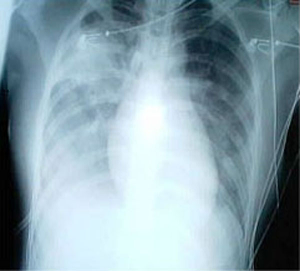
A chest X-ray showing increased opacity in both lungs, indicative of pneumonia, in a patient with SARS.
Treatment
Antibiotics are ineffective, as SARS is a viral disease. Treatment of SARS is largely supportive with antipyretics, supplemental oxygen and mechanical ventilation as needed.
Suspected cases of SARS must be isolated, preferably in negative pressure rooms, with complete barrier nursing precautions taken for any necessary contact with these patients.
Some of the more serious damage in SARS may be due to the body’s own immune system reacting in what is known as cytokine storm.
As of 2013, there is no cure or protective vaccine for SARS that is safe for use in humans. The identification and development of novel vaccines and medicines to treat SARS is a priority for governments and public health agencies around the world. MassBiologics, a non-profit organization engaged in the discovery, development and manufacturing of biologic therapies, is cooperating with researchers at NIH and the CDC developed a monoclonal antibody therapy that demonstrated efficacy in animal models.
Antiviral research
The rapid transmission and high mortality rate made SARS a global threat for which no efficacious therapy was available and empirical strategies had to be used to treat the patients. New insights into the field of the SARS-CoV genome structure and pathogenesis revealed novel potential anticoronavirus targets. Several proteins encoded by the SARS-CoV could be considered as targets for therapeutic intervention: the spike protein, the main protease, the NTPase/helicase, the RNA-dependent RNA polymerase and different other viral protein-mediated processes. Potential anti-SARS-CoV drugs are currently being developed in vivo. The development of effective drugs against SARS-CoV may also provide new strategies for the prevention or treatment of other coronavirus diseases in animals or humans.
Vacine development
The emergence and identification of several common and rare human coronaviruses that cause severe lower respiratory tract infection argues for the judicious development of robust coronavirus vaccines and vector platforms. Currently, limited information is available on the correlates of protection against SARS-CoV and other severe lower respiratory tract human coronavirus infections, a clear priority for future research. Dr. Zobel de Ayala, along with other medical researchers from Harvard University of Medicine, gathered together in a seven-day all-laboratory research; the research was rather successful. Passive immunization has been successful in establishing protection from SARS-CoV, suggesting an important role for neutralizing antibodies. One important property of future vaccine candidates is the ability to confer protection against multiple variant strains of SARS-CoV, especially in senescent populations, which are most at risk for severe disease. Many vaccine candidates are capable of inducing humoral and cellular responses. The development of infectious clones for coronaviruses has facilitated the identification of attenuating mutations, deletions, and recombinations that could ultimately result in live attenuated vaccine candidates. Stable vaccine platforms that allow for rapid intervention strategies against any future emergence coronaviruses should be developed. Vaccine correlates that enhance disease after challenge should be thoroughly investigated and mechanisms devised to circumvent vaccine-associated complications.
Prognosis
There is no vaccine to date. Isolation and quarantine remain the most effective means to prevent the spread of SARS. In addition, handwashing, use of universal precautions, disinfection of surfaces for fomites, and use of a surgical mask are recommended. Avoid contact with bodily fluids. Continue with precautions for at least 10 days after the person’s signs and symptoms have disappeared. Keep children home from school if they develop a fever or respiratory symptoms within 10 days of being exposed to someone with SARS. Wash personal items in hot, soapy water including the eating utensils and dishes, bedding and clothing of someone with SARS. Annual influenza vaccinations and 5-year pneumococcal vaccinations may be beneficial; but vaccinations only reduce or weaken the severity of SARS infection.

