THE MECHANISM OF PHYSIOTHERAPEUTIC PROCEDURES ACTION (COMPRESSES, CUPS, HOT-WATER BOTTLE, MUSTARD PLASTERS, ICE BAG) ON THE BODY.
BED REST, SERIOUSLY ILL AND AGONAL PATIENTS CARE.
CONCEPT OF REANIMATION. TECHNIC OF THE SIMPLEST REANIMATING MEASURES.
Physical therapy or physiotherapy (sometimes abbreviated to PT or physio) is a health care profession primarily concerned with the remediation of impairments and disabilities and the promotion of mobility, functional ability, quality of life and movement potential through examination, evaluation, diagnosis and physical intervention carried out by physical therapists (known as physiotherapists in most countries) and physical therapist assistants (known as physical rehabilitation therapists or physiotherapy assistants in some countries). In addition to clinical practice, other activities encompassed in the physical therapy profession include research, education, consultation, and administration. Definitions and licensing requirements in the vary among jurisdictions, as each state has enacted its own physical therapy practice act defining the profession within its jurisdiction, but the American Physical Therapy Association (APTA) has also drafted a model definition in order to limit this variation, and the APTA is also responsible for accrediting physical therapy education curricula throughout the United States of America. In many settings, physical therapy services may be provided alongside, or in conjunction with, other medical or rehabilitation services.
Physical therapy involves the interaction between therapist, patients or clients, other health care professionals, families, care givers, and communities in a process where movement potential is assessed and diagnosed and goals are agreed upon.Physical therapy is performed by a therapist and sometimes services are provided by a physical therapist assistant (PTA) acting under their direction. Physical therapists and occupational therapists often work together in conjunction to provide treatment for patients. In some cases, physical rehabilitation technicians may provide physiotherapy services.
PTs are healthcare professionals who diagnose and treat individuals of all ages, from newborns to the very oldest, who have medical problems or other health-related conditions, illnesses, or injuries that limit their abilities to move and perform functional activities as well as they would like in their daily lives. PTs use an individual’s history and physical examination to arrive at a diagnosis and establish a management plan and, wheecessary, incorporate the results of laboratory and imaging studies. Electrodiagnostic testing (e.g., electromyograms and nerve conduction velocity testing) may also be of assistance. PT management commonly includes prescription of or assistance with specific exercises, manual therapy, education, manipulation and other interventions. In addition, PTs work with individuals to prevent the loss of mobility before it occurs by developing fitness and wellness-oriented programs for healthier and more active lifestyles, providing services to individuals and populations to develop, maintain and restore maximum movement and functional ability throughout the lifespan. This includes providing services in circumstances where movement and function are threatened by aging, injury, disease or environmental factors. Functional movement is central to what it means to be healthy.
Physical therapy has many specialties including sports, wound care, EMG, cardiopulmonary, geriatrics, neurologic, orthopaedic and pediatrics. PTs practice in many settings, such as outpatient clinics or offices, health and wellness clinics, rehabilitation hospitals facilities, skilled nursing facilities, extended care facilities, private homes, education and research centers, schools, hospices, industrial and this workplaces or other occupational environments, fitness centers and sports training
facilities.
Physical therapists also practice ion-patient care roles such as health policy, health insurance, health care administration and as health care executives. Physical therapists are involved in the medical-legal field serving as experts, performing peer review and independent medical examinations.
Education qualifications vary greatly by country. The span of education ranges from some countries having little formal education to others having doctoral degrees and post doctoral residencies and fellowships.
Physicians like Hippocrates and later Galenus are believed to have been the first practitioners of physical therapy, advocating massage, manual therapy techniques and hydrotherapy to treat people in 460 BC. After the development of orthopedics in the eighteenth century, machines like the Gymnasticon were developed to treat gout and similar diseases by systematic exercise of the joints, similar to later developments in physical therapy. The earliest documented origins of actual physical therapy as a professional group date back to Per Henrik Ling, “Father of Swedish Gymnastics,” who founded the Royal Central Institute of Gymnastics (RCIG) in 1813 for massage, manipulation, andexercise. The Swedish word for physical therapist is sjukgymnast = someone involved in gymnastics for those who are ill. In 1887, PTs were given official registration by Sweden’s National Board of Health and Welfare. Other countries soon followed. In 1894 four nurses in Great Britain formed the Chartered Society of Physiotherapy.[15] The School of Physiotherapy at the University of Otago in New Zealand in 1913, and the United States’ 1914 Reed College in Portland, Oregon, which graduated “reconstruction aides.”
Modern physical therapy was established towards the end of the 19th century due to events that had an effect on a global scale, which called for rapid advances in physical therapy. Soon following American orthopedic surgeons began treating children with disabilities and began employing women trained in physical education, massage, and remedial exercise. These treatments were applied and promoted further during the Polio outbreak of 1916. During the First World War women were recruited to work with and restore physical function to injured soldiers, and the field of physical therapy was institutionalized. In 1918 the term “Reconstruction Aide” was used to refer to individuals practicing physical therapy. The first school of physical therapy was established at Walter Reed Army Hospital in Washington, D.C., following the outbreak of World War I. Research catalyzed the physical therapy movement. The first physical therapy research was published in the United States in March 1921 in “The PT Review.” In the same year, Mary McMillan organized the Physical Therapy Association (now called the American Physical Therapy Association (APTA). In 1924, the Georgia Warm Springs Foundation promoted the field by touting physical therapy as a treatment for polio Treatment through the 1940s primarily consisted of exercise, massage, and traction. Manipulative procedures to the spine and extremity joints began to be practiced, especially in the British Commonwealth countries, in the early 1950s Around this time when polio vaccines were developed, physical therapists have become a normal occurrence in hospitals throughout North America and Europe. In the late 1950s, physical therapists started to move beyond hospital-based practice to outpatient orthopedic clinics, public schools, colleges/universities health-centres, geriatric settings (skilled nursing facilities), rehabilitation centers and medical centers. Specialization for physical therapy in the U.S. occurred in 1974, with the Orthopaedic Section of the APTA being formed for those physical therapists specializing in orthopaedics. In the same year, the International Federation of Orthopaedic Manipulative Physical Therapists was formed, which has ever since played an important role in advancing manual therapy worldwide.
Compression Wraps 
Compression wraps are bandages that help control swelling. They are most often.
How to apply:
1. Wash your hands with warm water and soap for at least 15 seconds. Rinse
with water and towel dry.
2. Unwrap the compression wrap to remove it. If you have a wound dressing that
was under the wrap, change the dressing as directed.
3. Check your wounds for signs of infection. If you have any of these signs, call
your doctor or nurse right away:
C Skin around the wound is more red, swollen, or feels hot
C Wound smells bad
C Pus drainage
C Temperature above 100.5F
4. Check the rest of the skin on your leg and foot for any blisters, red spots, open
areas or other signs of problems. If you find any new problems, call your
doctor or nurse.
5. Remove the compression wrap from the package or use a clean wrap.
6. Start at the base of the toes and wrap around and up your leg to just below the
knee. Put the wrap on so the edges overlap about half of the bandage. This
keep the pressure even and prevents bare patches where some of the foot or
leg may poke through.
7. Keep the wrap smooth. Any creases or wrinkles in your wraps may irritate
your skin. If you need to use more than one wrap to cover your leg, lap the end
over each other for about 6 inches and continue to wrap. The ends should hold
in place.Page 2
8. Wrap so the bandage is snug but not too tight. You should be able to slide a
finger under the layers of the wrap.
9. Use the clips or velcro the end of the wrap to hold it in place.
10. Wash your hands again with warm water and soap. Rinse and towel dry.
11. Loosen the wraps if you have pain, numbness or tingling in your toes or feet.
A mustard plaster is a poultice of mustard seed powder spread inside a protective dressing and applied to the chest or abdomen to stimulate healing. In times past and present, the mixture was spread onto a cloth and applied to the chest or back. The mustard paste itself should never make contact with the skin. Applied externally, black mustard is used in the treatment of bronchial pneumonia and pleurisy.
Mustard oil irritates mucous membranes; therefore, excessive internal use has been known to cause stomach problems and kidney irritation. Breathing the vapors of a mustard plaster can trigger sneezing, coughing, asthma attacks, or eye irritation. Leaving a mustard plaster on the bare skin for too long will lead to burning, blisters, or potentially even ulcers. A mustard plaster should never be left on for longer than 30 minutes. The actual mustard paste never comes in direct contact with the skin, just the cloth on which it is spread.
A typical mustard plaster recipe includes powdered mustard (amounts vary from recipe to recipe) and flour combined with water or egg white. This is then spread on a layer of cotton or flannel cloth and placed on the body. Some old sources suggest that the mustard powder be blended with egg white rather than water to prevent blistering of the skin.
Mustard plasters should not be used on children under the age of 6. Black mustard should not be used in patients with ulcers, venous problems, or kidney disease.
Mustard plasters are common in Russia and other Post-Soviet states, and France (paper sinapism of Dr Paul Jean Rigollot). It is a common belief there that mustard plasters stimulate the immune system, relieve pain and also have an anti-inflammatory effect. They are often used to treat common colds, runny noses, rheumatism and problems with the respiratory system.
Warming compress
WET TOWEL COMPRESS: A wet towel, folded and placed inside a plastic bag (a
large heavy zip-lock bag works well for this…be sure to leave the bag “unzipped” while heating) heated in the microwave for 2-4 minutes or until hot. Leaving the hot wet towel inside the plastic bag, remove it from the microwave, press all air out of the bag and “zip” or close it up. This heated towel can be applied on a sore area of the body. It works well, is economical and easily available in most homes. Be careful, however, the towel can become very hot using the microwave and you may want to wrap it with another towel or cloth as a buffer before applying it to the skin. This method worked well for an elderly woman I was taking care of for a few years. She was afraid to use an electric heating pad for many reasons and would only use this method. Her skin was very sensitive and I had to place a dry towel between the plastic bag containing the moist heated towel and her skin. The heated towel was good for about 20-30 minutes and theeeded to be reheated.
WET WASHCLOTH COMPRESS: A wet washcloth can be folded and placed in warm-hot water and used for sore areas that are small in size. During childbirth, we often use wet washcloth compresses soaked in warm-hot water or warm-hot comfrey leaf tea on the perineal area during the pushing stage of labor. This reduces painful, stretching-buring sensations, relaxes the perineum and vaginal opening allowing tissue to stretch as the baby’s head is crowning. It also keeps the blood circulation in the tissues, preventing tearing while it is stetching open and gives the mother a focal point on where to concentrate her labor efforts when we tell her to “push into the warm cloth”. Again, don’t make the water or tea too hot, just hot enough to hold in your hands comfortably. The perineal area and external genitalia is sensitive and you do not want to burn this area. Because the cloth is smaller, it will need to be replaced much more often. I often use two cloths and alternate their use so I always have a warm one available at all times, replacing the cooled one back into the warm liquid.
MICROWAVE COMPRESS: A tubelike microwavable compress can be purchased at most drugstores. These are reusable, reheatable compresses that are heated in the microwave and applied to sore areas, such as a sore neck. They are filled with a variety of substances such as heat-holding beads or sometimes uncooked rice or treated corn. Many have a looped handle on each end to grasp with hands. These may be used wet or dry, depending upon manufacturer instructions. To make a homemade microwavable compress, obtain a large men’s tube sock at any department store and fill with uncooked dry rice. The cuff end can be sewed or tied shut to keep the rice from spilling out. Do not use safety pins as most microwaves do not work well with metal objects. I do not recommend wetting these homemade compresses, however, I have never tried using them wet so I am not sure if wetting them would cause the rice to cook and soften – which you don’t want to do. Microwave the homemade compress for a few minutes (depending upon your microwave) and apply to the sore area that needs the heat. The uncooked dry rice is excellent for holding heat, is inexpensive, easy to work with and can be obtained at any supermarket. For a larger homemade compresses, 2 terrycloth wash cloths or hand towels, or squares of heavy flannel that can be sewed together and filled with uncooked rice and heated in the microwave. Adjust the time in the microwave accordingly, depending on size.
In place of using a rice sock (they can be quite heavy if they are large enough to retain heat well), you can use old fashioned oats in place of the rice in a homemade sock. Herbs can be added if desired, but these are not necessary. An oat sock smells clean and sweet when heated, retains the heat well and weighs considerably less. You can sew small loops of elastic at the ends so the person using it can either hold it in place if she wants or cloth ties or belts may be used to hold it in place. For more heat retention, the heated sock can be placed in a removable flannel sheath that velcros at one end and the elastic loops slip through.
Ice and heat for injuries
Ice and heat are often used in treating injuries.
· Icing may be used along with compression, elevation, bracing, and/or support when treating acute injuries.
· Nonsteroidal anti-inflammatory drugs (NSAIDs) can produce a similar effect to icing. However, they may delay healing with acute injuries (like sprains, strains, and fractures). If your doctor recommends medicine, make sure you are aware of the right dosage and when to take it, and if there are any side effects.
· The use of ice and heat is just one part of a treatment program. Even if symptoms are relieved, there is usually a need for exercises to restore flexibility and joint motion, strength, general fitness, and sport-specific skills.
· Effects of ice: Decreases circulation, metabolic activity, and inflammation and numbs the skin.
· Benefits of ice: Decreases pain, swelling, inflammation, and muscle spasm/cramping. Best used after exercise or after pain-producing activity.
· Risks of ice: Prolonged use can cause frostbite.
· Methods for applying cold therapy: Ice packs, ice bath/ice whirlpool, ice massage. (See “Options for applying ice.”)
· Immediately before physical activity
· If area of icing is numb
· When the pain or swelling involves a nerve (such as the ulnar nerve or “funny bone”)
· If the athlete has sympathetic dysfunction (an abnormality of nerves that control blood flow and sweat gland activity)
· If the athlete has vascular disease (such as poor circulation due to blood loss, blood vessel injury, compartment syndrome, vasculitis, blood clots, or Raynaud disease)
· If there is skin compromise (such as an open wound; a wound that has not healed; skin that is stretched, blistered, burned, or thin)
· If the athlete has cold hype
· long to use ice
· Two to 3 times per day (minimum); up to once per hour.
· Duration varies with technique; usually 20 to 30 minutes per session. (See “Options for applying ice.”)
· Ice may continue to be useful in treatment as long as there is pain, swelling, inflammation, or spasm. There is no need to switch to heat after 48 hours or alternate between ice and heat.
·
·
· row long to use ice
· Two to 3 times per day (minimum); up to once per hour.
· Duration varies with technique; usually 20 to 30 minutes per session. (See “Options for applying ice.”)
· Ice may continue to be useful in treatment as long as there is pain, swelling, inflammation, or spasm. There is no need to switch to heat after 48 hours or alternate between ice and heat.
1. Ice packs are best for icing larger areas of pain, swelling, or spasm (like a swollen knee, deep thigh bruise, muscle strain, shoulder tendonitis, or neck or back spasm).
· Small cubes or crushed ice in plastic bag.
· Bag of frozen vegetables (such as frozen peas).
· Reusable commercial ice pack or circulating “cryocuff” (made specifically for therapeutic icing). Do not use blue ice packs directly on the skin; they are colder than frozen water and can cause frostbite
Place on the affected area for at least 20 minutes per session. Hold in place with a towel, elastic wrap, or shrink-wrap.
2. Ice bath/ice whirlpool is used to reduce swelling in peripheral joints (such as with ankle sprain, wrist sprain, or severe shin splints).
· Bucket or tub with mixture of ice and water
Immerse affected area for 20 to 30 minutes per session. Do not use an ice bath if there is an open wound, bleeding, or a skin infection.
3. Ice massage is used to reduce superficial, well-localized inflammation (for example, tendonitis of the hand, wrist, or elbow; heel or elbow bursitis; ganglion cyst; apophysitis; or irritation of a growth plate).
· Ice cube or frozen ice cup (made by freezing water in a paper or Styrofoam cup)
Rub ice in a circular pattern over the affected region for 8 to 10 minutes per session.
Ø Figure 3-5-4 Place the moist towel on the area being treated.
Ø
Ø
Ø Figure 3-5-5 Wrap the hot, moist towel with a waterproof pad and secure.
Ø SKILL 3-5
Ø Applying Moist Heat
Ø 11. Application of moist heat for a longer period of time may damage the client’s skin and may predispose the client to edema formation from circulatory congestion.
Ø 12. The client may feel chilled when the warm compress is removed. Dry the area completely to prevent chilling.
Ø 13. The basins and thermometer can be used again. Proper disposal of all other equipment reduces the transmission of microorganisms.
Ø 14. Reduces the transmission of microorganisms and gets the equipment ready for use again.
Ø 15. Reduces the transmission of microorganisms.
Ø 16. The condition of the client’s skin and any signs of heat sensitivity should be assessed and documented.
Ø 17. Communicates the findings to the other members of the health care team and contributes to the legal record by documenting the care given to the client.
Ø
Ø 11. If it is tolerated, leave the compress in place for approximately 30 minutes and then remove it.
Ø 12. Dry the affected area with sterile towels if there is an open wound and with clean towels if there is no open wound.
Ø 13. Properly dispose of all single-use equipment according to hospital protocol.
Ø 14. Clean the bath basin and thermometer. Return the sterile basin to the appropriate place for resterilization.
Ø 15. Remove gloves if they were worn and wash your hands.
Ø 16. Reassess the condition of the client’s skin.
Ø 17. Record the procedure. Note the condition of the client’s skin and the length of the application. Report any abnormal findings to the physician.
Ø > EVALUATION •
Ø Evaluate post-treatment for decreased swelling, decreased edema, decreased muscle tension, and improved circulation depending on the purpose of the treatment. • Evaluate skin post-treatment for any heat damage such as excess redness, swelling, or blistering.
Ø > DOCUMENTATION
Ø Document the treatment and the results of treatment. • Document the condition of the skin. • Document the purpose and type of moist heat application. • Record the results of the treatment and the client’s tolerance. • Document client teaching.
Ø Applying Cold Treatment
Ø KEY TERMS >
Ø Cold application
Ø Cold compresses
Ø Cold therapy
Ø ASSESSMENT
Ø 1. Ascertain the client’s sensation of hot and cold changes at the site where cold therapy is to be administered. Certain areas of the skin have sensitivity to temperature variations, while other areas may not be as sensitive; the perineal areas are very sensitive.
Ø 2. Assess if decreased circulation is present at the site where cold therapy will be applied such as areas with wounds and damaged tissue present because cold application may cause further tissue damage.
Ø 3. Check the client’s systemic temperature. If larger areas are exposed to cold, the total body temperature may be decreased. If the client is cooled too rapidly with extreme cold, a reverse chilling effect may occur, increasing body metabolism and defeating the cooling effect.
Ø 4. Assess age. Tolerance to cold varies with individuals and is related to age, thinner layers of skin, or general sensitivity to cold.
Ø > OVERVIEW OF THE SKILL
Ø Cold therapy is used to decrease blood flow to an area by promoting vasoconstriction and increased blood viscosity. These changes facilitate clotting and control bleeding. Cold decreases tissue metabolism, reduces oxygen consumption, and decreases inflammation and edema formation. Cold therapy has a local anesthetic effect by raising the threshold of pain receptors. It causes a decrease in muscle tension. Cold is used to reduce fever. Sources of cold include ice packs, ice bags, cold collars, or commercial cold packs. If the client’s systemic temperature is elevated, cooling blankets or cooling tepid sponge baths can be used. Moist cold compresses or immersion of a body part can be used for large areas of acute inflammation or swelling. Cooling the extremity decreases blood flow and may also decrease pain and suppress inflammation.
Ø
Ø CHAPTER
Ø 3 Patient Care and Comfort
Ø > PLANNING
Ø Expected Outcomes:
Ø 1. The client will experience decreased bleeding.
Ø 2. The client will have decreased inflammation and/or edema.
Ø 3. The client will experience decreased pain or discomfort.
Ø Equipment Needed:
Ø (Select equipment depending on the treatment chosen and supplies available.) • Pan for cold soak • Ice or ice bag • Gauze or towel • Water bottles or reusable containers if used for one client only • Compresses (if moist cold) consisting of gauze dressing, iced or chilled solution, and a container of the appropriate size for the body part • Commercially prepared ice pack (see Figure 3-9-2) • Disposable ice pack • Tape, elastic wrap, or bandage (see Figure 3-9-3)
Ø > CLIENT EDUCATION NEEDED:
Ø 1. Report any pain or lack of sensation (if cold is being used as local anesthetic then decreased sensation is expected).
Ø 2. Report chilling from overexposure since this would increase general metabolism. This can especially happen if the client has a large area of the body exposed to cold; therefore, it may be necessary to add increased covers to the rest of the body.
Ø 3. Teach the client the basics of cold application, why it is used, and how long the treatment will remain in place (20–30 minutes).Modify the therapy for home use, and teach the client how to apply the treatment at home, if applicable.
Ø 4. Teach clients the reason for cold therapy vs. hot therapy. Estimated time to complete the skill: 20 minutes for application plus preparation time of 5–10 minutes
Ø IMPLEMENTATION—ACTION/RATIONALE
Ø ACTION RATIONALE
Ø 1. Reduces the transmission of microorganisms.
Ø 2. Provides baseline data for post-treatment comparison.
Ø 1. Wash hands.
Ø 2. Assess the client’s sensation and skin color at the site of planned application.Determine if any tissue damage is present. Assess for bleeding or wound drainage (see Figure 3-9-4).
Ø
Ø
Ø Figure 3-9-2 Ice pack
Ø
Ø
Ø Figure 3-9-3 Elastic wrap
Ø
Ø
Ø Figure 3-9-4 Assess the skin at the site of planned application for color, sensation,wounds, or skin irritation.
Ø
Ø
Ø Figure 3-9-5 Assess for circulatory impairment or neuropathy before beginning treatment.
Ø
Ø
Ø Figure 3-9-6 Wrap the cold pack in a towel.
Ø SKILL 3-9
Ø Applying Cold Treatment
Ø 3. Cold causes vasoconstriction and decreased metabolism and can cause tissue damage in people with impaired circulation and sensation.
Ø 4. A physician’s or qualified practitioner’s order is needed in most situations of cold treatment, and the client should be informed of the reason for the application of cold.
Ø 5. If air is removed from the bag, the bag will be easier to mold to the client’s body.The bag is wrapped to prevent injury to the client’s skin or exposed tissue because direct cold can cause damage.
Ø 6. Easier to mold to the client’s body.The collar is wrapped to prevent injury to the client’s skin.
Ø 7. When the pack is squeezed or kneaded, an alcohol-based solution is released, creating the cold temperature.The pack cannot be used again.
Ø
Ø 3. Determine the diagnosis. Identify whether the client has a history of circulatory impairment or neuropathy (see Figure 3-9-5).
Ø 4. Check the physician’s or qualified practitioner’s order and the reason for the application of cold.
Ø 5. If using an ice bag with moist gauze or towels, fill the bag three-fourths full with ice and remove the remaining air from the bag. Close the bag. Check for leaks.Wrap the bag in a towel or protective cover and place it on the affected area. If cold soaks are being applied, use the appropriate-size basin for the body part to be soaked.
Ø 6. If an ice collar is used, fill the collar threefourths full with ice and remove the remaining air from the collar before closing the collar. Check for leaks. Place the collar in a protective cover and around the client’s neck.
Ø 7. If a disposable cold pack is used, activate the pack according to the manufacturer’s directions, wrap the pack in a towel (see Figure 3-9-6), and place it on the affected area (see Figure 3-9-7). Some packs come with covers. Secure pack in place with tape, elastic wrap, or bandage (see Figures 3-9-8, 3-9-9, and 3-9-10). Dispose of the pack after the treatment.
Ø CHAPTER
Ø Patient Care and Comfort
Ø 8. Signs of intolerance to cold are pallor, blanching, mottling, or numbness of the skin.
Ø 9. Longer application can cause tissue damage, especially because the client’s pain sensation is decreased in the presence of cold. A reflex vasodilatation occurs after 20–30 minutes, thereby negating the therapeutic effect of the cold treatment.
Ø 10. The client’s skin should be assessed, and any signs of cold changes and intolerance should be documented.
Ø 11. Reduces the transmission of microorganisms.
Ø 8. Assess the client’s skin periodically for signs of cold intolerance or tissue damage.
Ø 9. If the client can tolerate the cold, leave the cold application in place for approximately 20–30 minutes at approximately 15°C (59°F).
Ø 10. Reassess the condition of the client’s skin or exposed tissue.
Ø 11. Wash hands.
Ø
Ø Figure 3-9-7 Place the cold pack on the affected area.
Ø
Ø Figure 3-9-9 This cold pack is properly wrapped in a towel and secured with tape.
Ø
Ø Figure 3-9-8 Use tape to secure the cold pack to the affected area.
Ø
Ø Figure 3-9-10 Elastic wrap may be used to hold bulky cold packs in place. > EVALUATION •
Ø Evaluate decreased bleeding, inflammation, and/or edema. • Evaluate pain level. • Evaluate tissue integrity, especially for blanching, redness, or cold burns.
Ø > DOCUMENTATION
Ø CHAPTER
Ø Patient Care and Comfort
Ø . Signs of intolerance to cold are pallor, blanching, mottling, or numbness of the skin.
Ø Longer application can cause tissue damage, especially because the client’s pain sensation is decreased in the presence of cold. A reflex vasodilatation occurs after 20–30 minutes, thereby negating the therapeutic effect of the cold treatment.
Ø The client’s skin should be assessed, and any signs of cold changes and intolerance should be documented.
Ø Reduces the transmission of microorganisms.
Assess the client’s skin periodically for signs of cold intolerance or tissue damage.
If the client can tolerate the cold, leave the cold application in place for approximately 20–30 minutes at approximately 15°C (59°F).
Reassess the condition of the client’s skin or exposed tissue.
Ø. Wash hands.
Ø
Ø Figure 3-9-7 Place the cold pack on the affected area.
Ø
Ø Figure 3-9-9 This cold pack is properly wrapped in a towel and secured with tape.
Ø
Ø Figure 3-9-8 Use tape to secure the cold pack to the affected area.
Ø
Ø Figure 3-9-10 Elastic wrap may be used to hold bulky cold packs in place. > EVALUATION •
Ø Evaluate decreased bleeding, inflammation, and/or edema. • Evaluate pain level. • Evaluate tissue integrity, especially for blanching, redness, or cold burns.
Ø > DOCUMENTATION
Ø • Record the procedure and the client’s response to the treatment such as whether bleeding was stopped, inflammation decreased, edema decreased. • Record the client’s skin condition after the procedure. • Record the length
Ø • Record the procedure and the client’s response to the treatment such as whether bleeding was stopped, inflammation decreased, edema decreased. • Record the client’s skin condition after the procedure. • Record the length of time of application.
APPLYING DRY HEAT
Dry heat can be used to enhance circulation, promote healing, reduce swelling and inflammation, reduce pain, reduce muscle spasms, and increase systemic temperature. Different types of equipment are used to apply dry heat to body surfaces, specific areas, and the entire body.
These can be divided into the following categories:
1. Body surfaces. Equipment used to apply heat to any body surface includes disposable instant hot packs, gel-filled hot packs, aquathermia pads, electric heating pads, and hot water bags or bottles. Aquathermia pads are waterflow rubber heating pads with tubing and a reservoir control unit, sometimes called aqua pads, k-pads, t-pump, orhydrocalculator. Hot water bags or bottles should be used only by clients at home because bags and bottles cannot be cleaned properly to meet universal standards.
2. Specific areas. Equipment used to apply heat to specific areas includes heat lamps or infrared lamps (generally for the abdomen, perineum, or the chest), heat cradles (generally for the lower extremities), and diathermy (generally for deep heat treatment, which utilizes electrical energy that is changed to heat).
3. Entire body. Equipment used to heat the entire body to treat cases such as hypothermia includes thermal blankets and infant radiant warmers, which are discussed in Skill 3-8. The principles and precautions are similar in most types of heat application.
• Equipment determined by type of heat treatment:
disposable gel-filled packs , aquathermia pad, heating pad, hot water bottle (generally
used only in home setting if at all), heat lamp or heat cradle, hot blankets, or hot air patient warming system
• Protective cover to be used between heat source and patient
• Electrical source for pads
• Timer or clock
1. Check the physician’s or qualified practitioner’s order and the purpose of the heat treatment.
2. Determine if there are any underlying problems that may affect the use of heat treatment such as decreased sensation; decreased mentation; or a history of diabetes mellitus, bleeding disorders, peripheral vascular disease, or peripheral neuropathy.
Heat should not be used over areas of scarring.
3. Wash hands.
4. Check the skin for lotions or ointments and remove if present.
5. Explain the reasons for heat treatment, the expected outcomes, any potential complications, and the necessity to alert the nurse of heat intolerance.
6. Gather equipment and complete as follows:
For a disposable heat pack:
• Activate the pack according to the manufacturer’s directions. Some packs must be
heated in boiling water, others can be heated by microwave, and some require
bending and chemical activation.
• Wrap the pack in a towel or protective covering (some manufacturers include cover). Do not use pins.Use tape if needed to secure the towel.
Discard after use.
For a heating pad:
• Note: Heating pads are generally not used in hospital facilities.
• Place a formed cover, usually a flannel, over the pad.Towels should not be used. Pins should never be used. If it is necessary to secure the cover, use tape.
• Instruct the client not to lie on the heating pad.
• Turn on the switch to low and place the heating pad on the affected area.The nurse
may increase heat after the client adjusts to the heat. Instruct the client not to adjust the heat.
· Effects of heat: Increases circulation, metabolic activity, and inflammation.
· Benefits of heat: Improves compliance of soft tissues; relieves pain and spasm. Heat is most useful in warming up stiff or scarred soft tissues before stretching or exercise; heat may also be useful in relieving pain or spasm associated with neck or back injuries.
· Risks of heat: May increase swelling and inflammation; using heat for too long or at temperatures that are too high can cause burns.
· Methods for applying heat: Hot packs, hydrocollator; hot bath/whirlpool.
· After physical activity
· If the area is numb
· If there is an open wound or burn
· Immediately after an acute injury
· If body temperature is elevated from fever or heat stress
Basics of cardiopulmonary resuscitation
Resuscitation is a complex of actions, which prevent irreversible changes and restore vital functions of an organism in a state of clinical death. A person, who conducts these actions is called a rescuer.
The final goal of resuscitation is to bring back life of full value to a patient after clinical death. This task might be realized only with immediate, professional and sequential measures. Nevertheless always care about your own safety, as your duty is to help, not to increase number of victims. Pay attention to the conditions in which clinical death appeared, make sure you are not in danger, use gloves and eyewear if they are available. Of course final decision about priorities belongs to you, but every biological liquid contacting your skin and mucosa is a potential infection source. If you feel unwilling to perform rescue breaths mouth-to-mouth or physically it is impossible to ventilate the patient for some reason at least do chest compressions – according to the modern level of resuscitation knowledge blood flow is the most important target of CPR.
The first stage of resuscitation is basic life support. It is conducted by a rescuer who is not obligatory a health care professional, but a witness who acquired basic life support skills. After clinical death is stated (try not to evaluate respiration (B) and heart action (C) more than 10 seconds) a rescuer should immediately start basic life support (optimal position of patient who is on the flat surface lying supinely).
Pic. 2.1 ABC check
Successful cardiopulmonary resuscitation is based on three pillars:
I. Airways (A). To make upper airways free use triple method of Peter Safar: 1. Open the mouth of the patient and clean the oral cavity, if necessary, from foreign bodies and liquids such as vomit, sputum, false jaws, blood cloths, etc (using your finger or forceps with surgical drape).
Pict.2.2.Cleaning of the oral cavity with the finger, wrapped iapkin
2. Title the head backwards (remember, that in case of each trauma patients we always suspect neck injury, so titling and sharp neck moves should be rather avoided). After this in most cases upper airways become conductive (soft palate and tong are not blocking air passage any more).
Pict.2.3. Backwards head titling (neck extension).
3 . Thrust the jaw forward. In all cases this part of Safar method provides final air passage.
Pict.2.4 Jaw thrusting forward and up. Pic. 2.5 Oropharyngeal airway
You can also use simple airway adjuncts such as oropharyngeal and nasopharyngeal airways.
II. Breathing (B). Respiratory support in conditions of BLS is usually mouth-to-mouth ventilation. If only there is a chance use devices for pre-hospital ventilation: pocket resuscitation masks of different types or at least handkerchief. Place closely your mouth over that of the patient and make a normal exhale (volume 600-800 ml). Remember to keep the airways conductive using the methods described above; use fingers of your free hand to close the nostrils of the patient. In case of correct ventilation chest rises and falls silently. Repeat this action one more time.
III. External heart massage (C- circulation). Standing aside the patient (on your knees), place your hands in the center of the chest: heel of the hand in the middle of the lower part of sternum or between lower and middle thirds of sternum. Don’t loose precious time looking for anatomic landmarks, as soon as possible start heart massage. Pay attention to your fingers – they should not lean on the chest, otherwise you will break the ribs during compressions. The frequency of compressions should be 100 per minute. It means that until you haven’t provided airways with endotracheal tube you make 30 compressions per every 2 breathes. Heart massage is extremely important: do it correctly as its efficiency (and thus cerebral blood circulation) depends on your technique. Use the most developed muscles of your body – back muscles, keep your elbows straight and compress the chest with the power of your trunk, not upper limbs. Every compression should be 5-6 cm deep. After each compression don’t forget to let chest recoil completely.
Pict.2.7 If there are two rescuers they perform rescue breaths and compressions by turn, but always 2 to 30.
Sings of effective resuscitation actions are pupils contraction, normalization of the skin color, appearance of peripheral pulse synchronized with the massage, sometimes even possibility of blood pressure measurement. Sometimes heart action restores even during BLS.
Second stage is advanced life support, which is provided by health-care professionals in hospitals with the usage of medicine, diagnostic and therapy equipment. The main ideas of ALS are: determination of cardiac arrest type (shockable/nonshockable rhythm), pharmaceutical and electric treatment, usage of advanced artificial ventilation (if available also devices for heart massage) and therapy of reversible clinical death reasons.
When resuscitation team works functions of rescuers must be divided in order to gain maximal efficiency. After CPR started the main purpose is to decide whether the defibrillation is necessary or not, other words to monitor the type of cardiac arrest: shockable rhythms are ventricular fibrillation (VF) and pulseless ventricular tachycardia (VT without pulse); nonshockable rhythms are asystole (A) and pulseless electrical activity (PEA) of the heart. Without energetic resources VF and pulseless VT quickly change into PEA and asystole, so to shorten the time between arrest and defibrillation paddles visualization (apex-sternum position) should be used even before the electrodes will be placed on the chest. For the first defibrillation use the dose of 360 Joules for monophasic defibrillators (old models) and 150-200 Joules for biphasic defibrillators (modern devices). Subsequent shocks might be of the same (200 J) or escalated energy (150-360 J) – the efficiency of energy increase is not proved, so it depends on you and local standards.
Pict.2.8 Electrical defibrilator.
Cardiac arrest types:
1. Asystole – flat line on ECG
2. Ventricular fibrillation – chaotic contractions of myocardial fibers visualized on ECG as waves of different shape and amplitude (high, medium and low).
3. PEA – different ECG rhythms, including normal, but combined with the lack effective systole (pulse).
Simultaneously a venous access attempt should be done, as after third defibrillation administration of adrenaline (1 ml of 0,1% solution =1 mg followed by 20 ml of 0,9% NaCl solution) and amiodarone (6 ml of 5% solution = 300 mg with 5% Glucose solution, total volume – 20 ml) are required. Adrenaline administration should be repeated every 3-5 minutes in the same dose. In case of PEA and asystole adrenaline should be given from the moment of intravascular access achievement. Atropine is not any more included into the official algorithms of ALS if the cardiac arrest is not caused by vagal effect. However according to actual Ukrainian standards single administration of Atropine is still recommended (3 ml of 0,1% solution=3 mg, followed by 20 ml of 0,9% NaCl solution).
If, however, venous access attempts are unsuccessful within 2 minutes you should think about alternatives, such as intraosseous access. As for the medicine delivery via the tracheal tube – it is no longer recommended. Central venous line insertion is a prerogative of the most skilled and competent members of the team. Previously it was thought, that triple doses of resuscitation drugs given through endotracheal tube or through the needle in the crico-thyroid membrane will be effective, but according to the actual recommendations such way of admission is unpredictable and thus caot be an alternative. Intracardiac delivery of drugs nowadays has rather historical value: in most developed countries it is not practiced any more.
Mechanical ventilation is much more effective than the mouth-to-mouth one. There are different types of devises for respiration (carrying and stationary) and numerous devises which play the role of connector between patient’s airways and apparatus for artificial pulmonary ventilation (ventilation masks, laryngeal masks and tubes, combitubes, endotracheal tubes, etc). In case of CPR the endotracheal tube with cuff is an absolute golden standard, as it allows asynchronous ventilation/massage and protects patient from aspiration (cardiac arrests are mostly sudden, so there is always a risk of regurgitation, aspiration and thus aspiration syndrome development). Under control of direct laryngoscopy it’s possible to clean upper airways with electric or pneumatic suction device and what is even more important – to intubate the trachea.
During the CPR think about reversible cardiac arrest reasons and try to treat them: there are easy mnemonic schemes of 4 H and 4 T for this purpose. So, the reversible causes of the clinical death are:
hypoxia tension pneumothorax
hypovolemia tamponade (cardiac)
hypothermia toxins
hypo/hyper electrolytic and metabolic disorders thrombosis
Treat them with: oxygen and artificial ventilation in case of hypoxia; crystalloids and colloids in case of hypovolemia; warming (including warm infusions) in case of hypothermia and proper electrolyte infusions in case of electrolytes and metabolic disorders (for example use 5-10 ml of 10% calcium chloride solution if you suspect hyperkalemia or hypocalcemia caused by dialysis, hemolysis, massive tissue damage, etc.). Use needle thoracocentesis for tension pneumothorax, needle pericardiocentesis for cardiac tamponade, antidotes and detoxification methods for toxic agents and thrombolytic therapy for thrombosis (if required).
The third stage of resuscitation is post-resuscitation care provided also in intensive treatment unit.
On the first stage of this care check again the patient’s condition: monitor constantly condition of cardiovascular and respiratory systems, measure blood pressure and central venous pressure, evaluate CNS state (reflexes, neurological deficiency), perform laboratory tests (take blood and urine samples, liquor if necessary), etc. Well-planned, comprehensive examination allows us to identify homoeostasis disorder and choose optimal treatment. After main parameters are stabilized central nervous system becomes your main concern: protect the brain from hypoxia by all available means, because hypoxic damage of neurons is usually irreversible. To achieve this purpose you should:
– provide adequate oxygenation (however excess of oxygen is no longer recommended, so keep blood saturation at the level 94-98%); hyperventilation might be used in case of brain oedema (first 12-24 hours of artificial ventilation);
– decrease metabolic needs of the CNS by craniocerebral hypothermia (give saline solution with the temperature 2ºC in order to lower body temperature to 32-34ºC for 12-24 hours) or by continuous narcosis (sodium thiopental 3-5 mg/kg, diazepam 0,2 mg/kg, neuroleptics, etc.);
– control blood glucose level (avoid hyperglycaemia over 10 mmol/l; hypoglycaemia caot be accepted at all);
– additionally prescribe antihypoxants like sodium oxybate (20-40 mg/kg every 4 hours), cytochrome C (0,5 mg/kg i/v); antioxidants like α-tocopheryl acetate (500 mg i/v), B-vitamins (2-3 ml), ascorbic acid (5 ml of 5% solution 3 times a day); calcium antagonists like verapamil (2 ml 3 times a day), magnesium sulphate (5-10 ml of 25% solution i/v every 4 hours with blood pressure control);
– improve cerebral perfusion with haemodilution (give crystalloids to get hematocrit 0,3-0,35 l/l), relative hypertension (20-30% over the normal level), solutions influencing rheological properties of the blood and microcirculation (rheopolyglucin, 2 ml of 0,5% curantil solution, heparin 5000 units every 4 hours, etc);
– treat cerebral oedema with mannitol (1g/kg), furosemide solution 10 mg i/v 3 times a day), dexametazon solution (8 mg every 4 hours);
– use hyperbaric oxygenation from the 5-th day of post-resuscitation care (totally 10 procedures);
– give nootropic drugs and neuroprotectors (piracetam, cerebrolysin, aminalon, etc.) The most common reasons of acute respiratory failure:
a. airways obstruction (tongue, vomit, foreign bodies)
b. inhibition of respiratory center (opiates. anesthetics)
c. disorders of breathing biomechanics (convulsions, myasthenia, tension pneumothorax or hemothorax)
d. restrictive disorders (massive pneumonias, shock lung syndrome, pneumothorax or hemothorax).
The most common reasons of primary brain death: acute vascular disorders (subarachnoid hemorrhage, hemorrhagic and ischemic strokes, brain dislocation).
Patients with a predictably high risk of sudden death should be constantly under vital functions monitoring. In case of compensation failures medical personnel of the department should intrude with a treatment directed at correction of disorders and intensive care. That is why an in-hospital sudden death should be an exception. Never the less hospital stuff should be prepared to provide immediate life support.
Here is the list of equipment for in-hospital resuscitation:
1. Portable manual respirator
2. Oxygen supply (cylinder)
3. Electric suction device with suction catheters
4. Electrocardiograph, defibrillator, tonometer
5. Mouth-gag, tongue forceps, clips
6. Set of face masks and airways
7. Laryngoscope with a set of tubes
8. Set for conicotomy and for pericardiocentesis
9. Solutions of adrenaline, atropine, sodium bicarbonate, lidocaine, steroids, colloids and crystalloids
10. Infusion sets, i/v catheters, syringes of different sizes
11. Bandages, medical napkins, antiseptic solutions.
Peculiarities of in-hospital resuscitation
Patient is usually lying in bed.
The most common reasons of primary brain death: acute vascular disorders (subarachnoid hemorrhage, hemorrhagic and ischemic strokes, brain dislocation).
Patients with a predictably high risk of sudden death should be constantly under vital functions monitoring. In case of compensation failures medical personnel of the department should intrude with a treatment directed at correction of disorders and intensive care. That is why an in-hospital sudden death should be an exception. Never the less hospital stuff should be prepared to provide immediate life support.
Here is the list of equipment for in-hospital resuscitation:
I
The most common reasons of primary cardiac arrest:
a. acute cardiac failure (coronary disease, myocardial infarction, rhythm disorders, sudden coronary death)
b. acute obstruction of main vessels (pulmonary thromboembolism)
c. acute and intense deficiency of blood volume (significant blood loss, dehydration)
d. acute decline of peripheral vessels resistance (acute suprarenal failure, anaphylactic shock, somatogenic collapse in case of acute intoxication, orthostatic medication collapse)
The most common reasons of acute respiratory failure:
e. airways obstruction (tongue, vomit, foreign bodies)
f. inhibition of respiratory center (opiates. anesthetics)
g. disorders of breathing biomechanics (convulsions, myasthenia, tension pneumothorax or hemothorax)
h. restrictive disorders (massive pneumonias, shock lung syndrome, pneumothorax or hemothorax).
The most common reasons of primary brain death: acute vascular disorders (subarachnoid hemorrhage, hemorrhagic and ischemic strokes, brain dislocation).
Patients with a predictably high risk of sudden death should be constantly under vital functions monitoring. In case of compensation failures medical personnel of the department should intrude with a treatment directed at correction of disorders and intensive care. That is why an in-hospital sudden death should be an exception. Never the less hospital stuff should be prepared to provide immediate life support.
Here is the list of equipment for in-hospital resuscitation:
12. Portable manual respirator
13. Oxygen supply (cylinder)
14. Electric suction device with suction catheters
15. Electrocardiograph, defibrillator, tonometer
16. Mouth-gag, tongue forceps, clips
17. Set of face masks and airways
18. Laryngoscope with a set of tubes
19. Set for conicotomy and for pericardiocentesis
20. Solutions of adrenaline, atropine, sodium bicarbonate, lidocaine, steroids, colloids and crystalloids
21. Infusion sets, i/v catheters, syringes of different sizes
22. Bandages, medical napkins, antiseptic solutions.
Peculiarities of in-hospital resuscitation
Patient is usually lying in bed.
1. To get a firm surface [efficiency of chest compressions depends on this] lay under the patient spinal board or move him/her on the floor.
2. Duration of the first stage should be minimal (5 to 7 minutes) as the beginning of advanced life support in intensive care unit is more important.
3. In witnessed primary cardiac arrest caused by ventricular fibrillation during first 20-30 seconds precardiac thump might be effective (so called “mechanical defibrillation”).
4. Sometimes tracheostomy or conicotomy might be necessary in case of upper airways obstruction (laryngospasm, stenosis of larynx, foreign body in the glottis).
The most favorable resuscitation prognosis is connected with primary respiratory arrest and the most unfavorable – with the primary cerebral death.
Drowning: types and resuscitation.
There are different types of drowning thanatogenesis.
True drowning. Most victims under the water according to a reflex stop breathing. But after some time due to hypercapnic stimulation of the respiratory center they unwillingly begin to make respiratory movements. Liquid gets to the lungs: fresh water, which is hyperosmolar to plasma, diffuses easily through blood-air barrier into the blood, thus increasing it’s volume. Extra 1500-2000 ml of water in addition to hypoxia lead to cardiac arrest. At the same time osmotic hemolysis (caused by rapid lowering of plasma osmolarity) and hyperpotassemia are also cardiac arrest factors.
In case of salt water true drowning fluid part of blood according to the osmotic gradient moves from bloodstream into the bronchi and trachea. This way surfactant is being destroyed and pulmonary edema begins.
Dry drowning. 8-10% of victims in the moment of water aspiration have reflexive vocal cords closure. This prevents further water entering into the lower airways. The cardiac arrest is caused by hypoxia.
Syncopal drowning happens in 5 % of cases. Due to fear, immersion in cold water, injury of reflexogenic zones caused by falling primary cardiac arrest appears. It is called “syncope”. Those victims have gray color of skin and there is no water in their airways.
Peculiarities of resuscitation.
1. In case of true drowning early respiratory support is the most important. Right after airways management (head titling, oral cavity cleaning) rescue breathing should be provided. Don’t waste victim’s precious time on shaking out the water by pressing the abdomen or lowering the head: the amount of water inside is not that dangerous and you efforts are hopeless and unnecessary. The only thing you really can achieve by these actions is vomiting and aspiration of gastric contents is much more dangerous, than aspiration of water.
2. During resuscitation of the patient, who drowned in sweet water, 10 % solution of calcium chloride is used (5 or 10 ml).
3. All patients who were drowning should be transported to the ICU and observed most carefully (few days).
4. To avoid secondary drowning (fulminant pulmonary edema causing death) patients with true drowning should receive respiratory support with positive end expiratory pressure.
5. On the third stage of resuscitation patients with true drowning in sweet water should receive solution of sodium bicarbonate in order to prevent renal failure (renal tubules are being blocked by hemoglobin, which accumulates due to hemolysis) and diuretics.
6. On the third stage of resuscitation patients with true drowning in salt water should receive hypotonic infusions (in order to correct hyperosmotic hypohydration).
In case of dry drowning and syncopal drowning prognosis is favorable even after prolonged clinical death, unlike drowning in sweet water. In case of drowning in cold water (0ºC) biological death is not stated until the body becomes warm, resuscitation lasts much longer, than in conditions with normal temperature.
Resuscitation in case of mechanical injuries (fall from a height, car accidents).
1. Clinical death might be caused by severe injuries incompatible with life or reflex cardiac arrest. It is obvious, that resuscitation will be successful in second case, but not first. If it is a witnessed cardiac arrest precardiac thump might be effective.
2. In trauma cases you should be careful with the neck of the patient: always suspect backbone injury until it is not excluded with additional methods of diagnostics (computer tomography diagnostics, X-ray diagnostic). Don’t title the head backwards: in such cases it’s enough to thrust the jaw forward.
3. If the patient has fractures of facial skeleton or injuries of face soft tissues mouth-to mouth respiration might be ineffective or even impossible, however mouth-to-nose respiration might be useful. Transportation, if it’s possible, should be in a save position with head turned aside.
4. Constantly examine patient’s condition: injuries, which seem to be unimportant at the beginning sometimes change into life threatening; this is why examinations should be repeated and careful. Transportation of severe patients sometimes ends with decompensation of main vital systems.
Electric trauma and lightning stroke.
1. After electric injury or lightning stroke clinical death might happen due to primary respiratory arrest (spasms of respiratory muscles, respiratory center damage), ventricular fibrillation or cerebral affection (in last case vital sings are minimal, so there were cases, when patients were buried “alive” and “raised”; this could be the reason of a superstitious believes that for bringing back life after lightning stroke victim should be buried).
2. As soon as possible break the contact between victim and electricity source. Still, remember that your own safety is of not less importance.
3. In most cases electric defibrillation will be necessary (so called shockable rhythms).
4. Even patients who seem to be stable according to their vital parameters should be admitted to the ITU and observed most carefully for few days. There is always a risk of sudden cardiac arrest due to violation of myocardial excitability and conductivity during first 24 hours after electric trauma or lightning stroke.
Mechanical asphyxia.
1. In case of mechanical asphyxia first of all provide the patency of airways (clear the oral cavity, throat, larynx with your finger or any available equipment (clamp, forceps, aspirator): with laryngoscopy or without it and then decide what type of respiratory support patient needs.
2. If there is no chance to treat the obstruction of upper airways using usual methods or tries were unsuccessful urgent conicotomy (3.3) or tracheostomy (3.4, in hospital conditions) are the only ways of rescuing patient’s life.
3. Never ever try to push “deeper” the foreign body you can’t take out!
Mouth-to-mouth ventilation.
Indications: respiratory arrest or ineffective patient’s breathing, when there is no respiratory apparatus.
Necessary equipment: napkin or handkerchief; artificial airway, gloves- if available.
Procedure: First of all free upper airways – open the mouth of the patient and clear oral cavity as mentioned above: turn the head aside, open the mouth and remove vomit, blood cloths, foreign bodies with a finger. Then title head backwards and thrust the jaw forward. To make mouth-to-mouth ventilation less unpleasant you can put a napkin or a piece of bandage on the mouth of the victim. Close patient’s nostrils with your [fingers], press your mouth against the mouth of the patient and make a forced expiration. Inhaling the air observe the chest: if it’s moves according to your respiratory efforts breathing is effective. However if chest is not rising check again airways patency: thrust the jaw placing your fingers over its angle and moving them forward (lower teeth should overlap upper teeth). Your aim is to exhale nearly 600-800 ml of air with the frequency 10 times per minute (2 breathes to 30 compressions) during CPR. If it is respiratory arrest alone you can make 15-20 breathes per minute.
Chest compressions (External heart massage).
Indications: cardiac arrest (clinical death).
Necessary equipment: doesn’t need any; gloves if they are available.
Procedure: place the patient on the firm flat surface in supine position; if you have an assistant one of you should provide airways patency and breathing, another – chest compressions. Staying on your knees aside the patient place one of your hands in the middle of the chest and cover it with another one. Fingers should not touch the chest, otherwise while compressions you will break the ribs. Frequency of compressions should be 100 per minute*, depth – 5-6 cm. Keep your elbows straight and use mainly mussels of your back (weight of the body): thus you will exhaust slower. In case of effectively provided CPR you might observe constriction of pupils, normalization of skin color, pulse on peripheral vessels, sometimes it’s even possible to measure blood pressure.
Pic. 2.11 Chest compressions.
Heart punction.
There are two types of heart punction: punction of heart cavity (previously used for adrenaline injection or in case of air embolism) and pericardial punction performed for extraction of blood in case of haemopericardium.
Indications: air embolism, haemopericardium.
Necessary equipment: needle 7-10 cm, syringe 10-20 ml.
Procedure: find the forth intercostal space and puncture the skin with a saline filled syringe 1-1,5 cm to the left from the sternum border. Needle should be directed over the fifth rib sugittally and a bit to the middle, all the time control the needle position pulling the plunger back. At a depth of 4-5 cm you will feel a kind of resistance – wall of the right ventricle, after that expect appearance of blood in the syringe – sing of the needle located in the left ventricle. Control the hub with your left hand and push the plunger with the right hand to infuse the medicine from the syringe (or aspirate the air case of air embolism).
Pic. 2.12 Place of chest punction for drug administration
Electric heart defibrillation
Indications: ventricular fibrillation, ventricular tachycardia without pulse.
Necessary equipment: defibrillator, electrode paste (electricity conductive gel).
Procedure: evaluate the rhythm during CPR as soon as possible. After stating “shakable rhythm” defibrillate immediately. According to the actual CPR recommendations heart massage should be interrupted only for a moment of defibrillation itself and continued during paddles placement and charging. So while 2 rescuers are continuing CPR the third should place the paddles in sternum-apex position (previously putting on them layer of electrode gel), choose the correct mode on the defibrillator and press charge button. Begin with 150-200 J in case of biphasic defibrillator and 360 J in case of monophasic. Make sure neither you nor your colleagues are contacting the patient or equipment, otherwise you might get injured. At the moment of defibrillation remove the source of the oxygen from the patient and stop the infusion. After defibrillator is loaded press “defibrillate” button on the paddle and at the moment defibrillation is over restart the massage. Put paddles on their place without crossing or contacting them in the air. In two minutes check the rhythm and vital sings again and if necessary – repeat the defibrillation with a greater voltage (300-360J).
Paddles placement: a) one electrode is placed in front of the heart (anterior position) and another one on the back, behind the heart, between the scapula (posterior position); b) apex-sternum position: one electrode is placed over the heart and another one on the right side of the sternum
Using of main types of medicines. Physical treatment.
Applying Moist Heat
SKILL 3-5 SKILL 3-5
KEY TERMS
Heat application
Hot soaks
Moist heat
application
Warm compresses
Ø OVERVIEW OF THE SKILL
Ø Heat application is used to promote vasodilatation, increase capillary permeability, decrease blood viscosity, increase tissue metabolism, and reduce muscle tension. Moist heat can be in the form of immersion of a body part in a warmed solution or water. It can also be accomplished by wrapping body parts in dressings that are saturated with warmed solution.
Ø > ASSESSMENT
Ø 1. Assess the area to receive heat treatment for circulation. Heat increases circulation; adequate vasculature must be present to be effective.
Ø 2. Assess the skin sensation and integrity around the area to be treated. Heat treatment cannot be used over areas of blisters, burns, or redness indicative of burning.
Ø 3. Assess for open wounds that may be affected by the treatment. Moist heat provides an ideal climate for the growth of microorganisms. Moist heat should be applied to open wounds only with orders from a physician or qualified practitioner.
Ø > DIAGNOSIS
Ø 1.4.1.1 Altered Tissue Perfusion
Ø 9.1.1 Pain
Ø 1.6.2.1.2.2 Risk for Impaired Skin Integrity
Ø > PLANNING
Ø Expected Outcomes:
Ø 1. If heat treatment is being used to decrease muscle tension or alleviate pain, then the client will experience a decrease in pain and tension.
Ø 2. If heat treatment is being used to increase circulation, then circulation will improve as demonstrated by color or assessment for blanching.
Ø 3. If heat treatment is being used to decrease edema, then the client will experience a decrease in swelling in the area being treated.
Ø Equipment Needed (see Figure 3-5-2):
Ø • Aqua heat pad •
Ø Commercial heat pack •
Ø Solution for heat treatment •
Ø Gauze and waterproof pads •
Ø Examination gloves •
Ø Sterile glove if open wounds •
Ø Towel
Ø SKILL 3-5
Ø Applying Moist Heat
Ø > CLIENT EDUCATION NEEDED:
Ø 1. Teach client that overuse of heat can cause tissue damage and burn the skin. 2. Applying heat longer than the prescribed time can cause reflex vascular constriction.
Ø 3. Moist heat can cause damage faster than dry heat.
Ø 4. Do not use moist heat over scarred or exposed tissue unless treatment is specifically for those areas.
Ø 5. If electric heating systems are used, avoid high settings. Teach the caregiver to watch so that the client at home doesn’t fall asleep with the unit in place.Warn the client to stay awake when a heating unit is in place.
Ø 6. Teach the client not to use the wrong heat source—placing lower legs on a space heater for warmth, for example. Estimated time to complete the skill: Depends on condition and use. Usually heat is applied for 20–30 minutes, but the nurse may not need to be in attendance the entire time.
Ø
Ø Figure 3-5-2 Aquathermia pad
Ø IMPLEMENTATION—ACTION/RATIONALE
Ø ACTION RATIONALE
Ø 1. Provides baseline information for comparison assessments. Because scar tissue may be heat sensitive or insensitive, this area should be avoided, if possible, when the compress is applied. Any open wounds should be avoided unless the treatment is specific for these areas, such as body immersion for debridement.
Ø 2. Sensation is often impaired in peripheral vascular disease, diabetes and especially in peripheral neuropathy. People with impairments in sensation may not be able to identify when the compresses are too hot.The risk of burns is greater with moist heat than with dry heat.The clients history and medical diagnosis may alert you to other problems.
Ø 3. A physician’s order is required.
Ø 4. Reduces the transmission of microorganisms.
Ø 1. Assess the client’s skin for areas of redness, breakdown, or scar tissue. If open wounds are involved, carefully assess the open wounds.
Ø 2. Determine the client’s condition, medical diagnosis, and any history of diabetes mellitus or impairments in sensation.
Ø 3. Check the physician’s order and the reason for the warm compress.The reason for the compress should be explained to the client.
Ø 4. Wash hands.
Ø CHAPTER 3
Ø Patient Care and Comfort
Ø 5. Sterile saline is used to prevent any contamination of the wound. A temperature above 113°F will cause further injury.
Ø 6. Protects the client’s bed and clothing.
Ø
Ø 5. Warm the container of sterile saline or tap water by placing it in a bath basin filled with hot tap water. Sterile saline should be warmed to 105°–113°F. If you are using a commercial compress, follow the manufacturer’s directions for heating the compress.
Ø 6. Place a waterproof pad under the body area that needs the warm compress (see Figure 3-5-3).
Ø 7. A thin layer of petroleum jelly may be placed on the client’s skin in the area to be treated.Do not put petroleum jelly on an open wound or use with oxygen therapy.
Ø 8. Pour the sterile saline into the sterile basin. Soak an appropriate-size piece of gauze or a towel, wring out the excess saline, and place it on the affected
Ø9. If air is removed from the bag, the bag will be easier to mold to the client’s body.The bag is wrapped to prevent injury to the client’s skin or exposed tissue because direct cold can cause damage.
Ø 10. Easier to mold to the client’s body.The collar is wrapped to prevent injury to the client’s skin.
Ø 11. When the pack is squeezed or kneaded, an alcohol-based solution is released, creating the cold temperature.The pack cannot be used again.
Ø
Ø 12. Determine the diagnosis. Identify whether the client has a history of circulatory impairment or neuropathy (see Figure 3-9-5).
Ø13. Check the physician’s or qualified practitioner’s order and the reason for the application of cold.
Ø 14. If using an ice bag with moist gauze or towels, fill the bag three-fourths full with ice and remove the remaining air from the bag. Close the bag. Check for leaks.Wrap the bag in a towel or protective cover and place it on the affected area. If cold soaks are being applied, use the appropriate-size basin for the body part to be soaked.
Ø 15. If an ice collar is used, fill the collar threefourths full with ice and remove the remaining air from the collar before closing the collar. Check for leaks. Place the collar in a protective cover and around the client’s neck.
Ø 16. If a disposable cold pack is used, activate the pack according to the manufacturer’s directions, wrap the pack in a towel (see Figure 3-9-6), and place it on the affected area (see Figure 3-9-7). Some packs come with covers. Secure pack in place with tape, elastic wrap, or bandage (see Figures 3-9-8, 3-9-9, and 3-9-10). Dispose of the pack after the treatment.
Ø CHAPTER
Ø Patient Care and Comfort
Ø . Signs of intolerance to cold are pallor, blanching, mottling, or numbness of the skin.
Ø Longer application can cause tissue damage, especially because the client’s pain sensation is decreased in the presence of cold. A reflex vasodilatation occurs after 20–30 minutes, thereby negating the therapeutic effect of the cold treatment.
Ø The client’s skin should be assessed, and any signs of cold changes and intolerance should be documented.
Ø Reduces the transmission of microorganisms.
Assess the client’s skin periodically for signs of cold intolerance or tissue damage.
If the client can tolerate the cold, leave the cold application in place for approximately 20–30 minutes at approximately 15°C (59°F).
Reassess the condition of the client’s skin or exposed tissue.
Ø. Wash hands.
Ø
Ø Figure 3-9-7 Place the cold pack on the affected area.
Ø
Ø Figure 3-9-9 This cold pack is properly wrapped in a towel and secured with tape.
Ø
Ø Figure 3-9-8 Use tape to secure the cold pack to the affected area.
Ø
Ø Figure 3-9-10 Elastic wrap may be used to hold bulky cold packs in place. > EVALUATION •
Ø Evaluate decreased bleeding, inflammation, and/or edema. • Evaluate pain level. • Evaluate tissue integrity, especially for blanching, redness, or cold burns.
Ø > DOCUMENTATION
Ø • Record the procedure and the client’s response to the treatment such as whether bleeding was stopped, inflammation decreased, edema decreased. • Record the client’s skin condition after the procedure. • Record the length of time of application.
APPLYING DRY HEAT
Dry heat can be used to enhance circulation, promote healing, reduce swelling and inflammation, reduce pain, reduce muscle spasms, and increase systemic temperature. Different types of equipment are used to apply dry heat to body surfaces, specific areas, and the entire body.
These can be divided into the following categories:
1. Body surfaces. Equipment used to apply heat to any body surface includes disposable instant hot packs, gel-filled hot packs, aquathermia pads, electric heating pads, and hot water bags or bottles. Aquathermia pads are waterflow rubber heating pads with tubing and a reservoir control unit, sometimes called aqua pads, k-pads, t-pump, orhydrocalculator. Hot water bags or bottles should be used only by clients at home because bags and bottles cannot be cleaned properly to meet universal standards.
2. Specific areas. Equipment used to apply heat to specific areas includes heat lamps or infrared lamps (generally for the abdomen, perineum, or the chest), heat cradles (generally for the lower extremities), and diathermy (generally for deep heat treatment, which utilizes electrical energy that is changed to heat).
3. Entire body. Equipment used to heat the entire body to treat cases such as hypothermia includes thermal blankets and infant radiant warmers, which are discussed in Skill 3-8. The principles and precautions are similar in most types of heat application.
• Equipment determined by type of heat treatment:
disposable gel-filled packs , aquathermia pad, heating pad, hot water bottle (generally
used only in home setting if at all), heat lamp or heat cradle, hot blankets, or hot air patient warming system
• Protective cover to be used between heat source and patient
• Electrical source for pads
• Timer or clock
1. Check the physician’s or qualified practitioner’s order and the purpose of the heat treatment.
2. Determine if there are any underlying problems that may affect the use of heat treatment such as decreased sensation; decreased mentation; or a history of diabetes mellitus, bleeding disorders, peripheral vascular disease, or peripheral neuropathy.
Heat should not be used over areas of scarring.
3. Wash hands.
4. Check the skin for lotions or ointments and remove if present.
5. Explain the reasons for heat treatment, the expected outcomes, any potential complications, and the necessity to alert the nurse of heat intolerance.
6. Gather equipment and complete as follows:
For a disposable heat pack:
• Activate the pack according to the manufacturer’s directions. Some packs must be
heated in boiling water, others can be heated by microwave, and some require
bending and chemical activation.
• Wrap the pack in a towel or protective covering (some manufacturers include cover). Do not use pins.Use tape if needed to secure the towel.
Discard after use.
For a heating pad:
• Note: Heating pads are generally not used in hospital facilities.
• Place a formed cover, usually a flannel, over the pad.Towels should not be used. Pins should never be used. If it is necessary to secure the cover, use tape.
• Instruct the client not to lie on the heating pad.
• Turn on the switch to low and place the heating pad on the affected area.The nurse
may increase heat after the client adjusts to the heat. Instruct the client not to adjust the heat level.
Generally the highest setting is not used and is blocked from use by taping the control
in place.
• Set a timer and remove the pad after 20 minutes.
• Clean appropriately after use.
For a hot water bottle:
• Hot water bottles are usually used only in home care settings.
• Fill the bottle with tap water, tighten the cap, turn the bottle upside down, then open
the cap and empty.
• Fill the bottle or bag with hot water (40.5°–46°C or 105°–115°F). Fill bag only
two/thirds full, expel any air from top, and secure cap. Wipe off excess moisture.
• Cover with protective cover or towel. Never use pins.Tape may be used.
• Keep the bottle in place for 20–30 minutes.
7. Wash hands.
APPLYING COLD TREATMENT
Cold therapy is used to decrease blood flow to an area by promoting vasoconstriction and increased blood viscosity. These changes facilitate clotting and control bleeding. Cold decreases tissue metabolism, reduces oxygen consumption, and decreases inflammation and edema formation. Cold therapy has a local anesthetic effect by raising the threshold of pain receptors. It causes a decrease in muscle tension. Cold is used to reduce fever. Sources of cold include ice packs, ice bags, cold collars, or commercial cold packs. If the client’s systemic temperature is elevated, cooling blankets or cooling tepid sponge baths can be used. Moist cold compresses or immersion of a body part can be used for large areas of acute inflammation or swelling. Cooling the extremity decreases blood flow and may also decrease pain and suppress inflammation.
EQUIPMENT NEEDED:
(Select equipment depending on the treatment chosen
and supplies available.)
• Pan for cold soak
• Ice or ice bag
• Gauze or towel
• Water bottles or reusable containers if used for one client only
• Compresses (if moist cold) consisting of gauze dressing, iced or chilled solution, and a container of the appropriate size for the body part
• Commercially prepared ice pack
ACTION
1. Wash hands.
2. Assess the client’s sensation and skin color at the site of planned application.Determine if any tissue damage is present. Assess for bleeding
or wound drainage
3. Determine the diagnosis. Identify whether the client has a history of circulatory impairment or neuropathy.
4. Check the physician’s or qualified practitioner’s order and the reason for the application of cold.
5. If using an ice bag with moist gauze or towels, fill the bag three-fourths full with ice and remove the remaining air from the bag. Close the bag. Check for leaks.Wrap the bag in a towel or protective cover and place it on the affected area. If cold soaks are being applied, use the appropriate-size basin for the body part to be soaked.
6. If an ice collar is used, fill the collar threefourths full with ice and remove the remaining air from the collar before closing the collar. Check for leaks. Place the collar in a protective cover and around the client’s neck.
7. If a disposable cold pack is used, activate the pack according to the manufacturer’s directions, wrap the pack in a towel, and place it on the affected area. Some packs come with covers. Secure pack in place with tape, elastic wrap, or bandage. Dispose of the pack after the treatment.
8. Assess the client’s skin periodically for signs of cold intolerance or tissue damage.
9. If the client can tolerate the cold, leave the cold application in place for
approximately 20–30 minutes at approximately 15°C (59°F).
10. Reassess the condition of the client’s skin or exposed tissue.
11. Wash hands.
Administering an Intramuscular injection
1.Wash hands and put on clean gloves.
2.Close door or curtains around bed and keep gown or sheet draped over client.
Identify client.
3.Select injection site.
• Inspect skin for bruises, inflammation,
edema, masses, tenderness, and sites
of previous injections.
• Use anatomic landmarks.
4.Select needle size: Assess size and weight of client and site to be used.
5.Assist client into a comfortable position:
• For vastus lateralis, lying flat or supine with knee slightly flexed.
• For ventrogluteal, lying on side or back with knee and hip slightly flexed.
• For dorsogluteal, lying prone with feet turned inward or on side with upper knee and hip flexed and placed in front of lower leg.
• For deltoid, standing with arm relaxed at side or sitting with lower arm relaxed on lap or lying flat with lower arm relaxed across abdomen.
• Distract client by talking about an interesting subject.
6.Use antiseptic swab to clean skin at site.
7. While holding swab between fingers of nondominant hand, pull cap from needle.
8.Administer injection:
• Hold syringe between thumb and forefinger of dominant hand like a dart
• Spread skin tightly or pinch a generous section of tissue firmly—for cachectic patients.
• Inject needle quickly and firmly (like a dart) at a 90° angle.
• Release the skin.
• Grasp the lower end of the syringe with nondominant hand and position dominant hand to the end of the plunger. Do not move the syringe.
• Pull back on the plunger and aspirate to ascertain if needle is in a vein. If no blood appears, slowly inject the medication.
9. Remove nondominant hand and quickly withdraw the needle. Apply pressure with the antiseptic swab.
10. Apply pressure. Certain protocols suggest gentle massage action.
11. Assist the client to a comfortable position.
12. Discard the uncapped needle and syringe in a specified biohazard sharps container.
13. Remove gloves and wash hands.
Administering a Subcutaneus injection
1. Wash hands and put on clean gloves.
2.Close door or curtains around bed and keep gown or sheet draped over client.
Identify client.
3. Select injection site.
• Inspect skin for bruises, inflammation, edema, masses, tenderness, and sites
of previous injections and avoid these areas.
• Use subcutaneous tissue around the abdomen, lateral aspects of upper
arm or thigh, or scapular area.
4. Assist client into a comfortable position:
• Relax the arm, leg, or abdomen.
• Distract client by talking about an interesting subject or explaining what you are doing step by step.
5. Use antiseptic swab to clean skin at site. While holding swab between fingers of nondominant hand, pull cap from needle.
6. Administer injection:
• Hold syringe between thumb and forefinger of dominant hand like a dart.
• Pinch skin with nondominant hand.
• Inject needle quickly and firmly (like a dart) at a 45° to 90° angle.
• Release the skin.
• Grasp the lower end of the syringe with nondominant hand and position dominant hand to the end of the plunger. Do not move the syringe.
• Pull back on the plunger to ascertain that the needle is not in a vein. If no blood appears, slowly inject the medication. (Aspiration is contraindicated with some medications; check with the pharmacy if you are unclear.)
7. Remove hand from injection site and quickly withdraw the needle. Apply pressure with the antiseptic swab. Do not push down on the needle with the swab while withdrawing it, as this will cause more pain.
8. Apply pressure. Some medications should not be massaged. Ask the pharmacy if you are unclear.
9. Assist the client to a comfortable position.
10. Discard the uncapped needle and syringe in a disposable needle receptacle.
11. Remove gloves and wash hands.
Warm Soaks and Sitz Baths
SKILL 3-4
SKILL3-6
KEY TERMS
Episiotomy care
Nonpharmacological pain relief
Perineal care
Sitz bath
Vasoconstriction
Vasodilation
Wound healing
Ø OVERVIEW OF THE SKILL
Ø The application of warm soaks encourages increased circulation and helps distribute the body’s healing elements to a specific area. In addition to encouraging healing, warm soaks also provide nonpharmacological pain relief and help to localize infection. Sitz baths are a method used to apply warm soaks to the perineum.
Ø > ASSESSMENT
Ø 1. Assess the client for conditions such as circulatory problems, decreased sensation, age, or diagnosis that may require alteration to the treatment plan. 2. Assess the ability of the client to participate in treatment. For example, ascertain whether a client will be able to maintain a position for 15–20 minutes to determine whether any alterations to the procedure are necessary.
Ø 3. Assess the area of injury for drainage, edema, or redness. The injured area should not have increasing edema at the time of heat application. Determines the appropriateness of the procedure.
Ø 4. Assess the availability of appropriate equipment and clean hot water to determine whether changes to the procedure will be necessary.
Ø 5. Warm soaks are used frequently in many people’s homes. Assess each client’s experience of using warm soaks before teaching to determine the type and amount of client teaching needed.
Ø > DIAGNOSIS
Ø 9.1.1 Pain Edema and/or Muscle Spasm
Ø 1.6.2.1.2.2 Risk for Impaired Skin Integrity
Ø > PLANNING
Ø Expected Outcomes:
Ø 1. Client will experience decrease in pain.
Ø 2. Affected area will heal.
Ø Equipment Needed:
Ø Sitz Bath:
Ø Portable •
Ø Portable sitz bath (see Figure 3-6-2) •
Ø Towels •
Ø Peri-care equipment
Ø > CLIENT EDUCATION NEEDED:
Ø 1. Do not begin using warm soaks until the initial injury phase is over, usually the first 12–24 hours
Ø CHAPTER 3
Ø Patient Care and Comfort
Ø 4. Allows client to sit in the water.
Ø
Ø 4. For stand-alone model, fill basin with water (see Figure 3-6-3).
Ø 5. Pad the seat with a towel (see Figure 3-6-4).
Ø 6. Always use universal precautions when assisting with perineal care treatments. Have client remove and dispose of peri-pad using universal precautions.
Ø 7. Ensure that the floor is dry. Assist client to the bathroom if necessary.
Ø 8. Have client sit in the basin (see Figure 3-6-5). For toilet insert model, demonstrate how to unclamp the tubing to start the water flow.
Ø 9. Cover the client’s lap for warmth and modesty (see Figure 3-6-6).
Ø 10. Ensure that the client can reach the call button. Instruct the client to call before standing up.
Ø 11. After 20 minutes (or sooner if client is finished), help the client dry the area by gently patting with clean towels.
Ø 9. Provides client comfort and privacy.
Ø 10. Water may splash over the floor, creating a slipping hazard.
Ø 11. Warm soaks should last no longer than 20 minutes to prevent rebound vasoconstriction.
Ø 5. Provides client comfort.
Ø 6. Prevents infection.
Ø 7. Prevents injury from falling.
Ø 8. Water flow is soothing and helps cleanse the area.
Ø
Ø Fill the sitz bath basin with warm water.
Ø
Pad the seat of the sitz bath with a towel for comfort.
Ø
Ø Have the client sit in the basin.
Ø
Cover the client’s lap with a towel or blanket for warmth and modesty.
Ø SKILL 3-6
Ø Warm Soaks and Sitz Baths
Ø 12. Prevents congestion and decreases swelling.
Ø 13. Prepares equipment for the next use.
Ø
Ø 12. Assist client to bed. Encourage client to lie flat or elevate hips for 20 minutes.
Ø 13. For toilet insert model, empty remaining water into toilet. Rinse basin and bag and allow to air dry.Then clean according to institutional policy. For stand-alone model, empty water from drain tap into basin (see Figure 3-6-7). Clean according to institutional policy.
Ø
Open the drainage tap to allow waste water to empty into a basin for disposal.
Ø 1. Client experienced decrease in pain.
Ø 2. Affected area is healing as indicated by improved appearance of the injured area.
Ø 3. Client does not report a negative reaction to the procedure.
Ø > DOCUMENTATION
Ø Follow the guidelines for documentation in your institution.
Ø • Record the date and time of each warm soak. • Document any variation from expected, individual needs and preferences. • Document improvement or decline in the client’s condition.
1.1. These rules are designed in accordance with Article 4 of the Law of Ukraine “On Medicines”, subparagraph 8.9 point 8 paragraph 4 of the Ministry of Health of Ukraine, approved by the President of Ukraine on April 13, 2011 № 467, as well as to strengthen quality control drugs produced (manufactured) in pharmacies.
1.2. These Rules apply to all entities engaged in production (manufacturing) of medicines in pharmacies.
1.3. Key terms used in these Rules:
active pharmaceutical ingredient (drug substance, the active substance, substance) (hereinafter – API or active ingredient) – any substance or mixture of substances intended for use in the production of pharmaceuticals and during this use is its active ingredient. Such compounds have pharmacological or other direct effect on the human body, in the finished forms of medicines are used to treat, diagnose or prevent disease, to change the state, structures or physiological functions, care, treatment and relief of symptom
aseptic unit – complex production facilities, which includes a gateway, aseptic assistant, facilities for water injection, filling, sealing and sterilization produced (manufactured) drugs;
assistant – a separate, specially equipped manufacturing facilities in the pharmacy for the production (manufacturing) medicines;
Validation – actions according to the principles of good manufacturing practice show that certain methods, process, equipment, raw material, activity or system actually give the expected results;
production (manufacturing) of medicines in pharmacies – individual production (manufacturing) of drugs prescribed by doctors, the order (s) of health care settings and production (manufacturing) vnutrishnoaptechnoyi workpiece;
industrial premises – premises in pharmacies, which produced (manufactured), controlled, packed, marked, stored and dispensed medicines. All production facilities pharmacies that manufacture (manufacturing) drugs should exclude counter-productive flows; vnutrishnoaptechna blank – concentrated solutions and intermediates that are used for production (manufacturing) extemporaneous preparations and extemporaneous preparations made (made) in reserve for repetitive specifications;
vnutrishnoaptechnyy control – a set of preventive measures and the types of controls that adopt quality and safety of drugs is carried out directly in the pharmacy;
State Pharmacopoeia of Ukraine – a legal act which contains general requirements for medicines, pharmacopoeial monographs as well as quality control methods for medicinal products (hereinafter – SPU);
radionuclide – Any system incorporating a fixed parent radionuclide from which daughter radionuclides, which are extracted by elution or by any other method and used in a radiopharmaceutical product; active agents (substances) – biologically active substances that can change the status and function of the body or have a prophylactic, diagnostic or therapeutic action and used for the production (manufacturing) finished drugs; Excipients – additional materials required for production (manufacturing) finished drugs;
extemporaneous preparations (hereinafter – ELZ) – medicines manufactured (made) in a pharmacy on prescription for a particular patient or on request (demand) health care setting, and vnutrishnoaptechni piece. In extemporaneous preparations are also medicines made (made) in reserve for repetitive specifications;
contamination – making unwanted impurities chemical or microbiological nature or foreign substances in (on) starting materials, intermediate products, finished products or API during the manufacturing process, sampling, packaging or repackaging, storage and transportation;
concentrated solutions (concentrates) – a source solutions of drugs in much higher concentrations than those substances prescribed in the recipes in calculating the appropriate dilution to the recipe concentration (hereinafter – concentrates);
dosage form – given medicinal product is convenient to use the state at which the desired therapeutic effect;
drug – any substance or combination of substances (one or more APIs and excipients), which has properties and is intended for the treatment or prevention of disease in humans, or any substance or combination of substances (one or more APIs and excipients) which can be designed to prevent pregnancy, restoration, correction or modification of physiological functions in humans through pharmacological, immunological or metabolic action, or to establish a medical diagnosis;
drug – drug in the form of a dosage form. This product is ready, packed, packed, labeled with a specific medical purpose and set the expiration date;
Medicines produced (made) of stock – extemporaneous preparations made (made) in advance that remain in reserve ready to issue a prescription or order;
quality control methods (hereinafter – MKYA) – approved in due course regulatory documentation that defines quality control methods for medicinal products establishes qualitative and quantitative indicators of the product and their permissible limits, requirements for packaging, labeling, storage conditions, transportation, shelf life, that were approved at state registration (re-registration) of medicinal product;
semi – vnutrishnoaptechni blanks mixtures of two or more substances in those relationships, and that in words, which are often produced (manufactured) in pharmacies;
radionuclide precursor – Any other radionuclide produced for the radio-labeling of another substance prior to its use;
radioactive drugs – drugs used in clinical practice due to their properties of ionizing radiation;
Radionuclide set – any drug that must be reconstituted or combined with radionuclides in the final radiopharmaceutical product, usually prior to its administration;
Radiopharmaceutical drug – any drug that is in a ready for use, contains one or more radionuclides (radioactive isotopes) included for medical purposes;
batch of medicine – a certaiumber of similar products (drug) produced (manufactured) in the pharmacy of a number of raw materials according to process instruction in a single technological process from one load per unit capacitive equipment. An indication of the series is uniform;
technological instruction – an internal document of the entity that belongs to the category of manufacturing instructions, which defines technological methods, technological tools, norms and standards of production (manufacturing) medicines, quality control procedures and that sets the quality and quantity of medicines, their permissible limits, the requirements for their packaging, labeling, storage conditions, expiry date;
clean room (zone) – the room in which is controlled by the concentration of suspended particles in the air and viable microorganisms and is constructed and used in such a way to minimize their income, education and keeping indoors.
1.4. Made (made) in pharmacies medicines must meet SPU, legal acts Ministry of Ukraine, including this Regulation.
1.5. Each batch produced (manufactured) according to process instruction.
1.6. For the production (manufacturing) drugs may be used only in Ukraine registered or permitted for use MoH Ukraine active agents (substances), excipients, primary packaging materials that meet the requirements defined in the SPU, MKYA, technological instructions and other regulations.
1.7. Production (manufacturing) radiopharmaceuticals, given their short shelf life, can be carried in pharmacies in health care settings for their own use.
1.8. Made (made) in pharmacies medicines not subject to state registration and their implementation entities that implement drugs, except medical institutions, is prohibited. Implementation of medicinal products (made) in pharmacies permitted through pharmacies and divisions of the entity which owns the pharmacy that they produce (manufacture), with the conditions of storage, delivery and transportation.
List vnutrishnoaptechnoyi piece that produces (makes) pharmacy, coordinates regional office Derzhliksluzhby Ukraine.
1.9. All drugs produced (manufactured) in pharmacies by prescription or with health care facilities, as well as vnutrishnoaptechna blank, packaging medicines, concentrates and intermediates subject vnutrishnoaptechnomu control according to SPU and these Rules.
1.10. Results of monitoring must be logged in magazines in the form provided in Appendices 1 – 8 of these Regulations. Magazine pages numbered proshyvayutsya, signed and stamped fastened sub facility management (if any). At the end of the calendar year term storage magazines – six months.
II. General requirements for production (manufacturing) medicines in pharmacies
2.1. An entity that operates the production (manufacturing) medicines in the pharmacy must provide:
compliance of logistics, availability of production and support facilities for the production (manufacturing) treatment and storage of raw materials, vnutrishnoaptechnoyi blanks, concentrates, intermediate products, medicinal products (made) in reserve, all produced (manufactured) drugs according to their physical and chemical properties and requirements of SPU and other regulations;
quality system of medicines that includes precautions quality control requirements for employees, premises and equipment, documentation, active ingredients (substances) and excipients, packing process;
compliance with sanitary norms and rules, sanitary and epidemiological regime nd these Regulations;
implementation of all quality control produced (manufactured) drugs;
serviceability and accuracy of all measuring equipment through regular metrological verification in accordance with the law;
input control as active substances (substances) and excipients, packaging materials in accordance with the law;
the presence of an authorized person;
Having a plan for urgent action to remove if necessary from circulation produced (manufactured) drugs and their subsequent disposal or destruction of, in particular, the period of validity has expired;
proper storage conditions produced (manufactured) drugs;
SPU availability, technological instructions and other regulations of the Ministry of Health of Ukraine, which regulate the production (manufacturing) and quality control of medicines in pharmacies;
regular conduct samoinspektsiy which is a component of quality assurance;
consideration of claims for manufactured (made) and sold drugs in accordance with written procedures;
systematization reported adverse reactions and side effects of drugs to identify substandard medicines and prevent similar occurrences.
2.2. The production (producing) drugs for oral and topical use can use dugs, if specified in the prescribing physician for individual production (manufacturing).
2.3. Dosage forms that consist of some solid dry particles of varying degrees zdribnenosti produced (made) in pharmacies must comply with the requirements of Article «5.N.1.1. Extemporaneous sterile medicines “SPU.
2.4. Medicinal products for newborns and infants:
2.4.1. Technology of production (manufacturing) treatment for newborns and infants should ensure their quality in accordance with regulations.
2.4.2. All formulations for newborns and infants must be sterile, regardless of the instructions the doctor who prescribed recipe.
2.4.3. Production (manufacturing) treatment performed under aseptic conditions (or using a laminar box) according to SPU and other regulations of the Ministry of Health of Ukraine rules technology appropriate dosage forms.
2.4.4. Solutions for internal use for newborns and infants masoob’yemnym way to prepare sterile water or purified water for injection under aseptic conditions without the addition of stabilizers or preservatives.
2.5. For the production (manufacturing) intravenous infusion of drugs, injectable drugs that caot be heat sterilized should use “sterile water for injection.”
2.5.1. For the production (manufacturing) eye drops, which are subject to further heat sterilization must use “purified water in containers.”
2.5.2. For the production (manufacturing) drops and lotions that caot be sterilized, using “purified water” or sterile “water for injection”.
2.5.3. All active substances (substances) should be kept to a breach of integrity in their original containers in areas (zones) for storage after opening containers – in designated clean areas (zones) that can be fitted in assistant – in shtanhlasah to be clean (cleaned and sterilized) and appropriately marked.
2.5.4. All shtanhlasah of active substances (substances) and excipients contained in the storage area should mention their name, country, name of manufacturer, batch number of the manufacturer, the number of certified laboratory analysis, expiry date, date of completion and signature of the person shtanhlaca that it filled.
2.5.5. All shtanhlasah of active substances (substances) and excipients in assistant shall be the date of completion, signatures of persons who have completed and verified the identity of the substance.
In the logbook identifying drugs is recorded in accordance with Annex 4 to this Regulation.
2.5.6. In shtanhlasah of active substances (substances) and excipients containing moisture should indicate the percentage of moisture on cylinders with liquids (hydrogen peroxide solution, formaldehyde solution, ammonia solution, etc.) – the actual content of the active ingredient.
2.5.7. Shtanhlasy with solutions, tinctures and semi liquid if necessary provided with normal kraplemiramy or samplers. The number of drops in a given volume is determined by weighing and indicated on shtanhlasi. Small quantities of liquid drugs that are listed in the standard specifications drops should vidmiryuvaty empirical kraplemirom (ocular pipette), calibrated by the corresponding liquid.
2.6. All the utensils used in the production (manufacture) drugs must clean in accordance with Annex 2 to the Regulations on the sanitary epidemiological regime pharmacies, approved by the Ministry of Health of Ukraine of 15 May 2006 № 275, registered in Ministry of Justice of Ukraine on May 31, 2006, № 642/12516, sterilized, corked and stored in tightly closed cabinets. Shelf life of sterile utensils used in the production (manufacture) sterile medicinal products is not more than three days. For packing injection, intravenous infusion of drugs should be used containers and cover that comply with SPU and technical documentation on them. Shelf life of sterile utensils (including containers) used for the production (manufacturing) and packaging of medicines under aseptic conditions – no more than 24 hours. Results sterilization recorded in the logbook sterilizing drugs, auxiliary materials, utensils, etc. in the form provided in Appendix 6 to these Rules.
2.7. Manufacturing (production) batch vnutrishnoaptechnoyi blanks and medicines in store pharmacies in accordance with technological instructions.
2.8. Released medicines manufactured (made) in the pharmacy only after checking their quality and to permit the realization of the Agent, and in its absence – other employees referred to in section V of these Regulations.
2.9. The production (manufacture) medicines in pharmacies entity provides:
2.9.1. Conduct quality control of existing substances (substances), medicinal plant raw and auxiliary materials, namely, associated documents, invoices, certificates of quality products, information about registration status or the presence of an individual decision MOH in the cases provided for in Article 17 of the Law of Ukraine “On Medicines means “no prohibition of state quality control of medicines obtained on a series of medications, compliance with these documents for visual and organoleptic characteristics, the presence of” opinion on the quality of “accredited or certified laboratories for active ingredients (substances) that are used in pharmacies for production (manufacturing) and parenteral dosage forms of drugs used in ophthalmic practice, as well as narcotic drugs, psychotropic substances and precursors. On imported drugs must have an opinion on the quality of imported to Ukraine medication.
2.9.2. Continuously monitor the content of all prescriptions and orders medical institutions entering the pharmacy, correct design, compatibility of ingredients that make up the medicines prescribed doses matched according to age of the patient.
2.9.3. Implementation of mass production (manufacturing) drugs that are produced (manufactured) in reserve for previously developed and approved in due course technological instructions.
2.9.4. Control the quality of medicines in accordance with regulations of the Ministry of Health of Ukraine.
2.9.5. Microbiological control of air sampling, purified water and water for injection, flushing of facilities and equipment, hands and clothing of personnel directly involved in the technological process of production (manufacturing) medicines, pharmaceutical glassware and produced (manufactured) drugs that should be conducted in the manner planned oversight once a quarter. This bacteriological control of the above objects is made subdivisions of the State Sanitary and Epidemiological Service of Ukraine for free.
III. Preparation and quality control of purified water and water for injection
3.1. Preparation of water in the production (manufacture) of drugs is carried out according to the requirements of SPU (Amendments 1 and 4) to purified water and water for injection, which entered into force in accordance with the April 1, 2004 by the Ministry of Health of Ukraine of 31 December 2003 № 637 and from 1 May 2011 by the Ministry of Health of Ukraine dated March 23, 2011 № 162:
3.1.1. Purified Water – is water for production (manufacturing) drugs, except those to be sterile and pyrogen-free if no other orders and approvals of the competent Authority.
Purified water must be obtained from drinking water according to the SPU, use freshly made or within three days after its receipt if stored in sealed containers, which are made of materials that do not alter the properties of water and protect it from foreign particles and microbiological kontaminatsiy.
3.1.2. Water for injection – water used as a solvent in the production (manufacture) of medicines for parenteral use (water for injection «in bulk»), or for the dissolution or dilution of substances or medicinal products for parenteral use before using (sterile water for injection).
Water for injection is obtained from drinking water or purified water according to the SPU. Water for injection, used for the production (manufacturing) parenteral drugs that are subject to further heat sterilization must comply with SPU (supplement 1), “Water for injections» «in bulk». It should be used freshly prepared or stored no more than 24 hours in sealed containers, which are made of materials that do not alter the properties of water and protect it from mechanical, microbiological kontaminatsiy.
3.1.3. For the production (manufacturing) intravenous infusion of drugs, injectable drugs that caot be heat sterilized should use sterile water for injection that meets the requirements of SPU (supplement 1) “sterile water for injection.”
For preparation of eye drops, which are subject to further heat sterilization must use water that meets the requirements of SPU (supplement 1) “Purified Water in containers.”
3.1.4. Purified Water (each cylinder, while presenting water pipeline – in every workplace) must withstand the test in terms of “purity test” SPU (supplement 1) “Purified Water« in bulk ».
3.1.5. Water for injection, intended for production (manufacturing) parenteral, ophthalmic drugs, medicines for neonates and infants, which are subject to further heat sterilization (each cylinder, while presenting water pipeline – in every workplace) should be checked for indicators “purity test” according to SPU (supplement 1), “Water for injections« in bulk »a set for particular pharmacy periodicity.
3.1.6. Sterile water for injection (each series), which is used for the production (manufacturing) sterile medicinal products not subject to further heat sterilization, should be checked by the “purity test” according to SPU (supplement 1) “Water for others’ sterile injections “except indicators” dry residue “,” Sterility “,” Bacterial endotoxins. “
3.1.7. Purified Water in containers (each series) should be checked by the “purity test” according to SPU (supplement 1) “Purified Water in containers,” except indicators “Cuhyybalance”, “Microbiological purity.”
3.1.8. Results of monitoring of water recorded in the logs recording the results of control of purified water “in bulk”, purified water containers, water for injection «in bulk» and sterile water for injection form in accordance with Annexes 1 and 2 to these Regulations.
3.1.9. Quality control of purified water and water for injection in terms of “purity test” according to SPU (supplement 1) “Purified Water” and “Water for Injection” once a quarter certified laboratories.
IV. Requirements for facilities and equipment
4.1. Recruiting and pharmacies must operate in accordance with the processes performed in them and shall be made in order to prevent negative effects on the quality of produced (manufactured) drugs. The room must be adapted to the activities to minimize the risk of defects.
4.2. Location of production facilities shall meet the sequence of operation of the production process and requirements to the level of cleanliness, prevent cross technological, material and human flows.
4.3. Should be taken to prevent the entrance to the party workers. Areas of production (manufacturing), storage and quality control of existing substances (substances) and finished medicines should not be used as a passage for workers that they are not working.
4.4. An entity shall take measures to validate processes that occur in production facilities and MKYA raw and manufactured (made) drugs.
4.5. An entity shall provide the necessary staff and the area of manufacturing and service-domestic premises pharmacies:
pharmacy with the production (manufacture) sterile medicines should have the following individual production facilities: assistant – not less than 20 square meters. m, to obtain purified water – at least 8 square meters. m, for washing and sterilizing dishes – at least 8 square meters. m, a separate room – cabinet pharmacist analyst, or other workplace pharmacist assistant in intelligence, equipped with the necessary set of equipment and instrumentation, reagents according to the process instruction;
pharmacy with the production (manufacture) of sterile medicinal products in aseptic conditions must have all of the above premises and additionally equipped with the following production facilities: to obtain water for injection – an area of 8 square meters. m (possible combination with room for obtaining purified water), aseptic assistant with the gateway – from 13 (10 +3) square. m, for sterilization produced (manufactured) drugs on April 10. m, facilities for controlling marking and hermetic sealing of drugs – from April 10. m;
for pharmacies that produce (manufacture) under aseptic conditions only eyedrops presence in aseptic unit separate facilities for sterilization produced (manufactured) drugs for controlling marking and hermetic sealing of drugs is not mandatory;
these areas may be reduced production facilities with the necessary performance by the entity of legal requirements regarding quality produced (manufactured) drugs;
service and domestic premises for employees (staff room, dressing room (possibly overlapping personnel and dressing room), toilet), a separate room or closet for storing household and other equipment;
room / area for storage of active substances (substances), vnutrishnoaptechnoyi workpiece (concentrates, intermediate) produced (manufactured) drugs, auxiliary materials, packaging, etc.;
other facilities, the number and composition of which is determined by the entity
4.6. Production area pharmacies with the production (manufacture) drugs should be provided facilities and equipment for the proper production (manufacturing) and storing drugs (production equipment, laboratory equipment, instrumentation, cabinets, shelves, refrigerators, safes, etc.) and technical means for continuous control of temperature and humidity. Do not place in industrial premises equipment and facilities that are not related to work performed in them. Equipment and materials used for all operations must comply intended use.
4.7. Placement and use of equipment must be carried out so as to minimize the risk of errors and ensure effective cleaning and maintenance in order to avoid contamination and any adverse impact on the quality of medicines.
4.8. Production area pharmacies should be subject to wet cleaning using detergents and disinfectants under Chapter 5 Instructions sanitary epidemiological regime pharmacies, approved by the Ministry of Health of Ukraine of 15 May 2006 № 275, registered in the Ministry of Justice of Ukraine on May 31, 2006 , № 642/12516.
4.9. Production area pharmacies that manufacture (manufacturing) medicines should be equipped with ventilation system-driven or mixed natural ventilation system with mechanical tidal to achieve proper air purification according to the DBN V.2.2-10-2001 ‘Buildings. Healthcare. “
4.10. Dressing room must be equipped to ensure the maintenance and preservation of personal and clothing workers in accordance with the requirements of paragraph 3.5.12 point 3.5 of Section III of licensing conditions of the business of drug production, wholesale and retail sale of medicines approved by the Ministry of Health of Ukraine October 31, 2011 № 723, registered with the Ministry of Justice of Ukraine on December 7, 2011 under number 1420/20158.
4.11. Requirements for premises and equipment of pharmacies that manufacture (production) of sterile medicines:
production (manufacturing) sterile medicinal products should be carried out in clean rooms (zones) in aseptic conditions;
aseptic unit shall consist of a gateway, aseptic assistant, facilities for water injection, filling, sealing and sterilization of medical products. Possible combination of aseptic packaging and assistant;
aseptic room unit should be as isolated from other buildings pharmacy rationally interrelated for pryamopotochnyh workflows and reduce traffic flow of drugs in the process of production (manufacturing), equipped with locks that protect the outside air aseptic assistant from contamination;
windows in aseptic assistant should be tightly closed;
aseptic unit must be equipped with ventilation system dominated by the flow of air to the recovery, which provides at least 10-fold air exchange per hour. The ventilation air must be considered: the size of premises, equipment and staff who are in it, and have appropriate filters;
access to the aseptic unit should have only identified employees pharmacies.
4.12. Industrial premises where manufactured (made) radiopharmaceutical products for use in the health care setting should exclude cross process streams.
V. Requirements for Employees
5.1. Pharmacies that manufacture (production) of drugs should be staffed workforce with appropriate special education and meet the unique qualifications.
5.2. Employees who are directly involved in the production (manufacturing) and quality control of radiopharmaceuticals must have completed higher education on a specialty “pharmacist”, “engineer radiohimik”, “engineer-radio” appropriate training, qualifications, experience and undergo a medical examination and get admission in accordance with the Procedure for a special audit to provide individuals access to perform specific work at nuclear facilities with nuclear materials, radioactive waste and other sources of ionizing radiation, approved by the Cabinet of Ministers of Ukraine of 25 December 1997 № 1471 (as amended by Resolution Cabinet of Ministers of Ukraine dated March 15, 2006 № 284).
5.3. Pharmacy workers engaged in production (manufacturing) drugs when hiring and periodically subject to mandatory prophylactic medical examinations in accordance with the law.
5.4. Pharmacy workers engaged in production (manufacturing) drugs must meet requirements for personal hygiene in accordance with Chapter 6 Instructions sanitary-epidemiological regime pharmacies, approved by the Ministry of Health of Ukraine of 15 May 2006 № 275, registered in the Ministry Justice of Ukraine on May 31, 2006, № 642/12516.
5.5. Head of the entity must provide ongoing training of employees in accordance with regulations.
5.6. Head of Pharmacy, his deputies, the Agent must have all kinds of quality control vnutrishnoaptechnoho produced (manufactured) drugs in the absence of pharmacist analytics to ensure its implementation.
5.7. Pharmacist Analyst exercise regular supervision over the process of production (manufacturing) medicines and instructing persons who take part in it.
5.8. Responsible for the proper organization of the aseptic units and production (manufacturing) of sterile medicines are leaders / managers of pharmacies that conduct annual briefings and validation of knowledge workers aseptic units on the production (manufacturing) medicines.
VI. Vnutrishnoaptechnyy quality control of medicinal products (made) in pharmacies
6.1. By vnutrishnoaptechnoho control ELZ according to SPU include: writing, questionnaire, organoleptic, physical, chemical and control during tempering in accordance with regulations.
All ELZ made (produced) by prescription for a particular patient or to order medical institutions, must be organoleptic (visual), writing, questionnaire monitoring and control when dispensing. They are not usually subject to physical and chemical control, they are trained under the supervision of a responsible person.
Physical and chemical controls must be ELZ made (produced) by prescription for a particular patient or orders health care facilities that contain potent, poisonous, narcotic drugs, psychotropic substances and ELZ for newborns and infants.
Hydropathy
General indications:
Cold treatments (30 ° C) are general tonic. They increase metabolism, resistance, tone the cardiovascular and nervous systems.
Hot tubs (above 39 ° C) placing increased demands on the heart muscle, they are used very rarely. They are shown in renal colic, in some acute poisoning (a pair of gasoline), their use requires caution (overdose).
Warm baths (36-38 ° C) are shown in various diseases of the joints, peripheral nerves (radiculitis, plexitis, neuritis), myositis, chronic poisoning by salts of heavy metals – mercury, lead (especially warm sulfide).
Procedures neutral temperature (33-36 ° C) shown in fatigue, insomnia, nervous instability processes vehetonevrozah, thyrotoxicosis.
Thermotherapy
General indications. Thermotherapy used for subacute and chronic lesions of the musculoskeletal system (joints, muscles, bones), female reproductive organs, diseases and traumatic lesions of peripheral nervous system (radiculitis, plexitis, neuritis, neuralgia), stomach ulcer and duodenal intestine without shilnosti to bleeding and not acute, soldering processes in abdominal injury various consequences. The most effective use of heat treatment in subacute processes and in the initial stage of chronic disease, when there were gross anatomical irreversible changes in the affected organ (tissue).
Contraindications. Acute infectious diseases, cancer, tuberculosis, acute suppurative disease of the CNS sensitivity disorders, pregnant all the terms degrees of atherosclerosis.
Phototherapy
Visible infrared
Indications: chronic inflammation of the joints, female genital organs, liver and biliary tract, pleura, urinary tract, nasal cavities, nerves and muscles.
Contraindications: neoplasms, bleeding tendency, heart disease with circulatory failure, marked atherosclerosis and hypertension, tuberculosis, diseases of the central nervous system in violation sensitivity.
Ultraviolet rays
Indications. General ultraviolet irradiation of prophylactic used to prevent light starvation phenomena, the lack of natural sunlight (Far North, underground work), to enhance the body’s resistance to infection, prevention of rickets. General irradiation with therapeutic purposes used in metabolic disorders, abrasions, psoriasis, eczema, neurodermatitis, diseases of the joints. Local irradiation used in erysipelas, neuralgia, neuritis, radiculitis, infectious nonspecific polyarthritis, bronchial asthma, chronic and subacute inflammations of the female reproductive organs, trophic skin ulcers, bedsores, to treat wounds, infiltrate, hematomas. Applying erythema dose of ultraviolet rays account for their analgesic,protyzapalu, desensitizing effect.
Contraindications: acute tuberculosis, cancer, heart failure, marked atherosclerosis, hypertension, total exhaustion, anemia, hyperthyroidism, kidney failure from their function, nervous system disorders of sensitivity.
Ultrasound
Using ultrasound in body injected drugs (proteins, hydrocortisone, aminophylline) – phonophoresis. There are local impact directly on the affected area and segmental – the reflex zones.
Electropathy
Galvanization and electrophoresis
Indications: to reduce the pain of neuralgia, neuritis, neuromyositis, plexitis, radiculitis, to enhance the regenerative process if there is a violation of nerve conduction paths in traumatic, infectious, toxic neuritis, plexitis, paresis, paralysis to action on the central nervous system functions with nervousness, hypertension stage I-II, ulcers, sleep disorders, the effects of contusions, concussions, vascular disorders in the region of the brain and extremities to improve metabolism and trophic tissue for resorption of infiltrates in chronic and subacute inflammatory processes in the joints, pelvic organs and so on.
Choosing ion electrophoresis dictated its pharmacological action. For example, the
electrophoresis of novocaine used for pain with neuritis, neuralgia, sprains, slaying;
electrophoresis calcium – in diseases autonomic nervous system, calcium metabolism, to improve the trophic bones, increasing the ability of blood to coagulate bleeding:
electrophoresis iodine – atherosclerosis of cerebral vessels, traumatic neuritis, some inflammatory processes in the pelvis, postoperative adhesions, scars.
Contraindications: widespread eczema, dermatitis, erysipelas, malignant tumors, atherosclerosis in severe form, cardiovascular failure II-III degree, idiosyncrasy DC.
Pulsed current of low frequency and low voltage
Current with a rectangular pulse shape – readings – neurosis with sleep disorders, phantom-pain, astenoneurotic syndrome at initial cerebral, hiccups, etc.
AC high frequency
Darsonvalization – readings – neuralgia, hair loss, skin paresthesia, trophic ulcers, wounds that heal poorly, anal fissures, hemorrhoids – contraindications – malignant tumors and susceptibility to bleeding.
Inductothermy – indications – acute and chronic inflammatory processes, especially those located deep in the tissues and organs, pneumonia, arthritis, radikulity, plexitis, neuritis, spastic colitis, acute nephritis – contraindications – malignant tumors, acute inflammatory processes (risk of exacerbation) , bleeding tendency, sensory disturbances (burn hazard), purulent processes.
The electric field of ultrahigh frequency – indications – acute inflammations, boils, carbuncles, gidradenity, sinusitis, sinusitis, chronic tonsillitis, otitis, osteomyelitis, freezing allspupeniv – contraindications – malignant tumors, bleeding tendency.
Microwave therapy – indications – the same as in the UHF-therapy as well as bursitis, myositis, periarthritis, disease ligamentous system and tendon.
Constant electric field of high voltage
Franklinization – readings – neurotic, accompanied by headache and insomnia.
Elektroaerozolinhalyatsiya
testimony – asthma, bronchitis, acute and chronic inflammation of the lungs
Factors influencing the effect of drugs Drug substance interacts with the molecules in the cells of all people at the same biophysical and chemical laws, but the ultimate pharmacological effect is determined by the biological properties of cells and tissues that different people are different.
Dependence of drugs on the state of organism
Individual properties of the organism depends on hereditary, genetic characteristics and properties, gained in utero and personal life.
An important factor that determines the pharmacological response of the body of a functional system of the body is to change the sensitivity and responsiveness to their drugs.
Sensitivity of living systems is determined threshold concentration or dose that begins to cause primary minimum specific effect.
Reactivity is determined by the response of living systems to the threshold dose and higher doses of substances close to the doses that cause maximal specific effect. With further increase in dosage is determined by load capacity of organisms to toxic effects.
Pharmacological effect depends on the functional state of the body. For example, CNS depression caused by anesthetic or hypnotic drugs, sensitivity to stimulating substances is significantly reduced, so they prescribe the maximum doses.
In the action of drugs relevant age and sex of the patient. Yes, children characterized by high sensitivity to emetic and expectorant agents. The body of the child is less resistant to drugs that inhibit or excite the nervous system. In elderly and old age is the metabolic disorders, weakened the function of various organs and systems, which leads to changes in response to drugs. As already noted the important role of drugs has sex. Yes, women are more sensitive to strychnine, nicotine, than men. Affect the action of drugs physiological condition: pregnancy, menopause, menstruation alter the sensitivity of the female body to drugs.
When state psychomotor agitation sensitivity to drugs, which inhibit the central nervous system (sedatives, anesthesia, sedatives, etc.)., Reduced, because these funds administered to patients in the maximum or close to it doses.
In pathological conditions change sensitivity and reactivity of the organism to the drugs. Patient body reacts differently to drugs than is healthy.
For the action of drugs matter diet. For example, in the treatment of insulin and other drugs that lower blood sugar in the diet reduces the amount of carbohydrates. When using monoamine oxidase inhibitors (Nialamide) consume cheese, bananas, beer, ie foods that contain precursors of biogenic amines (tyrosine, tyramine) are not desirable – a violation deamination latter may occur hypertensive crisis.
Effect of drugs depends on the condition of the body. Nervous and mental activity reduces the effect of sedatives and hypnotics. Physical activity reduces efetkoronarorozshyryuvalnyh of angina.
Stagnation in the liver reduces the action of digitalis drugs introduced through the mouth, because slowing their absorption.
Cardiac glycosides are effective in myocardial hypertrophy. Antipyretic remedies are more effective at elevated body temperature.
Dependence of drugs from external conditions.
On the effect of drugs affecting various parts of exterior design (type, color, packaging, annotation, etc.).. Thus, the psychological factor positive attitude of the patient to the drug plays a relevant therapeutic role. Positive feedback pharmacist about the drug gives the best therapeutic effect. Compliance with medical and pharmaceutical deontology is a patient confidence in the success of treatment.
On the effect of drugs affecting the ambient temperature. For example, ergotamine, causing serotonin in rats fever only at temperatures above +30 ° C., and at a lower – hypothermia.
Influences on the effect of drugs as radiant energy. Against radiotherapy Corazol loses its antidote properties to barbiturates.
On the pharmacological effect of drugs influencing meteorological conditions (barometric pressure, wind, humidity, temperature, etc.).. For example, the effectiveness of the tonic and stimulant of resorptive activity increases in cold and windy weather, through reflex irritation stimulates the central nervous system and vice versa when hot weather comes a stronger effect inhibiting agents.
Seasonal fluctuations affect the action of drugs not only meteorological factors, but the fact that the body is changing hormonal activity, its richness in vitamins, proteins, salts, and others.
Hronofarmakolohiya – direction of pharmacology that studies the interaction of drugs with the systems of the body depending on biorhythm, physiological and pathological their activity. Knowledge hronofarmakolohiyi necessary for setting the time of day, time of year, when the drug substance will show the best pharmacological effect.
Knowledge of daily, seasonal and other biorhythms in the person gives an individual chart for her drug use and choose an optimal dose. Hronofarmakolohiya enhances the effectiveness and safety of medicines.
Hronoesteziya – the sensitivity of the body, depending on the time of day, year, etc..
Hronoerhiya – reactivity of the system depending on the time of day, and so on.
The main regulatory mechanism is hronofarmakolohiyi CNS, which changes the function of the endocrine system and with it inherited biorhythms affect cells and tissues. As a result of this integration of signals going to change the “fate” of the drug in the body and changes in receptor sensitivity and reactivity of cells and tissues.
Hronofarmakodynamika studying dependence pharmacological effect of the drug on diurnal and other rhythms, namely the sensitivity to substances at different times of day.
DS – disorders biorhythms. They occur when working at night, moving from one time zone to another and diseases.
Repeated administration of drugs
The effects of most drugs taken by vyzchaneni intervals does not change. The action of certain drugs with repeated administration may decrease or increase.
In cases when the second injection drugs begins decreasing specific actions speak about addiction. This phenomenon is characteristic of all living organisms, ranging from microorganisms and simple man. To indicate the addictive effects used a number of synonyms: stability, tolerance, resistance, load capacity, adaptation.
1. Habituation – decreased activity of drugs during their repeated administration (tolerance) observed when using analgesics, antihypertensives, laxatives and other remedies, may be associated with a decrease in the absorption of drugs, increasing the speed of inactivation and increased the intensity of their removal from the body. In addiction to get the effect must increase the dose of a substance or replaced by another. But we should consider the possibility of cross-addiction to substances similar in chemical structure. There are slow and fast addictive. Slow habituation develops with prolonged repeated intake of laxatives, alcohol, barbiturates. The mechanism of induction is addictive these substances several NADP-H2-dependent enzymes endoplazmatych aqueous reticulum liver. Increasing the number of these enzymes accompanied by accelerated destruction of drugs in their re-introduction. Habituation, which develops quickly – tachyphylaxis, rapid reduction reaction of the drug with repeated administration at short intervals (ephedrine with rhinitis).
2. Cumulation – increase the effect of a number of substances with repeated administration of the same dose. When the effect of the drug increases, it can cause side effects or toxicity. There are material and functional cumulation.
Material accumulation occurs with frequent administration of drugs that are tightly bound to blood proteins and is slowly eliminated from the body, then the drug accumulates (cardiac glycosides, including digitalis drugs, bromides, barbiturates, and other).
When functional accumulation in the body accumulates not the drug that is undergoing change, and its effects (delirium tremens attacks that occur in alcoholics from repeated use of alcohol).
3. Sensitization – Hypersensitivity to drugs or metabolite which is apparent allergic reactions. Many drugs interact with proteins change their body structure. The presence in the body such altered proteins accompanied by the formation of antibodies and the development of sensitization (increased sensitivity of pregnant uterus to oxytocin other hormones ovaries (estrone, estradiol). Much more likely with repeated administration of drugs is allergic sensitizatioature. Many drugs interact with proteins change their body structure. presence in the body such altered that were strangers, proteins accompanied by the formation of antibodies and the development of sensitization. Repeated administration of such drugs in the sensitized organism manifested allergic reactions.
4. Drug dependence (Passion) – a condition that develops when repeated interaction of drugs with the body during long-term use (weeks, months) of drugs that affect the mental activity (some stimulating, analgesic, sedative drugs) and are of particular state relief – euphoria leads to addiction, characterized by the need for continuous or periodic renewal receiving appropriate drug to feel its effects or to avoid unpleasant symptoms caused by the termination of exposure to the substance. This significantly changes the function of the central nervous system and metabolism. By re-introducing these substances (drugs and equal to them means) they become permanent members of metabolism in tissues of the brain. Taking a break from taking this drug is a violation of the permanent status of metabolism, in cases that reached far leads to the development not only of severe subjective symptoms of abstinence (abstinence), but significant functional disorders. There are mental and physical drug dependence.
Psychic drug addiction – a condition in which the body’s drug substance is fun, mental recovery and requires periodic or continuous administration of the drug to again feel pleasure or to avoid discomfort.
Physical drug addiction – is the condition of the body, which manifests intense physical disorders (decreased blood pressure, respiratory failure, pain, etc..), Which terminated with the introduction of the drug.
Psychotropic means causing psychological dependence that can be combined with physical (morphine and its analogs, barbiturates, alcohol).
The mechanism of drug dependence is that the second injection of the drug makes it a constant presence in the environment, the resulting changes metabolism of substances and hence functioning tissues. If no substance enters the body, disturbed equilibrium set of biochemical processes, develops a painful syndrome vidnyattya (abstinence), which can only be removed reentering substance in the body. To prevent the development of addiction to conduct outreach, dispensing such drugs from pharmacies carried out on account of the special forms (form prescription form number 3).
Pharmacogenetics studies the genetic determinism reaction sick people on drugs. The main goal is pharmacogenetics the role of hereditary factors in shaping the body’s response to drugs. Found that the main role in the action of drugs play genetic factors. Often the difference in the sensitivity of people to drugs linked to varying intensity of their metabolism by genetically determined differences in the activity of enzymes that ensure that process. At high enzyme activity metabolism related drugs is quick and so content in their blood and tissues does not reach therapeutic levels and vice versa.
Interaction of drugs
In clinical practice, often use combined effect of drugs, prescribed for taking several drugs. At the same drugs can interact with each other, changing the expression of the main character and effect.
Types of interaction of drugs – physico-chemical, pharmaceutical and pharmacological.
Physico-chemical interaction – direct interaction between the drugs, which have a chemical or physical nature (physical – adsorption, chemical – drug molecules react with each other to form new substances, non-toxic). Reactions chemical interaction – oxidation, neutralization, substitution.
Pharmaceutical interactions. In some cases, possible incompatibility pharmaceutical drugs, in which the process of making preparation and storage, as well as mixed in the same syringe the interaction of components and such changes occur, in which the drug is unsuitable for treatment. This therapeutic activity of drugs decreases or disappears completely. Pharmaceutical incompatibility of drugs may be associated with chemical, physical and physico-chemical properties of substances.
Pharmacological interaction of drugs. There pharmacokinetic and pharmacodynamic interaction type.
Pharmacokinetic interaction type – change the pharmacokinetics of a drug in the presence of another. Changes may occur at various stages:
a) the stage of absorption of drugs, the penetration of drugs can vary for different reasons. Thus, in the gastrointestinal tract may bind drugs adsorbent (activated charcoal, white clay) or anion exchange resins to form with appropriate compounds or complexes (tetracycline antibiotics with ions of iron, calcium, magnesium). This interaction prevents the absorption of drugs and therefore reduces their effect. On the absorption of drugs in the gastrointestinal tract affects its functional state. With increased intestinal peristalsis decreases absorption of substances and vice versa. Imposing drugs can affect functional status. Yes, cholinomimetics increase peristalsis and thereby reduce the absorption of cardiac glycosides, whereas holinoblokatory reduce peristalsis and promote absorption. Inhibition of enzyme activity can also affect the absorption of drugs. Yes, diphenineblocks folatdekon’yuhazu and the absorption of folic acid, which can lead to its failure in the body. Some drugs (Almagel) form a layer on the surface of the gastric mucosa, which may decrease the absorption of drugs;
b) during transport blood proteins. In this case, one substance displaces another from the same binding sites on albumin levels. For example, anti-inflammatory drugs – (phenylbutazone, indomethacin) release from the complex with blood proteins indirect anticoagulants (coumarin), it increases the concentration of available anticoagulants and may cause bleeding. Phenylbutazone and salicylates increase the blood concentration of free hypoglycemic agents (butamid) and can cause hypoglycemic comma;
c) the stage of biotransformation. There are drugs that increase (induce) the activity of microsomal liver enzymes (phenobarbital, hrizeofulvin, diphenine), against the background of these substances biotransformation of many drugs proceeds more rapidly, and this reduces the severity and duration of the effect;
d) during withdrawal. By varying the reaction urine can increase or decrease the degree of ionization of the substances. The lower the degree of ionization of substances, the higher their lipophilicity. A highly substance poorly reabsorbed and excreted by the kidneys.
Pharmacodynamic interaction reflects the interplay type drugs, which are based on the features of their pharmacodynamics – localization and mechanisms of action, andyihholovnyh effects. If interaction occurs at the receptor level, it mainly relates to agonists and antagonists of different types of receptors. This one substance can strengthen or weaken the effect of another. The ability of one substance exacerbate the effects of another is called synergism, and reduce the effects – antagonism.
Chemical and physico-chemical interaction of drugs in the body environments often only used in overdose or acute poisoning. Adsorbing properties of slow absorption of drugs from the intestine and reduce toxic effects. In case of overdose of heparin administered his antagonist – protamine sulfate, which inactivates heparin through electrostatic interaction with him.
Combined action of drugs
Under the combined influence of drugs understand the simultaneous administration of several drugs. If substances are combined in the same direction, reinforcing the effect of one another when they are called synergists, and the phenomenon of synergism. The combined administration of drugs synergist their pharmacological effects may occur in two forms.
Synergism – increasing effect of the simultaneous interaction of drugs.
Localization of these options is synergy:
a) direct (additive) – both substances have a common target, for example, the active center of the receptor (epinephrine and mezatona, ether and chloroform);
b) allosteric potentiation – one drug substance interacts with the allosteric center, resulting in easier interaction with other agents active center of the receptor;
c) indirect – two substances have different target (different receptors, atropine + adrenaline – expansion of the pupil of the eye; receptors and enzymes).
The magnitude of the effect – summation and potentiation. In summation value effect of the simultaneous action of two or more substances is the sum of effects that cause each substance when administered alone at the same dose. When potentiation effect size, while the introduction of drugs is greater than the sum of effects of each drug taken separately (ether + chloroform, chlorpromazine + ether)
In the spectrum of synergy can be full (when a substance plays all other effects) or partial (when the drug substance is only part of the effects that occur when entering another matter).
Antagonism – reduction with simultaneous action of two or more substances.
Localization of these options is antagonism:
a) competitive (direct) – both substances have a common target, including the same area one receptor – M-cholinomimetics (M-XM) and M-holinonoblokatory (M-HB).
b) non-competitive – substances have different targets within the same receptor complex.
c) indirect – when two substances have different target – different receptors (M-cholinomimetics and agonists – action on the heart).
In the spectrum of action:
a) complete – a substance reduces the effects of all other (magnesium sulfate and calcium chloride);
b) partial – in the presence of the drug reduces the effects of some other substance, and the value of some of them does not change (morphine and atropine – on the respiratory center opposite effect, while the action on the smooth muscle they are synergists).
Antagonism may be equilibrium – the interaction of two substances which have the same target and the same relationship to it. In this case, the predominant effect of the substance, the concentration of which is greater. Changing the ratio of concentrations can be achieved advantages of one or the other drug.
Nonequilibrium antagonism – when the action of one substance dominates the action of another. Increasing the concentration of the drug does not lead to the full restoration of its action.
Each effect drugs have latent period – the time from the introduction to the developing maximum therapeutic effect. The latent period is determined mainly by the introduction, the speed of absorption and distribution of drugs in tissues and organs. The duration of effect is caused by the speed of inactivation and excretion of substances. Effects of drugs depends on the physical condition of the body, the chemical structure and dose, route of administration, age and gender.
The compounds are readily soluble in water, quickly absorbed and show its effect faster. Alcohol solutions are absorbed faster than water or oil. The nature of drugs is largely dependent on the chemical structure. Yes, barbituric acid derivatives inhibit the activity of the central nervous system. Found that with increasing number of carbon atoms in the radical barbituric acid strength increases, but decreases the duration.
harmacological agent – a chemical compound, or the amount of substances obtained by chemical synthesis or extracted from natural objects, as well as new combinations of known substances which have a pharmacological effect. Effect of drug substances – depends on the dose. Yes, rhubarb extract in small doses acts as an astringent and used for diarrhea in large doses as a laxative – used for constipation.
Doses of drugs. Dose – amount of substance that acts on the body, expressed as mass (volume) of the drug substance (g, mg, ml) or weight (volume) of a substance per unit weight (mg / kg mg / kg).
There are the following doses of drugs: the average effective, most portable, single, daily, average therapeutic: Senior – single, daily, rate, shock, Fractional, toxic and deadly).
The maximum tolerated dose (LD o) – the highest dose of a substance that is introduced into the body and causes toxic effects.
Due to the existence of individual differences in pharmacological studies conducted on large populations of biological objects. In studying the quantitative relationship dose – effect – that determine the dose response, which causes the effect in 50% of the study population. This is the average dose, depending on which study the effect can be effective (ED 50).
The average effective dose – the amount of a substance that causes a certain effect in 50% of cases.
The ratio between the values of lethal and therapeutic doses indicates the degree of security agents. What is the ratio the more the drug substance safer.
Comparing effective and lethal dose can be determined by the danger of the drug therapeutic index (TI).
where TI – therapeutic index; LD 50 – dose of a substance that causes death in half of test animals, ED 50 – dose that causes the effect in 50% of cases. These results are obtained in experiments on animals, then extrapolate to the patient.
The interval between the minimum active and least toxic dose is called the breadth of therapeutic action. The ratio of average toxic dose to the median effective is called the therapeutic index (LD 50/ED 50).
Particular attention should be paid to the calculation of the dose of the drug for children. There are several principles of calculating doses of drugs for children of all ages.
Due to differences in absorption, distribution, metabolism and elimination in children than adults, the proportional reduction in drug dose adult enough to calculate safe and effective dose for the child. If the dose for children is not specified, the calculation is made one of the methods that take into account the individual characteristics of the child (age, weight or body surface area). Any method of calculation (one of the methods described hereinafter, or as recommended by the manufacturer) dose for a child should not be higher than the dose for an adult.
Payment methods are focused on the statistical middle child with a predictable reaction to the introduction of the drug, enable practitioners with no background information about the individual characteristics of a particular child’s reaction to the drug to determine roughly approximate values doses of medicines for children.
Age group
Dose
State Pharmacopoeia (DB X edition) suggests, for the approximate calculation of doses for children, guided by the following method: drug dose for an adult is taken as unit. The dose for a child, regardless of age, decreases.
Higher single and daily doses of toxic and potent remedies for children are specified in the DB in a separate table, which the doctor must use.
Below given dose calculations based on age or body weight, while, as a rule, is understated dose.
Calculation of age:
Dose = adult dose
More precisely calculated dose to body weight:
Dose = adult dose
More adequate calculations that take into account body surface area:
Body weight (kg)
Approximate age
The body surface area, m 2
Percentage of adult dose
Individual dose for a child can be determined using dozys factor which takes into account the surface of the body weight of the child and its anatomical and physiological features. This single dose for adults transferred to 1 kg of body weight in kilograms.
For example: adult dose (D) – 0.5 g body weight (kg) 70 lbs. Age of child 8 years old, weight 32 kg child, DB (on the table) – 1.4. Calculation:
The dose for a child =
Calculation of doses of dozys-factor can be made only for products to which children are less sensitive to the experiences of clinical pediatrics (eg, cardiac glycosides).
A. Lenart developed methods of calculating children’s doses, taking into account age, body weight and the intensity of the reaction of the child on medication. Calculate the dose for a child as a percentage of the dose for an adult according to the formula:
K (%) = 2 years (age) + weight (kg)
where K (%) -% of the adult dose for a child of a certain age and weight.
At the same age and weight figures rounded: to reduce the six months, after six months – increased.
For example, for 3-year old child weighing 15 kg dose of 2×3 +15 = 21% of the adult dose.
When prescribing substances for which the child exhibits increased sensitivity coefficient K decreases, and in the case of drugs to which the child’s body responds less rapidly than adult (antipyretics, enzymes, vitamins, some sleeping, sulfonamides, some antibiotics), coefficient K increases by the same abstract value – number 12.
The dose for adults 0.5 – 100 (100%)
The dose for children three years x – 21 (21%)
The dose for children three years of 0.1 g (100 mg).


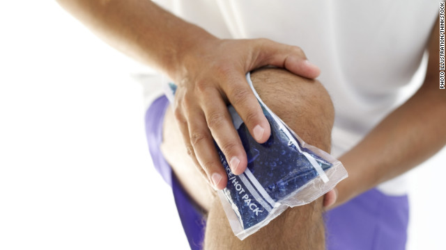


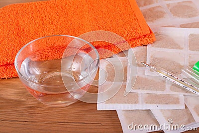