DIAGNOSTIC AND TREATMENT OF HEPATIC AND BILIARY SYSTEM DISEASES IN OUTPATIENT CONDITIONS. DIFFERENTIAL DIAGNOSIS OF JAUNDICE. PROPHYLACTIC MEDICAL EXAMINATION. SANATORIUM TREATMENT. MEDICAL AND LABOR EXPERTISE.
CHRONIC CHOLECYSTITIS
Cholecystitis (Greek, –cholecyst, “gallbladder”, combined with the suffix -itis, “inflammation”) is inflammation of the gallbladder, which occurs most commonly due to obstruction of the cystic duct with gallstones(cholelithiasis). Blockage of the cystic duct with gallstones causes accumulation of bile in the gallbladder and increased pressure within the gallbladder. Concentrated bile, pressure, and sometimes bacterial infection irritate and damage the gallbladder wall, causing inflammation and swelling of the gallbladder. Inflammation and swelling of the gallbladder can reduce normal blood flow to areas of the gallbladder, which can lead to cell death due to insufficient oxygen. Not everyone who has gallstones will go on to develop cholecystitis.
Actuality of problem. Disease of gall-bladder and bile ducts represent a widespread pathology of internal organs, that is observed in 10-15% of the population in the developed countries. Despite obvious success, achieved during last decades, in diagnosis and treatment of these diseases, still there are some unsolved questions and contradictions concerning their interpretation. First of all it touches upon the relations between gall-stone diseases, chronic gall-bladder disease and diskinetics of bile ducts. That’s why studying the diagnostic criteria of gall-bladder diseases and bile ducts are actual.
CLINICAL ANATOMY OF BILE DUCTS
Bile, produced by hepatic cells, enters intercellular bile channels, which are formed by bilious poles of two or more adjacent hepatic cells. The membrane of hepatic cells constitutes the wall of bile channels . Intercellular bile channels uniting at the periphery of hepatic particle, form larger perilobular bilious channels which fall in to interlobes bilious channels. These channels make shunt between each other, increase in their sizes, and turn into large lobe channels. On the lower liver surface, in the area of lumbar furrow, the left and right bile channels unite, and form general hepatic channel. Mirizi sphincter is located in the place of hepatic channels confluence. Mirizi sphincter regulates the bile reception into a gall-bladder and hinders its reverse flow into the liver. A general hepatic channel joins with gall-bladder, making-up a general bilious channel (choledochus). Lyutcens’ sphincter is situated in the place of gall-bladder neck transition into gall-bladder channel, and regulates the reception of bile from the gall-bladder into gall-bladder channel and choledochus. The distal part of general bilious channel is extended. In its wall there is the layer of smooth musculature – sphincter of hepato-pancreatis ampoule – sphincter Oddi, that resists bile flowing from a general bilious channel into a duodenum. Hepatic cells produce bile constantly, but into the duodenum it comes during digestion only (secretory pressure in the liver is constantly supported at a level not less than 300 mm of water post). Synchronous contraction and weakening of the gall-bladder, sphincters and channels is one of key moments of bilious system function during reception meal.

The Fig. 1. Anatomy of bile ducts
1– Branches of hepatic duct.
2 – Common hepatic duct and Mirizi sphincter/
3 – Cystic duct.
4 – Cystic duct.
5 – Common bile duct.
6 – Sphincter Oddi.
7 – Duodenum.
8 – Pancreatic duct.
P.S. :
· Common hepatic duct formed by junction of right and left hepatic bile ducts.
· Cystic duct connects common hepatic duct with gallbladder.
· Common bile duct formed by junction of cystic duct and common hepatic duct.
The Fig. 2. Gallbladder Anatomy
Regulation of bile excretion is carried out by hormones definite, such as gastrin, secretin, cholecystokinin which stimulate secretion of bile and empty the gall-bladder; somatostatin and vasoactive intestinal polypeptid do the opposite.
Belong to the most important components of bile – Water (82%), bile acids (12%), letsetin and other fosfolipdi (4%), non-estherified cholesterol (0,7%), and other components: biliroubin, proteins, electrolytes, slime. Bile presents by itself micellear solution. Bilious acids, cholesterol and fosfolipids, are components of micella. Concentration of bilious acids has to exceed the quantity of cholesterol in 10 times to ensure normal micellation. Bile acids in hepatic cells are synthesized from cholesterol in presence of enzyme 7a-gidrocsilase. Two bile acids are synthesized from cholesterol in the liver – cholic and chenodesoxycholic (“primary bilious acids”). Conjugation of primary bile acids with taourin and glycin, forming salts as a result takes place in hepatocytes. In the intestine salts of primary bilious acids turn into the secondary bilious acids ( desoxycholic and lithocholic). 90% bilious acids are absorbed from intestine into blood and enter the liver again.
The Fig. 3. The bile travels through cystic duct into the common bile duct, then unites with the pancreatic duct to empty bile into the duodenum.
Evacuation of bile from the gall-bladder in time and absence of bile stagnation play an important role in prevention of violations of bile colloid stability.
Chronic without gall-stone holetcystitis is polyetiologic inflammatory disease of gall-bladder wall, accompanied by deteriotation of its motoric function and absorbing ability, structure and bile properties.
Chronic acalculous holetcystitis (CAC) is observed more frequently in women than in men. Correlations, according to various authors ranges 1:3,- 1:4. CAC occurs considerably, than it is diagnosed. The diagnosis of chronic cholecystitis in most developed countries is put only in case of complicated gall-stone disease or obvious inflammatory process.
ETIOPATHOGENEZIS
To the basic causal factors of SAS belong development:
1) infection;
2) impairment of bile outflow ;
3) suppression of immune status;
4) presence in bile the agents, that irritate the mucous membrane of gall-bladder;
Today an infectious factor is not the main factor. Pathogenic staphylococci, streptococci, intestinal stick and others are more frequent reasons of of acute cholecystitis development. Reduction of immunity, presence of of chronic infection focuses, period after viral hepatitis, stagnation of bile at hypotonic discinesis . assists the development of infection in bilious ways. An infection can get into the gall-bladder by contact (from an intestine), or limfogenic or gematogenic way from any focus of chronic inflammation in the organism (parodontoz, chronic tonsillitis, pielit, etc.).
An inflammatory process in a gall-bladder can be formed without participation of infection. Any temporal or permanent violations of outflow of bile lead to the increase of osmotic pressure in a gall-bladder. Dilatation and edema of its walls appear and there can be breaks and heart attacks with the secondary aseptic inflammation.
One of the principal reasons of chronic holetsistis origin is the irritation of mucous shells by different agents. The products of grilling fats, chemical food additions, belong to them, abuse by rich food, that results permanent receipt of bilious acids, especially desoxycholates, which irrilate the mucus shell of gall-bladder.
CLASSIFICATION
I. BASEDON DEGREE OF: II. ON PHASE OF
DISEASE SEVERITY:
1) MILD FORM; 1) EXACERBATION
2) MODERATE; 2) ATTENUATION
EXACERBATION
3) SEVERE. 3) UNSTABLE REMISSION;
4) STABLE REMISSION.
III. ON PRESENCE OF COMPLICATIONS:
1) UNCOMPLICATED;
2) COMPLICATED BY PERICHOLECYSTITIS;
3) COMPLICATED BY CHOLANGITIS OR ANGIOHOLITIS;
4) COMPLICATED BY HEPATITIS (PANCREATITIS,GASTRITIS, DOUODENIT IS ETC.).
IV. ON PRESENCE OF CONCOMITANT SYNDROMES:
1) NEURASTHENIC; 4) SOLAR;
2) CARDIAC; 5) OBESITY;
3) HYPOTHALAMIC; 6) ALLERGIC.
CLINICAL MANIFESTATIONS
It is characterized by the presence of pain, dyspeptic syndrome and common displays.
(Algesic) Pain syndrome – is conditioned, mainly, by motive violations in the bilious system, and inflammatory process in the gall-bladder.
Characteristic pain in the right subcostal area with an irradiation to the neck, shoulder, right shoulder-blade, back, in the area of heart; arises after the reception of alcohol, rich, fried food and, is accompanied by nausea, bitter taste and dryness in the mouth.
At chronic acalculus choletcystitis, that runs on a background of hypertensive discinezis, pain is fit-like, the picture can be same like at colic, but intensity of pain and duration are less, than at bilious goal-stone illness. Hypotonic discinezis is observed in these patients more frequently. In these case the pain in the right subcostal area is permanent, aching, nonintensive, strengthening at violation of diets and shaking ride.
Dyspeptic syndrome – patients complain on feeling of heaviness in the right subcostal area , epigastriс area, stomach swelling, nausea, bitter taste in the mouth, violation of excrements. The excretion of bile at interdigestive period can cause bilious reflux, nausea, bitter taste in the mouth. The decrease of maintenance in the bile of bilious acids leads to violation of digestion , especially hydrolysis and suction of fats . Besides, bilious acids stimulate motoric of bowels, their deficit can be the reason of low pressured constipation, and excess of bilious acids – causes hologenic diarrhea.
The common displays are a weakness, headache, arising of body temperature to the subfebril numbers, periodic fever, pain in joints, in the area of heart, tachycardia.
At the objective inspection –subicterus of the sclerar, soreness at palpation in the projections of gall-bladder, positive Kerr’ symptom (appearance or strengthening of pain at pat on height of inhalation in the gall-bladder point – place of crossing of costal arc with the right direct muscle of stomach), Murphy’ symptom (pain at palpatsion in the right subcostal area during deep inhalation), phrenicus-symptom (symptom of Myusi — pain at pressure between the legs of right sterno-cleido-masteideus muscle). At joining of inflammation in bilious channels positive Ortner’ symptom (pain at pat on the edge of right costal arc).
DIAGNOSTICS (data of laboratory-instrumental methods )
At research of blood in the phase of exacerbation of chronic holetsistitis especially caused by pathogenic microflora, moderate leycotsitozis is exposed, neytrofilozis, insignificant acceleration of ESR. These changes are accompanied clinical by fever, and the positive results of bacteriological research of bile. There are forms of holetsistitis without inflammatory process marks, with normal results of general blood test more frequently today.
Duodenal probing as a method of diagnostics of cholecystitis lost its value, because it was proved, that leucocytes in bile – indicators of inflammatory process- are very quickly collapse. It is possible to define the character of discinezis which supports the inflammatory reaction of mucous shell. Therefore at biochemical research, clotting of bile is marked up, its specific gravity grows, PH changes into the sour side, violation of bilious acids constant and is reduction is observed, then they annoy the mucous shell of gall-bladder. This method is used, mainly, for microbiological research of bile, determination of microflora, that caused inflammation, research of its sensitiveness to antibiotics, exposure of presence lamblia. But results require careful interpretation, because bile to collect in sterile conditions practically is not possible and more frequent sowed microflora enters to a test tube from oral cavity, thin bowel. The positive results of bacteriological research must be compared with the clinics of disease. It is necessary to take in to account the character of pain (has permanent character, and appear after meals), rise of body temperature, the phenomena of intoxication, changes of general blood test (leycocytozis, increasing of ESR).
The Fig. 4. Duodenal probing
Real-time ultrasonography is the method of choice for diagnosing chronic holetcystitis.
The Fig. 5. The ultrasonography gallbladder
The longitudinal cut of gall-bladder is pear-like. Normally gall-bladder in length is 8-
The Fig. 6. USD gallbladder (
The Fig. 7. The ultrasonography of gates of liver: 1– cystic duct, 2 – common bile duct, 3 – portal vein, 4 – gallbladder, 5 – hepatic artery.
The Fig. 8. USD. Chronic acalculous holetcystitis
Mild form of cholecystitis is characterised by short acute period for 2-3 days, is easily treated by diets. Substantial changes of functional violation of gall-bladder are absent.
Moderate form of chronic cholecystitis runs with the presence of clear acute periods and periods of remission. Acute period proceeds 2-3 weeks, which is confirmed by clinical, laboratory-instrumental data. The changes of functional tests of liver are exposed (increasing the activity of transaminase due to AlT, moderate rise of timole test, alkaline phosphatase). The changes of biochemical tests are absent during remission.
Severe form of chronic cholecystitis is characterized by nonstoping relapsing passing, by absence of clear long-term remission. The clinic of chronic hepatitis, pancreatitis joins to the clinic of chronic cholecystitis. At USD of gall-bladder expose a presence of the periprocesses, considerable thickening of gall-bladder wall.
GALL-STONE DISEASE. CHRONIC CALCULOUS
Gall-stone disease (cholelythiasis) is polietiological disease when in the gall-bladder (cholecystolitiasis), and sometimes in outflow channels (choledoholitiasis) stones appear.
Gall-stones are crystalline structures which appear in abnormal bile. Stones are divided into cholesterol, pigmented and mixed. Cholesterol and mixed stones occur more frequently (in 90-95% of all cases of gall-stone disease) and consist of cholesterol monohydrate, mixtures of calcium salts, bilious acids and pigments. Pigmental stone consists of calcium bilirubinate .
The Fig. 9. Location gall-stones in bile ducts
The Fig. 10. Location of stone in cystic duct
The Fig. 11. Location of stone in gallbladder and common bile duct
Risk factors
The main risk factors of pigmental stone is demographic factor that is more frequently met at vegetarians and at persons, who live in countries with hot climate, where is high-frequency of hemolytic anemia, malaria and other tropical diseases. Pigmented stone are more frequent observed in eastern people. Gall-stone disease in the countries of Europe,
Obesity, high-caloric diet, medical treatment by clofibrate, hormonal contraceptives, starvation and diabetes mellitus became risk factors as a result of cholesterol excretion strengthening. Pregnancy influences on concrements formation in the gall-bladder -progesteron increases the saturation of bile with cholesterol, decreases motoric of gall-bladder; also most pregnant women considerably add in weight and it leads to the increasing of endogenous cholesterol synthesis. High frequency of cholesterol cholelitiasis is observed in patients with gyperlipidemia of IV type according to Fridrecson.
Decreasing of bilious acids secretion is observed in women of mature age with liver diseases under the influence of female sexual hormones. The excretion of cholesterol totally depends on the liver function: decreasing or violation of bile formation can be the reason of hypercholesterolemia.
One of factors – is violation of water-salt exchange, tissue acidosis, decreasing of liquid using.
The important role in development of Gall-stone disease belongs to the gall-bladder, state of mucous shell and evacuation function. In inflammation there is the considerable excretion of mucous by the mucous shell of gall-bladder that contains big amount of mucopolisacharides, glycoproteins. It leads to bile clotting, forms the conditions for the delay of cholesterol on mucous, where in future the center of gall-stone will be formed. In literature mucopolisacharides, glyocoproteins are known as factors of enucleating.
FACTORS, THAT ASSIST IN DEVELOPMENT OF GALL-STONE DISEASE
I. GENERAL FACTORS:
FEMALE SEX
OLD AGE,
GENETIC PREDISPOSITION
OBESITY
THE INCREASED CONSUMPTION OF LIPIDS,
EASILY ASSIMILATED PROTEINS,
CARBOHYDRATES, CHOLESTEROL,
DISTURBANCES OF LIPID’S EXCHANGE
MEDICAL FACTORS (RECEPTION OF LIPIDO-REDUCING PREPARATIONS, CONTRACEPTIVES), STARVATION
DISTURBANCES OF WATER-SALT METABOLISM,
II. LIVER FACTORS:
-REDUCTION OF HOLATO-FORMATION FUNCTION OF LIVER,
–RISE OF HOLESTEROL EXCREATION FUNCTION OF LIVER
-REDUCTION OF EXCRETION OF WATER AND ELECTROLYTES BY EPITHELIUM OF INTERHEPATIC BILIOUS CHANNELS,
-VIOLATION OF MICELLA-FORMATION.
III. GALL-BLADDER FACTORS:
-HYPOTONIC-HYPOKINETIC DISKYNESIS OF GALL-BLADDER
-PRESENCE OF INFECTION IN GALL-BLADDER, INFLAMMATORY CHANGES OF MUCOUS SHELL OF GALL-BLADDER
-VIOLATION OF COLLOID STABILITY OF BILE.
Cholesterol is badly dissolved in water; and normally is in soluble state because of phospholipids and bilious acids. The lithogenisity of bile, its possibility to form a gall-stone is conditioned by strengthening of synthesis of cholesterol, by the reduction of bilious acids secretion and phospholipids. All risk factors of gall-stone disease assist to one or few mechanisms of rise of bile’s litogenisity increase.
CLASSIFICATION
I stage-physical and chemical;
II stage – latent non symptomatic gall-stone carrier;
III stage – clinical, complicated.
At I stage the diagnosis is based on the results of special research of bile- physical and chemical violation of bile (cholesterol “flakes” and precipitation). Clinical, eсhocardiograffic symptoms of the disease are absent. The II stage – is characterized by lythogenic physical and chemical changes of bile, formation of gall-stones in bile outflow channels (more frequent in the gall-bladder),which do not show up clinically, and can be diagnosed at ultrasound research. III stage – symptomatic stage depends on location of gall-stones, their size, composition and quantity, activity of inflammation, functional state of bile out-flow system, and also on character and degree of other organs lesion.
ClinicAL HISTORY
The typical pictures of gall-stone disease are attacks of hepatic colic, as a result of moving concrements from gall-bladder to the channels. Pain is conditioned by spastic contraction of gall-bladder, rising pressure by in it. Pain arises up suddenly, localizes in the right subcostal area or very often in epigastric area, irradiates into the right hand, right shoulder-blade, is accompanied by nausea, repeated vomiting that does not bring facilitation. Pain is easily taken off by baralgin, spazmolgon. If temperature rises, it is possible to think about joining of inflammatory process. Soreness in the projection of gall-bladder is objectively determined and increased gall-bladder sometimes can be palpated. At inflammation there are positive symptoms of Merfi, Ker, Vasilenco, Schyotkin-Blyumberg. If stone even briefly obturates choledohus, patients complain of darkening of urine after colic, and also excrements can be discolored. At dyspeptic form dyspeptic violations and dull pain are marked only in the right subcostal area, concrements can be exposed only at planned inspection .
Fig. 12. This image shows localization of pain at the gall-stone disease
1– Pancreatic area
2 – Gallbladder
3 – Shoulder area
COMPLICATIONS
Development of acute and chronic cholecystitis, secondary pancreatitis is the most frequent complication of gall-stone disease. Severe complication is hepatic icterus with following cholestasis. It arises up at obturation of general bilious channel by the stone. At valve stone icterus can have intermittent character. In these case USR and ERCP are informative. Hydropsy of gall-bladder can develop as a result of obturation of gall-bladder’s channel. Then pain disappears, and at objective examination increased gall-bladder is palpated.
LABORATORY AND INSTRUMENTAL FINDINGS
The Duodenal sounding practically is not important, more over it is dangerous because may cause the attack of hepatic colic. If concrements are not present, but there is a suspicion on a lithogenisity of bile, the defined diagnostic value has biochemical research of gall-bladder bile. Research of maintenance of cholesterol, phospholipids, bilious acids is conducted.
USR is the method of direct diagnostics of gall-stone disease. Echographical concrements in gall-bladder are determined as hyperechogenic structures which make acoustic shades. Concrements are usually mobile; their position is changing when the possition of body is changed. It is considerably difficult to display concrements in bilious channels. Static B mode ultrasonography and oral cholecystography are also sensitive and specific.
Several diagnostic methods— Endoscopic retrograde cholangiopancreatography (ERCP).
Direct cholangiography by either ERCP or percutaneous transhepatic cholangiography has a small but definite incidence of sepsis and of failure. Ultrasound and computer tomography reliably detect ductal dilation as evidence of obstruction; however, obstructing stones often are associated with nondilated ducts.
Fig. 13. ERCP. Location of stone in common bile duct
Fig. 14. Cholecystitis can be seen on a cholangiogram. Radio-opaque dye is used to enhance the X-ray. Multiple stones are present in the gallbladder
Fig.
Fig. 16. USD. Chronic cholecystitis . Location of stone in gallbladder
Fig. 17. Computed axial tomography (CAT) scan
Fig. 18. Cholecystolithiasis. CT scan of the upper abdomen showing multiple gallstones
VIDEO: Galblader ultrasound appearance
in chronic cholecystitis and multiple gallstones
DISKYNESIS OF GALL-BLADDER AND BILE DUCTS
This is unpunctual, incomplete or excess contraction of gall-bladder and sphincters (Oddi, Lyutcens-Martinov, Miritsi).
Depending on an etiologic reason of the origins, there are two types of discinesis of gall-bladder and sphincters of bilious channels: primary and secondary.
Primary discinesis is caused by violation of the neurogumoral regulation of gall-bladder and sphincters activity, conditioned by the presence of neurosis, neuroendocrinal violations, diseases of thyroid gland, by changes of ovaries in the period of the sexual maturing or in a climacteric, period, vegetative and vascular dystonus, diencephalic syndrome.
Development of secondary dyskinesis is related to the presence of pathological reflexes and violation of motoric and secretory function of other organs of digestion tract. Secondary dyskinesis arises up on a background of chronic gastritis, gastroduodenitis, ulcerous disease, chronic diseases of intestines, accompanies organic diseases of gall-bladder such as chronic cholecystitis, congenital defect and development of gall-bladder, bilious channels.
Classification. Depending on the character of violations of gall-bladder motoric function and sphincter tone of hepato-pancreatic ampoule the following types of gall-bladder dyskinesis are distinguished:
1) hypertensive-hyperkinetic;
2) hypotensive-gypokinetic
3) mixed.
Diagnosis. Hypertensive-gyperkinetic dyskinesis occurs more frequently is met in persons with advantage of parasympatic nervous system, hypotensive-gypokinetic- with advantage of sympatic nervous system.
Main clinical symptoms:
1. Age of patient – more frequently are young women with mental and emotional lability.
2. Asteno-vegetative syndrome.
3. Pain syndrome (the character of pain in the right under-costal area depends on the type of dyskinesis: dull, unsteady – at gypomotoric; periodic, paroxysm-like, that sometimes achieves a bilious colic – at hypertensive dyskinesis).
4. Presence of dyspeptic syndrome (nausea, sometimes vomiting, feeling of bitter taste in the mouth, meteorism).
5. Atonic constipation (at hypotensive dyskinesis).
6. Alternation of constipation and diarrhea (at hypertensive dyskinesis).
Diagnosis is based on:
1. Normal laboratory indexes.
2. Informative method is the multi-moment duodenal probing that helps to delimit violation of motoric and evacuation function of gall-bladder and violation of sphincters of extrahepatic bilious channels.
Data of the multi-moment fractional probing :
a) hypertensive-gyperkinetic type- lengthening of period of first phase;lengthening of time of closed sphincter Oddi; reduction of duration B-phase at normal volume of gall-bladder bile, or protracted, interruption of portion B excretion;
б) gypotonic–gypokinetic type – shortening of closed sphincter Oddi phase, lengthening of phase B and increasing of gall-bladder bile volume.
At both dyskinesis microscopic research of bile does not show elements of inflammation are not shown.
Table 1
DATA of multi-moment duodenal probing
|
Phase |
Name |
Color |
Duration min. |
Quantity of bile |
|
I
|
Choledo-chus |
Lightly-yellow |
10-15 |
15-20 |
|
II |
Closed sphincter Oddi |
Lightly-yellow |
3-6 |
absent |
|
III |
portion A beginning of sphincter opening Oddi |
Lightly-yellow |
3-5 |
3-5 |
|
IV
|
portion “B” beginning and ending of sphincter opening Lyutcensa |
dark-olive |
20-30 |
30-50 |
|
V |
portion “C” of sphincter opening Miritsi |
stramineous |
10-20 |
10-30 |
The mechanical irritation of duodenum attends opening sphincter Oddi and excretion of lightly-yellow bile from the common bilious channel to duodenum (choledochus-phase). After entering through the probe of 40 ml 33% solution of magnesium sulfate into the duodenum, sphincter Oddi closes (phase of closed sphincter Oddi). Then sphincter Oddi opens and excretes lightly—yellow bile (portion “A”). Opening of sphincter of Lyotcensa -Martinova is characterized by appearance of darkly-brown viscid gall-bladder’s bile. Probing is ending by opening of Miritsi sphincter and excreting of amber-colored bile (portion “C”).
2. Data of ultrasonic research of retraction power of gall-bladder:
a) The hypertensive type- 40 minutes after the bile-stimulating breakfast -gall-bladder retracts more than 2/3 of volume
б) Hypertensive type – stretched, increased gall-bladder is observed at USR, emptying less than 1/3 of volume after 40 minutes of bile-simulative breakfast.
The Fig. 19. The ultrasonography of gall-bladder with two bends
MEDICAL TREATMENT
Principles: it has to be directed for removing of pain, dyskinetic violations, influence on an infection and inflammation, correction of digestion and exchanges violations.
During the acute period a frequent (4-5 per day) diet is applied with reduction of volume and caloric content of meal (diet № 5, 5а according to Pevzner).
Medicinal medical treatment:
I removing of pain syndrome – anesthetics and spasmolytic preparations
parenteral (no-spa – 2 ml 2 % solution, papaverin hydrochloride-2 ml 1% solution, galidor – 2 ml 1% solution, platifilin gydrotartrat -2 ml 0,2 % solution; at expressed pain syndrome simultaneously entering analgin – 2ml 50 % solution, baralgin – 5 ml, fortral – 1-2 ml 3 % solution, at necessity- promedol – 1ml 2 % solution.
II. To prevent vomiting and dyskinesis entering metoclopramid -2 to ml 0,05 % solution or
peroral domperidon (motilium)-10mg 3-4 times per day before meal, tzizaprid (coordinacs,
prepoulsid) – 10 mg 3 -4 times per day before meal.
III. Antibacterial therapy is provided, taking in to account the sensitiveness of microflora (according to data of bile sowing). It is necessary to prescribe those antibacterial preparations which enter into the bile in high concentration. They are: half-synthetic penicillin (ampicillin, ocsatsilin, ampiocs) – 0,25-
meal, tarivid-0,2 2 times per day); preparations of tetratsiclin (tetratsiclin 0,1 -0,25 g4 times per day, metatsiclin, rondomitsin 0,3 2 times per day, docsitsiclin or vibramitsin 0,1-
claritromitsin 0,25-0,5 2 times per day, eritromitsin
At parasitogenic lesion of bilious channels simultaneously to antibacterial preparations should be prescribed vermocs 1 pill 2-3 times per day, hlocsil
IV. During intoxication – intravenous infusion 5% solution of glucose 200-400 ml,
polidez 250-450 ml.
V. In acute phase, on height of inflammatory syndrome prescribing of holeretics is contra-indicated, and at hypertensive bilious dyskinesis – holicinetics is contra-indicated too.
In the attenuation of acute phase after cancellation of antibacterial preparations, holeretic preparations are prescribed:
1) that contain bilious acids (hologon 0,2-
2) synthetic holeretics, that have anti-inflammatory and antibacterial action (nicodin
0,5
3) holeretics of plant origin (holagogoum 1 capsoule 3 times per day, febihol
1-2 capsules 3 times per day before meal, holosas 1-2 tea -spoon 2-3 times per day, flamin
Holicinetics (prescribed with a purpose to stimulate excretion of bile , to strengthening its outflow via bilious channels, retractive of gall-bladder): magnesium the sulfate 20-25% solution 1 soupspoon inward on an empty stomach 3 times per day; carlovar salt 1 tea-spoon on glass of water at the 3O minutes before meal 3 times per day; csilit, sorbit as a 10% solution on 50-100 ml 2-3 times per day 20 minutes before meal,oil, olive’s , sea-buckthorn’s oils 1 soupspoon 3 times per day before meal.
VI. Physical therapy at attenuation of accute condition : at hypertensive discinezis- electrophoresis; at hypotensive –diodinamic current, impulsive current of low frequency, ultrasonic therapy, balneotherapy- pearly, carbonic baths, at hypertensive discinezis- radon, coniferous, sulphuretted hydrogen).
VII. Mineral water prescribes at attenuation of accute condition , and also during remission -“Borgomi”, “Trouscavetsca”, “Esentouci-17” – room temperature 0,5-1,5 glasses 3 times per day, time of usage depends on the gastric secretion. Sanatoria and health in the phase of remission- in balneological resorts of Trouscavets, Morshin, Gousyatin.
Duration of stationary medical treatment takes 12-14 days, ambulatory- not less than 2 months, and after that the patient is subjected to the clinical supervision.
MEDICAL TREATMENT OF GALL-STONE DISEASE
For patients declining surgical treatment or for whom surgical treatment is inappropriate, gallbladder calculi may sometimes be dissolved in vivo by giving bile acids orally for many months. Stones must not be calcified, the size of concrements should not exceed
The Fig. 20. Papilosphincterotomy
(chart)
Endoscopic retrograde sphincterotomy (ERS) is a therapeutic application of ERCP in which soft tissues and sphincter fibers of the papilla and intraduodenal duct are divided with electrocautery to permit release of ductal calculi into the duodenum. The duct is emptied successfully in 90% of attempted procedures. Mortality (0.3 to 1.0%) and morbidity (3 to 7%) rates are lower than those reported with surgical treatment. Immediate morbidity is caused by bleeding, pancreatitis, perforation, and cholangitis. Late complications include restenosis and reformed stones in open ducts.
The standard operation for gallbladder removal through a right subcostal or midline incision is open cholecystectomy. When performed electively during a period free of complications, the procedure is relatively safe, with a mortality rate of 0.1 to 0.5%. However, since its introduction in 1988, laparoscopic cholecystectomy has been the treatment of choice for symptomatic gallstones. This technique was popularized largely because of a shorter convalescence, decreased postoperative discomfort, and improved cosmetic results. The procedure entails the insertion of specialized surgical instruments and a video camera into the peritoneal cavity through multiple small incisions in the abdominal wall. After insufflation of the peritoneal cavity, the gallbladder is removed under video monitoring. Laparoscopic cholecystectomy is converted to an open procedure in approximately 5% of cases, usually because of an inability to identify the anatomy of the gallbladder or to manage a complication.
DURATION OF TREATMENT
1. Patients with dysfunctional disorders of biliary way should be treated and investigated ambulatory in average during 4 weeks.
EFFICIENCY CRITERIA AND REQUIREMENTS FOR TREATMENT RESULTS
1. Liquidation of clinical and laboratory-instrumental displays of illness, improvement of life quality.
Clinical supervision
Patients with dysfunctional disorders of biliary way
Not foreseen.
DURATION OF TREATMENT
chronic noncalculous cholecystitis
Duration of stationary treatment makes 12-14 days, ambulatory not less than 2 months, whereupon a patient is subject to a clinical supervision.
EFFICIENCY CRITERIA AND REQUIREMENTS FOR TREATMENT RESULTS
Main criteria of efficiency of treatment are decreasing or disappearance of symptoms of biliar dyspepsia, improvement of life quality.
Table 2
Health center treatment of patients with chronic noncalculous cholecystitis
|
Nosologic form |
Frequency of internist’s watching |
Reviews by examination other specialists
|
Diagnostic inspections |
Basic medical and health measures. |
|
Chronic noncalculous cholecystitis |
1-2 times per year. |
Surgeon-according to necessity,dentist-2 times per year |
Clinical blood analysis,urine analysis, feces – 1 time per year, duodenal probing, cholecystography, retromanoscopy, analysis of gastric content according to necessity. |
Regimen, diet, healthcare resort treatment – according to necessity, drug treatment. |
Table 3
Disability examination of patients with chronic noncalculous cholecystitis
|
Disease |
Character of expert conclusion |
Work recommendations |
|
|
Antiindications to work conditions |
Indications to work conditions |
||
|
Chronic noncalculous cholecystitis |
Temporary disabled on the period of sharpening (as a rule 8-12 days) with employment on the lines of TCC. At the frequent sharpening, expressed pain syndrome and impossibility of operative treatment – employment on the line of TCC or MSEC (III group ). Rarely (development of complications, heavy run) – stable loss of workability (II group of disability). |
Considerable and sometimes moderate physical loadings, big nervously-emotional loadings, general and local vibration, longlasted forced position of body, especially with tension of abdominal press muscles, impossibility of the diet regimen observance.
|
Moderate physical loadings. Professions of administrative, economic and intellectual character, which allows observance of the dietary regimen. |
Chronic liver pathology
Chronic liver pathology is one of the burning problems of modern gastroenterology. According to statistic datum in
CHRONIC HEPATITIS is taking an important place among disability and mortality reasons and they have tendention to increas. In patients with chronic hepatitis a big risk of this pathology transition in hepatocirrhosis or hepatocellular carcinoma exist. Hepatocirrhosis is one of the main reasons of inhabitants mortality. It takes 4 place in mortality reasons structure of men 40 years older.
It stipulates actuality of this pathology studying with aim of rapid prescription of etiological and pathogenical treatment and appearing of vital dangerous complications prevention.
Anatomy of the liver and portal venous system
VIDEO 1 VIDEO 2 (liver cellular structure)
Anatomy of the liver and portal venous system
The Fig. 21. Anatomy of hepatobile tract
Anatomy of the liver:
The liver is located in the upper right-hand portion of the abdominal cavity, beneath the diaphragm and on top of the stomach, right kidney and intestines.
Normal function of the liver:
The liver has a multitude of important and complex functions. Some of these functions are to:
· synthesize a number of proteins, including albumin;
· metabolize fats;
· the liver plays a major role in maintaining normal blood sugar levels;
· secrete bile that contains bile acids to aid in the intestinal absorption of fats and the fat-soluble vitamins A, D, E, and K;
· eliminate, by metabolizing and/or secreting, the potentially harmful biochemical products produced by the body;
· detoxify, by metabolizing and/or secreting, drugs, alcohol, and environmental toxins.
Chronic hepatitis – is an polyetiological inflammative liver disease, lasting longer than six months and characterized by progressive run, fibrosis developing and could cause hepatocirrhosis.
Etiological factors:
There are many possible causes including viruses infection, some drugs, alcohol, autoimmune hepatitis and metabolic disorders.
· Contagious viral hepatitis such as hepatitis B, hepatitis C and hepatitis D.
· Medicines.
· Toxins such as alcohol.
· Autoimmune hepatitis. This is a disease in which a number of liver cells are destroyed by the patient’s own immune system. Autoimmune hepatitis can also sometimes occur as acute hepatitis. The cause is unknown.
· Inborn metabolic disorders, such as Wilson’s disease (disorder of the body’s cooper metabolism) and haemochromatosis (disorder of the body’s iron metabolism).
Many cases are idiopathic, when unknown etiology.
Hepatitis B virus (HBV) and hepatitis C virus (HCV) are major causes of chronic hepatitis; 5 to 10% of cases of hepatitis B (with or without hepatitis D virus co-infection) and about 75% of cases of hepatitis C become chronic. Infection with hepatitis A virus or hepatitis E virus is not the cause chronic hepatitis.
PATHOGENESIS
The mechanism of chronicity is uncertain, but a direct cytopathic effect of the virus appears to be only minor, especially with HBV infection; instead, liver injury is largely caused by an immune-mediated host reaction to the infection. The role of hepatitis G virus in chronic hepatitis is not clear.
Various drugs can cause chronic hepatitis, including isoniazid, methyldopa, nitrofurantoin, and possibly acetaminophen. The pathogenesis varies with the drug and may reflect an altered immune response, cytotoxic intermediate metabolites, or genetically determined metabolic defects.
The rare Wilson’s disease may present as chronic hepatitis and should be considered in children and young adults with the disorder. Occasionally, ![]() 1-antitrypsin deficiency produces chronic hepatitis, although inactive cirrhosis is more common.
1-antitrypsin deficiency produces chronic hepatitis, although inactive cirrhosis is more common.
Overwhelming evidence points to immunologic mechanisms of hepatocellular injury in patients with autoimmune hepatitis, including coexisting clinical and serologic immune markers; an association with HLA-B8 and -DR3 haplotypes; extensive periportal infiltration with T lymphocytes and plasma cells; and positive response to therapy with corticosteroids or immunosuppressive drugs.
Chronic hepatitis classification (Los – Angeles 1994 )
According to etiology and pathogenesis:
1. Chronic viral hepatitis B
2. Chronic viral hepatitis D
3. Chronic viral hepatitis C
4. Indefinite chronic viral hepatitis (G)
5. Autoimmune hepatitis (type I – anti-sma and anti-ana; type II – anti-lkm-1;typeIII-anti-sla and etc.)
6. Drug-induced chronic hepatitis
7. Toxic hepatitis
8. Alcohol hepatitis
9. Cryptogenic hepatitis
According to clinical-biochemical and histological factors:
1. Level of activity, which is determined by difficulties of necrotic and inflammative process:
mild; (when ALT exceeds a norm in 3-5 times)
moderate; -«- 5-10
severe. -«- more 10
2. Stage, which determines distribution of fibrosis and hepatocirrhosis developing:
0 – fibrosis is absent;
1 – hardly (mild) determined fibrosis;
2 – moderate fibrosis;
3 – huge (severe ) fibrosis;
4 – hepatocirrhosis:
Note:
At viral hepatitis verification:
A-replication phase;
B-integration phase;
At unverificative hepatitis:
A-acute phase (cytolytic syndrome presence, cholestasis, autoimmune displays, liver insufficiency);
B-remision.
INTERNATIONAL CLASSIFICATION OF DISEASES OF 10TH REVISION
K73.2 Chronic active(lupoid hepatitis), non classified in other chapters
B 18 Chronic viral hepatitis
B 18.0 Chronic viral hepatitis B with d-antigen
B 18.1 Chronic viral hepatitis B without d-antigen
B 18.2 Chronic viral hepatitis C
K 70.0 Alcohol liver fat dystrophy
K 70.1 Alcohol hepatitis (acute, chronic)
K 70.2 Alcohol liver fibrosis and sclerosis
K 71.0 Toxic hepatitis
CHRONIC HEPATITIS CLINIC
The most commons syndromes and symptoms of chronic hepatitis
I. PAIN:
· Periodic light pressure or pain below the right ribs – enlarged liver
II. dyspepsia:
· nausea, poor appetite, changes in taste perception, belch, flatulence
III. Jaundice:
· icterus; ictery of sclerae (the white portions of the eyes) and skin
IV. ASTENOVEGETATIVE DISORDERS (headache,
lethargy, weakness)
IV. LOW–grade fever.
V. weight Loss
VI. ARTHRALGIA (aching muscles) and MYALGIA
VII. Hepatomegaly
VIII. Splenomegaly
X. SYNDROME OF SMALL LIVER SIGNS
· spider-like blood vessels in skin (spider angioma)
and palmar erythema (liver palms)
Clinical features vary. Many patients are asymptomatic, especially in chronic hepatitis C.
The Fig. 23. The liver palms
In the autoimmune variant chronic hepatitis manifestations often occur, especially in young women. These can affect virtually any body system and include acne, amenorrhea, arthralgia, ulcerative colitis, pulmonary fibrosis, thyroiditis, nephritis, and hemolytic anemia.
A minority of patients develop predominant cholestatic features suggesting primary biliary cirrhosis.
Laboratory Findings
These include evidence of active hepatocellular inflammation, with predominant aminotransferase elevations and variable bilirubin and alkaline phosphatase values.
In such cases, other laboratory clues to chronicity may aid the diagnosis (eg, low serum albumin and others).
Cholestatic laboratory features occasionally dominate.
Serologic “immune” markers are common in the autoimmune variant. HBV surface antigen or anti-HCV in serum indicates causation by HBV or HCV, respectively.
Table 4
Biochemical indexes of
|
syndrome |
index |
changes |
notes |
|
cytolitic |
ALT AST LDG(4,5 faction) GGTP Cholinesterase Ceruloplasmine Vit.B12, Fe |
Ý Ý N or Ý Ý Ý Ý Ý Ý |
Activity of ALT and AST could be N at CHC Increasing could be sign of hepatocirrhosis beginning. Level of increasing characterizes difficulty of sharpness. |
|
mesenchymal |
ESR Le C –reactive protein
Albumines
Globulines
Tymol test |
Ý Ý Ý
N or ß
Ý
Ý
|
Hypergammaglobulinemia is characterized for autoimmune hepatitis |
|
cholestatic |
Bilirubin (straight) Alkaline fosfatasa
cholesterol |
Ý
Ý
Ý |
|
|
Hepato-depresive |
Albumin
Cholesterol
Cholinesterasa
Protrombin
Prokonvertyn
Fibrynogen
Estrogens Aldosteron |
N or ß
N or ß
N or ß
N or ß
N or ß
N or ß
Ý Ý
|
|
Chronic viral B hepatitis has two phases, termed the replicative and integrative.
Table 5
Markers of chronic viral hepatitis
|
|
hepatitis |
Replicative phase |
Integrative phase |
|
|||
|
|
B |
HbeAg+HbsAg,HBV-DNA, DNA-P, anti-HBc IgM In liver cell – HBcAg |
HbsAg, anti-Hbe IgG, anti-HBc IgG In liver cell- HBcAg |
|
|||
|
|
D |
HbsAg, anti-HDV IgM, HDV-RNA, anti-Hbe IgM |
– |
|
|||
|
|
C |
HCV-RNA,anti-HCV IgM |
– |
|
|||
|
Table 6 Differentiating viral and alcoholic hepatitis
|
|
||||||
|
|
Viral hepatitis |
Alcoholic hepatitis |
|
||||
|
|
|
||||||
|
History |
Risk factors |
Significant alcoholic intake |
|
||||
|
Physical examination |
Mild hepatomegaly, extrahepatic stigmata not prominent |
Moderate to marked hepatomegaly, florid stigmata |
|
||||
|
Laboratory examination |
AST variable |
AST <300 |
|
||||
|
Liver biopsy |
Mononuclear cells |
Polymorphs |
|
||||
|
|
|
|
|
|
|
||
Immunological indexes:
AMA- antimitochondrial antibodies;
ANA- antinuclear antibodies;
ALKM- liver-kidneys microsomal antibodies;
ALM- antibodies to liver cells membranes;
SLA- antibodies to dissolve liver antigen;
SMA- smouthmuscle antibodies;
CIC- circulative immune complexes.
Histological criterion of chronic hepatitis is the combination of inflammative-cellular infiltration and various forms of hepatocellular degeneration and necrosis, presence of sclerotic changes. One can see widening of portal ways and its inflammative cellular infiltration.
The Fig. 24. The Histological criterion of chronic hepatitis: combination of inflammative-cellular infiltration and various forms of hepatocellular degeneration and necrosis.
The Fig. 25. The Histological criterion of chronic hepatitis: portal ways cellular infiltration
The Fig. 26. Echographic picture of normal liver.
Liver parenchyma structure is homogeneous with weak echosounds from its inner structures. Soundabsorption is normal. Innerliver veins walls are not visible practically.
The Fig. 27. Echographic picture of chronic hepatitis. Echosound of parenchyma is increased and heterogeneous, which is caused by large amount crossing of increased echosound small areas and areas with less echosound.
The Fig. 28. Echographic picture of chronic hepatitis
VIDEO: liver ultrasound appearance in chronic hepatitis
Treatment
Medical diet № 5
I. Etiological:
Standart therapy is provided by recombinant ( which are made by gen engineering) interferons (IFN-a). One useof such of preparations: aferon, velferon, intron, realdyron, reaferon, roferon.
Indications to interferon monotherapy :
1. Young age (not older than 40 years old).
2. Female
3. Normal weight
4. Increased level of GGTP, ALT in blood.
5. Presence of moderate active level of inflammative process and minimal level of fibrosis in liver bioptats.
6. The begining of infection at old age.
7. Short duration of infection process.
8. Active viruse replication, at which one can find DNA HBV(PVR) and HbeAg, anti-HCV IgM.
9. Low DNA HBV level (less,than 200 by method of PCR)
10. Not high RNA HBV level (determined by way of PCR)
11. Infication of HCV genotype 2 or 3.
Unindications to IFN monotherapy:
1. Decompensative hepatocirrhosis (because of hepatitis exacerbation risk at 60 % sick)
2. Autoimmune hepatitis or other autoimmune diseases.
3. Endogenous or exogenous immunsuppression.
4. Depression or other psychic disorders.
5. Other liver diseases, such as Vilson disease, haematochromatosis, a-antitrypsyn deficit.
6. Epylepsy.
7. Leucopenia(<2·109/l), trombocytepenia(<50·109/l)
8. Pregnance.
Viral hepatitis C:
IFN is prescribed of 3 mln. IU 3 times on a week underskin
Standard combinated therapy needs subcutaneous injection 3 mln. IU 3 times for a week in combination with peroral use of rybaviryn (body weight <75kg – 1000 mg per 24 hours, means 2 pills for 200 mg in the morning and 3 pills for 200 mg in the evening, body weight>75kg – 1200 mg per 24 hours,means 3 pills for 200 mg in the morning and 3 pills for 200 mg in the evening)
Viral hepatitis B:
IFN is used for 5 mln.IU per 24 hours or 10 mln.IU 3 times per week subcutaneous.
For sick, to whom therapy with IFN was unsuccessful or which have little bit chance for good therapy by IFN (for sick with normal ALT level or with PRECORE- mutant (without Hbe Ag) HBV with prenatal infication, immunesuppresion, which is caused by HIV infection or ny organs transplantation) supposed to prescribed lamivudyn 100 mg per 24 hours per os during 1 year.
P.S. Priority of combinative therapy on monotherapy by lamivudyn wasn’t found.
II Pathogenetic therapy
Autoimmune hapatitis
Prednisolon 30 mg per os in a day, dose is lowered to 10-15 mg in a day during few weeks; azatiopryn is often prescripbed for 50 mg per os in a day, which allows to low dose of glucocorticoids and to avoid their side effects. It’s necessary to provide functional liver tests every month. It is possible soon to reduce the symptoms, but biochemical remision comes in few month, and histological – in 18-24 month. If we stop to give glucocorticoids – recidivation becomes at 50-90% cases and the demand to repeat treatment will appear. During often recidivations it is prescript small doses of glucocorticoids or asatyopryn per 24 hours.
III Symptomatic therapy
Hepatoprotectors:
I. Plant origin:
Flavonoids:
sylimaryn, sylobinin, sylibor, karsyl, legalon, darsil
hepabene;hepatofalk planta;hofitol
II. Preparations, which have essential phospholipids:
essentiale, essentiale H, forte H, lipin, lioliv
III. Preparations, which have aminoacids:
Heptral(ademiotynin), glutargin
IV. Synthetic preparations:
tiatriazolin,tyoktacyd(tyoctov acid)
V. Preparations which have bile acids:
ursofalk, ursosan, ursohol.
DURATION OF TREATMENT
Duration of stationary treatment on the III – IV level of medical care makes 21-28 days with next treatment in C.R.H., ambulatory – during 3-6 month, up to the year, whereupon a patient is subjected to a clinical supervision.
EFFICIENCY CRITERIA AND REQUIREMENTS FOR TREATMENT RESULTS
Main criteria of efficiency of treatment are normalization of the common state of patient, biochemical, immunological indexes, functional state of liver, patient’s transformation in the compensated state, absence of complications, improvement of life quality.
Table 7
Health center system of patients with chronic cryptogenic hepatitis
|
Nosologic form |
Frequency of internist’s watching |
Reviews by doctors of other specialties |
Diagnostic inspections |
Basic medical and health measures. |
|
Chronic cryptogenic hepatitis |
2 times per year. |
Infectionist 2 times per year, oncolo-gist, neurolo-gist, and endocrinologist – according to necessity. |
General blood analysis with determination of thrombocytes,retyculocytes, blood groups, resus factor(Rh);general urine analysis, feces analysis on hidden blood,biochemical blood analysis: protein fractions, potassium, bilirubin (free and linked), cholesterol, betalipoproteids aminotransferases, activity of LDG (4,5 fractions), glucose, serum iron, alkaline phosphatase,bloods urea, creatinine, fibrinogen,prothrombine index; coprogramm;ultrasound of organs in abdominal cavity, EGDS; ЕKG;markers of virus hepatitis В,С, Д with help of immuneferment method and determination of DNA and (HBV) and RNA (HCV) by means of ПЦР –1- 2 times per year according to indications. |
Regimen, diet, healthcare resort treatment – according to necessity, drug treatment |
Table 8
Disability examination of patients with chronic cryptogenic hepatitis
|
Disease |
Character of expert conclusion |
Work recommendations |
|
|
Antiindications to work conditions |
Indications to work conditions |
||
|
chronic cryptogenic hepatitis |
Temporary disabled on the period of sharpening (as a rule 1,5 – 3 months) with employment on the lines of TCC. Direction on MSEC for establishment of III group, and sometimes II groups of disability. Rarely (development of complications, heavy flowing) stable loss of workability (II group of disability).
|
Considerable and sometimes moderate physical loadings, expressed nervously-emotional loadings, overheats of body, contact with poisons, impossibility of observance of the diet regimen.
|
Light physical loadings. Professions of administrative, economic and intellectual character, which allows observance of the dietary regimen. Rarely – work in specially made conditions, at home. |
Hepatocirrhosis
Hepatocirrhosis – it is chronic diffuse defect, which is characterized by violation of normal liver lobule architectonics due to fibrosis and making of structure – unnormal knots of regeneration, that explains development of liver functional insufficiency and portal hypertension.
Table 9
Causes of cirrhosis
|
Alcohol |
|
Viral hepatitis |
|
Metabolic |
|
Cholestatic |
|
Autoimmune |
|
Drug-induced |
|
Congestive caused by vein outflow from liver |
|
Cystic fibrosis |
Pathogenesis – unreturn process of fibrosis and pathological knots developing.
Classification
I. according to etiology:
viral
alcohol
toxic
conncted with born metabolism disorders
conncted with injury of
(primary and secondary biliar cirrhosis)
caused by vein outflow from liver (Badd – Chiari syndrome, veinocclusive disease, constructive pericarditis)
cryptogenic cirrhosis
II. according to morphological signs:
· active
nonactive
1. micronodule
macronodule
mixed (micro-, macronodule).
III. according to run:
subsharp (hepatitis – cirrhosis)
fastprogressive(active)
slowprogressive(active)
latent.
IV. Stage of disease ( according to Child-Pugh criterion)
A-compensation, B-subcompensation, C – decompensation.
V. Complications:
esophago-gastric bleeding
spontaneous bacterial ascites-peritonitis
trombosis of portak vein
hepatorenal syndrome
hepatocellular carcinoma.
Morphology:
A morphological change at hepatocirrhosis includes: hepatocytes necrosis and dystrophy, hepatocytes nodule regeneration, fibrosis, organs structure change, organs deformation.
The Fig. 29. Liver Lobule, Pig (
The Fig. 30. Shown is a mixed macronodular and micronodular cirrhosis.
Table 10
Criteria for Child-Pugh classification
|
Indexes |
A(compensative) |
B(subcompensative) |
C (decompensative) |
|
Blood bilirubin |
<35 |
35-55 |
>55 |
|
Albumin |
>35 |
30-35 |
<30 |
|
Protrombine index |
60-80% |
40-50% |
39% and ß |
|
Ascites |
– |
Good treated |
Bad treated(resistant ascites) |
|
Encephalopathy |
latent |
Minimal 1-2 stages |
Precoma, coma 3-4 stages |
|
Weight |
normal |
good |
Low(exhaustion) |
Each index of “A ” class is marked in 1 point ; each index of “B” class is marked in 2 points; each index of “C” class is marked in 3 points.
Class “A” answers to 5-6 points; class”B” – 7-10 points; class”C”- >10 points.
ClinicAL HISTORY
1. Astenic syndrome
2. Dyspepsia
3. Pain syndrome
4. Icterus
5. Skin itching
6. Portal hypertension
7. Oedema-ascetic syndrome
8. Hemorrhage syndrome
9. Injury of endocrine system
10. Increasing of body temperature
11. Hepatosplenomegalia
12. Small liver signs(vascular stars, liver palms)
Biochemical indexes of liver at cirrhosis – cytolitic, cholestatic, mesenchymal – inflammative, hepatodepresive syndromes.
The Fig. 31. Spider naevus in liver cirrhosis
The Fig. 32. The ascitis
Imaging Studies: abdominal ultrasonography, CT scan, or MRI.
The Fig. 33. Echographic picture of hepatocirrhosis. Echostructure of liver parenchyma uneven, caused by places of fibrosis, general echosound is increased soundconseption is not changed.
The Fig. 34. Echographic picture of hepatocirrhosis and presence of ascetic fluid in abdominal cavity
The Fig.
Treatment
Treatment of subcompensative hepatocirrhosis.
I. Specific therapy
a) at autoimmune and viral – corticosteroids (prednisolon 20-30 mg per 24 hours during 3-4 weeks with next low on 0,5 mg in every 3 days to support dose 7,5 – 10 mg per 24 hours; to 6 months). At viral hepatocirrhosis interferontherapy is used also.
b) Patients with autoimmune hepatocirrhosis (if there is no antiindications) imuran (asatiopryn) to 25 mg per 24 hours, metotrexat to 7,5 – 15 mg per 24 hours is prescribed.
c) At biliar hepatocirrhosis – ursodesoxycholiv acid (ursofalk) 15-20 mg/kg during 6 months and more; vit.D in dose 400 IU per 24 hours (osteoporosis)
d) At Vilson-Konovalov disease – D-penicylamin in connection with pyridoxine, antioxidants(vit.A, E, C, tokoferol-tryovit.)
e) At hematochromatosis – symptom bloodletting under patient selffeeling control, intramuscular injection of desferal – 500-1000 ml per 24 hours, preparations, which have Zn (gumet – 1 tablespoon 3 times per day.)
f) At a-antitrypsin insufficiency – hepatoprotectors and preparations, which make better breath function of lungs (aminophylin, ventolin).
II. It doesn’t depend from cause of beginning you have to use:
a) vitamintherapy, such as balanced vitamin complexes; ryboxyn, essenciale H during 1-2 months with repeating 2-3 times per year.
b) Hepatoprotective therapy ;
Syndrome of malabsorbtion, maldigestion treatment, normal intestine microflora renewal. Normal intestine flora reimplantation: (bifidumbacteryn, bificol, laktobacteryn, bifi-form, acydofilus, symbiter);
c) Protein change correction: (anabolic steroids, albumin intravenous injection,clean aminoacids mixture: poliamin, alvezin, aminoplasmol, aminosol-600, 800).
Lipoid change correction: (essenciale, lipofundyn, intralipid-i/v)
Carbohydrate change correction: (i/v glucose)
d) desintoxication :
– i/v glucose with vit. from group B, C, neodesis, polydesis;
– enterosorbtion: enterodesis – 5g 3 times per 24 hours, enterosgel – 15g 3 times/24 hours
III at oedema-ascitis syndrome
– bed regimen, sodium use – 0,5-2g/24 hours;
– veroshpiron 75-200mg/24 hours;
– at huge ascetic syndrome: veroshpiron 150-200mg/24hours + uregit 25-100 mg or furosemid 80mg – hypothyasid 100mg+ veroshpiron 200mg. If there is no effect – blood ultrafiltration.
IV for hypersplenism syndrome treating:
– prednisolon 20-40mg/24 hours during 2-3 months with next dose low;
– erythrocytes and thrombocytes mass transfusion.
V chronic liver encephalopathy therapy
– using less of protein to 50g in food ration, mostly animal origin;
– braking of amiakogen microflora in intestines (prescription of monomicin, canamicyn, metronidasol);
– lactulose (dufalac, normaze) 30ml 3-5 times a day after meals;
– hepasol 500ml i/v drops;
– ornicetyl 2-6g/24 hours i/m or 2g 1-2 times/24 hours i/v;
– glutamine acid i/v drops 300-500ml 1%solution or glutargin 50ml 2 times/24 hours i/v (2 pills 3-4 times a day per os
– aminosol 600( 800,KEO 30ml/kg/24 hours);
– metabolic acidosis correction (potassium diet, i/v 50-80ml 4% solution KCl in 500-100ml 5% solution of glucose;
VI Stop of bleeding from varicose extended veins
– severe bed regimen, ice on epigastria;
– i/v drop 1-
– ¯ of portal pressure: vasopresyn 20 U in 100-200ml 5% sol. of glucose, if necessary – repeat every 4 hours; somatostatin i/v stream 1 dose 250mkg and than i/v drop 250 mkg during 24 hours (3 mg in 12 hours),treatment duration from 24 to 120 hours;
– hemostatic therapy; i/v stream 400-600ml of freshiced plasma;
– decinon i/m or i/v 250-500mg; vikasol 1ml 1% of solution i/m;
– antihemophil plasma 100-150 ml i/v drop;
– CaCl or gluconat 10 ml 10% sol. i/v
Liver transplantation
With the dramatic improvement in results, liver transplantation became the recognized management for end-stage liver disease. A patient should be considered for liver transplantation when the diagnosis of irreversible end-stage liver disease is made. The selection of appropriate candidates out of a large number of patients with liver disease combined with the relative scarcity of available organs requires a strict individual assessment, which must to a certain extent be tailored to the cause of liver failure. Transplantation for hepatitis B remains controversial, and physicians should be aware of their transplant center’s policy when considering patients for referral. Exclusion of patients with contraindications to liver transplantation (Table ) allows the best use of scarce donor resources while maximizing patient benefit.
Liver transplantation: contraindications
|
1.Absolute |
|
2. Relative |
DURATION OF TREATMENT
Duration of stationary treatment on the III – IV level of medical care makes 28 days with next treatment in C.R.H., ambulatory (according to necessity) – during month, whereupon a patient is subjected to a clinical supervision.
EFFICIENCY CRITERIA AND REQUIREMENTS FOR TREATMENT RESULTS
Main criteria of efficiency of treatment are improvement of the common state of patient, biochemical, immunological indexes, functional state of liver, patient’s improvement of life quality.
Table 11
Health center system of patients with chronic liver cryptogenic cirrhosis
|
Nosologic form |
Frequency of internist’s watching |
Reviews by doctors of other specialties |
Diagnostic inspections |
Basic medical and health measures. |
|
Chronic liver cryptogenic cirrhosis |
3-4 times per year |
Infectionist, oncolo-gist, neurolo-gist, and endocrinologist – according to necessity. |
General blood analysis with determination of thrombocytes,retyculocytes, blood groups, resus factor(Rh);general urine analysis, feces analysis on hidden blood,biochemical blood analysis: protein fractions, potassium, bilirubin (free and linked), cholesterol, betalipoproteids aminotransferases, activity of LDG (4,5 fractions), glucose, serum iron, alkaline phosphatase,bloods urea, creatinine, fibrinogen,prothrombine index; coprogramm;ultrasound of organs in abdominal cavity, EGDS; ЕKG;markers of virus hepatitis В,С, Д with help of immuneferment method and determination of DNA and (HBV) and RNA (HCV) by means of ПЦР –1- 2 times per year according to indications. |
Regimen, diet, healthcare resort treatment – according to necessity, drug treatment. |
Table 12
Disability examination of patients with chronic liver cryptogenic cirrhosis
|
Disease |
Character of expert conclusion |
Work recommendations |
|
|
Antiindications to work conditions |
Indications to work conditions |
||
|
Chronic liver cryptogenic cirrhosis |
An individual estimation of capacity in dependence on the form of cirrhosis, activity and stage of process, complications. Rarely (stabilization of process in the early stage of cirrhosis) a capacity at the proper employment by TCC can be stored. Usually is stable disability (II group of disability, at stable compensation of process – III group of disability, sometimes, at progress of portal hypertension, presence of complications – I group of disability ) |
Constant and inconstant moderate physical loads. Considerable nerve-psychic loadings. Forced body position, especially with tension of abdominal press muscles. Contact with poisons, vibration, body overheating, and impossibility of the diet regimen observance.
|
Light physical loadings. Professions of administrative, economic and intellectual character, which allows observance of the dietary regimen. Rarely – work in specially made conditions, at home. |
JAUNDICE
Jaundice (also known as icterus; from the Greek word ίκτερος, attributive adjective: icteric) is a yellowish pigmentation of the skin, the conjunctival membranes over the sclerae (whites of the eyes), and othe rmucous membranes caused by hyperbilirubinemia (increased levels of bilirubin in the blood). This hyperbilirubinemia subsequently causes increased levels of bilirubin in the extracellular fluid. Concentration of bilirubin in blood plasma is normally below 1.2 mg/dL (<25µmol/L). A concentration higher than 2.5 mg/dL (>50µmol/L) leads to jaundice. The term jaundice comes from the French word jaune, meaning yellow.
Hyperbilirubinemia could be caused by
· increased bilirubin production
· decreased uptake into the liver cells
· impaired conjugation
· interference with the secretion of conjugated bilirubin.
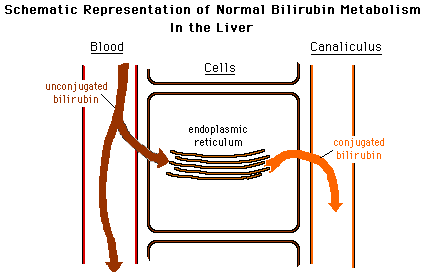
Here is a schematic diagram that represents normal bilirubin metabolism. Use this to compare with various abnormal states described below. Clicking on the diagrams will reveal larger versions of them.
Here are some examples of diseases in which hyperbilirubinemia is observed.
Hemolytic jaundice

· results in increased production of bilirubin.
· Here more bilirubin is conjugated and excreted thaormally, but the conjugation mechanism is overwhelmed, and an abnormally large amount of unconjugated bilirubin is found in the blood.
Gilbert’s disease

· may be caused by an inability of the hepatocytes to take up bilirubin from the blood.
· As a result, unconjugated bilirubin accumulates.
Physiological jaundice and Crigler-Najjar syndrome
· are conditions in which conjugation is impaired.
· Unconjugated bilirubin is retained by the body.
Dubin-Johnson syndrome
· is associated with inability of the hepatocytes to secrete conjugated bilirubin after it has been formed.
· Conjugated bilirubin returns to the blood.
Biliary obstruction
· by (for example) biliary calculi causes backup and reabsorption of conjugated bilirubin.
· Blood levels of conjugated bilirubin increase.
Jaundice is often seen in liver disease such as hepatitis or liver cancer. It may also indicate leptospirosis or obstruction of the biliary tract, for example by gallstones or pancreatic cancer, or less commonly be congenital in origin (e.g., biliary atresia).
Yellow discoloration of the skin, especially on the palms and the soles, but not of the sclera and mucous membranes (i.e. oral cavity) is due to carotenemia—a harmless condition[4] important to differentiate from jaundice.
Signs and symptoms
The conjunctiva of the eye are one of the first tissues to change color as bilirubin levels rise in jaundice. This is sometimes referred to as scleral icterus.
However, the sclera themselves are not “icteric” (stained with bile pigment) but rather the conjunctival membranes that overlie them. The yellowing of the “white of the eye” is thus more properly termed conjunctival icterus. The term “icterus” itself is sometimes incorrectly used to refer to jaundice that is noted in the sclera of the eyes, however its more common and more correct meaning is entirely synonymous with jaundice.
Jaundice is a sign of an underlying disease process. .
Common signs and symptoms seen in individuals with jaundice include:
· yellow discoloration of the skin, mucous membranes, and the whites of the eyes,
· light-colored stools,
· dark-colored urine, and
· itching of the skin.
The underlying disease process may result in additional signs and symptoms. These may include:
· fever,
· weakness,
· loss of appetite,
· headache,
· confusion,
· swelling of the legs and abdomen,
Iewborns, as the bilirubin level rises, jaundice will typically progress from the head to the trunk, and then to the hands and feet. Additional signs and symptoms that may be seen in the newborn include:
· poor feeding,
· lethargy,
· changes in muscle tone,
· high-pitched crying, and
· seizures.
Differential diagnosis
When a pathological process interferes with the normal functioning of the metabolism and excretion of bilirubin just described, jaundice may be the result. Jaundice is classified into three categories, depending on which part of the physiological mechanism the pathology affects. The three categories are:
![]()
Types of jaundice
|
Category |
Definition |
|
Pre-hepatic/ hemolytic |
The pathology is occurring prior to the liver. |
|
Hepatic/ hepatocellular |
The pathology is located within the liver. |
|
Post-Hepatic/ cholestatic |
The pathology is located after the conjugation of bilirubin in the liver. |
Pre-hepatic
Pre-hepatic jaundice is caused by anything which causes an increased rate of hemolysis (breakdown of red blood cells). In tropical countries, malaria can cause jaundice in this manner. Certain genetic diseases, such assickle cell anemia, spherocytosis, thalassemia and glucose 6-phosphate dehydrogenase deficiency can lead to increased red cell lysis and therefore hemolytic jaundice. Commonly, diseases of the kidney, such ashemolytic uremic syndrome, can also lead to coloration. Defects in bilirubin metabolism also present as jaundice, as in Gilbert’s syndrome (a genetic disorder of bilirubin metabolism which can result in mild jaundice, which is found in about 5% of the population) and Crigler-Najjar syndrome.
In jaundice secondary to hemolysis, the increased production of bilirubin, leads to the increased production of urine-urobilinogen. Bilirubin is not usually found in the urine because unconjugated bilirubin is not water-soluble, so, the combination of increased urine-urobilinogen with no bilirubin (since, unconjugated) in urine is suggestive of hemolytic jaundice.
Laboratory findings include:
· Urine: no bilirubin present, urobilinogen > 2 units (i.e., hemolytic anemia causes increased heme metabolism; exception: infants where gut flora has not developed).
· Serum: increased unconjugated bilirubin.
· Kernicterus is associated with increased unconjugated bilirubin, neonates are especially vulnerable to this due to increase permeability of the blood brain barrier.
Hepatocellular
Hepatocellular (hepatic) jaundice can be caused by acute or chronic hepatitis, hepatotoxicity, cirrhosis, drug induced hepatitis and alcoholic liver disease. Cell necrosis reduces the liver’s ability to metabolize and excrete bilirubin leading to a buildup of unconjugated bilirubin in the blood. Other causes include primary biliary cirrhosis leading to an increase in plasma conjugated bilirubin because there is impairment of excretion of conjugated bilirubin into the bile. The blood contains abnormally raised amount of conjugated bilirubin and bile salts which are excreted in the urine. Jaundice seen in the newborn, known as neonatal jaundice, is common iewborns as hepatic machinery for the conjugation and excretion of bilirubin does not fully mature until approximately two weeks of age. Rat fever (leptospirosis) can also cause hepatic jaundice. In hepatic jaundice, there is invariably cholestasis.
Laboratory findings depend on the cause of jaundice.
· Urine: Conjugated bilirubin present, urobilirubin > 2 units but variable (except in children). Kernicterus is a conditioot associated with increased conjugated bilirubin.
· Plasma protein show characteristic changes.
· Plasma albumin level is low but plasma globulins are raised due to an increased formation of antibodies.
Bilirubin transport across the hepatocyte may be impaired at any point between the uptake of unconjugated bilirubin into the cell and transport of conjugated bilirubin into biliary canaliculi. In addition, swelling of cells and oedema due to inflammation cause mechanical obstruction of intrahepatic biliary tree. Hence in hepatocellular jaundice, concentration of both unconjugated and conjugated bilirubin rises in the blood. In hepatocellular disease, there is usually interference in all major steps of bilirubin metabolism—uptake, conjugation and excretion. However, excretion is the rate-limiting step, and usually impaired to the greatest extent. As a result, conjugated hyperbilirubinaemia predominates.
The unconjugated bilirubin still enters the liver cells and becomes conjugated in the usual way. This conjugated bilirubin is then returned to the blood, probably by rupture of the congested bile canaliculi and direct emptying of the bile into the lymph leaving the liver. Thus, most of the bilirubin in the plasma becomes the conjugated type rather than the unconjugated type, and this conjugated bilirubin which did not go to intestine to become urobilinogen gives the urine the dark color.
Post-hepatic
Post-hepatic jaundice, also called obstructive jaundice, is caused by an interruption to the drainage of bile in the biliary system. The most common causes are gallstones in the common bile duct, and pancreatic cancer in the head of the pancreas. Also, a group of parasites known as “liver flukes” can live in the common bile duct, causing obstructive jaundice. Other causes include strictures of the common bile duct, biliary atresia, cholangiocarcinoma, pancreatitis and pancreatic pseudocysts. A rare cause of obstructive jaundice is Mirizzi’s syndrome.
In complete obstruction of the bile duct, no urobilinogen is found in the urine, since bilirubin has no access to the intestine and it is in the intestine that bilirubin gets converted to urobilinogen to be later released into the general circulation. In this case, presence of bilirubin (conjugated) in the urine without urine-urobilinogen suggests obstructive jaundice, either intra-hepatic or post-hepatic.
The presence of pale stools and dark urine suggests an obstructive or post-hepatic cause as normal feces get their color from bile pigments. However, although pale stools and dark urine are a feature of biliary obstruction, they can occur in many intra-hepatic illnesses and are therefore not a reliable clinical feature to distinguish obstruction from hepatic causes of jaundice.
Patients also can present with elevated serum cholesterol, and often complain of severe itching or “pruritus” because of the deposition of bile salts.
No single test can differentiate between various classifications of jaundice. A combination of liver function tests is essential to arrive at a diagnosis.
|
Table of diagnostic tests |
|||
|
Function test |
Pre-hepatic Jaundice |
Hepatic Jaundice |
Post-hepatic Jaundice |
|
Total bilirubin |
|
Increased |
|
|
Conjugated bilirubin |
|
Increased |
Increased |
|
Unconjugated bilirubin |
|
Increased |
|
|
Urobilinogen |
|
Decreased |
Decreased / Negative |
|
Urine Color |
|
Dark (urobilinogen + conjugated bilirubin) |
Dark (conjugated bilirubin) |
|
Stool Color |
|
Normal/Pale |
Pale |
|
Alkaline phosphatase levels |
|
Increased |
|
|
Alanine transferase and Aspartate transferase levels |
Increased |
||
|
Conjugated Bilirubin in Urine |
Not Present |
Present |
|
|
Present |
Present |
Absent |
|
Neonatal jaundice
Main article: Neonatal jaundice
Neonatal jaundice is usually harmless: this condition is often seen in infants around the second day after birth, lasting until day
Pathophysiology
Jaundice itself is not a disease, but rather a sign of one of many possible underlying pathological processes that occur at some point along the normal physiological pathway of the metabolism of bilirubin in blood.
When red blood cells have completed their life span of approximately 120 days, or when they are damaged, their membranes become fragile and prone to rupture. As each red blood cell traverses through the reticuloendothelial system, its cell membrane ruptures when its membrane is fragile enough to allow this. Cellular contents, including hemoglobin, are subsequently released into the blood. The hemoglobin is phagocytosed by macrophages, and split into its heme and globin portions. The globin portion, a protein, is degraded into amino acids and plays no role in jaundice. Two reactions then take place with the heme molecule. The first oxidation reaction is catalyzed by the microsomal enzyme heme oxygenase and results in biliverdin (green color pigment), iron and carbon monoxide. The next step is the reduction of biliverdin to a yellow color tetrapyrol pigment called bilirubin by cytosolic enzyme biliverdin reductase. This bilirubin is “unconjugated,” “free” or “indirect” bilirubin. Approximately 4 mg of bilirubin per kg of blood is produced each day.[13] The majority of this bilirubin comes from the breakdown of heme from expired red blood cells in the process just described. However approximately 20 percent comes from other heme sources, including ineffective erythropoiesis, and the breakdown of other heme-containing proteins, such as muscle myoglobin and cytochromes.
Hepatic events
The unconjugated bilirubin then travels to the liver through the bloodstream. Because this bilirubin is not soluble, however, it is transported through the blood bound to serum albumin. Once it arrives at the liver, it is conjugated with glucuronic acid (to form bilirubin diglucuronide, or just “conjugated bilirubin”) to become more water soluble. The reaction is catalyzed by the enzyme UDP-glucuronyl transferase.
This conjugated bilirubin is excreted from the liver into the biliary and cystic ducts as part of bile. Intestinal bacteria convert the bilirubin into urobilinogen. From here urobilinogen can take two pathways. It can either be further converted into stercobilinogen, which is then oxidized to stercobilin and passed out in the feces, or it can be reabsorbed by the intestinal cells, transported in the blood to the kidneys, and passed out in the urine as the oxidised product urobilin. Stercobilin and urobilin are the products responsible for the coloration of feces and urine, respectively.
Diagnostic approach

![]()
Biliary tract dilation due to obstruction as seen on CAT scan

Biliary tract dilation due to obstruction
Most patients presenting with jaundice will have various predictable patterns of liver panel abnormalities, though significant variation does exist. The typical liver panel will include blood levels of enzymes found primarily from the liver, such as the aminotransferases (ALT, AST), and alkaline phosphatase (ALP); bilirubin (which causes the jaundice); and protein levels, specifically, total protein and albumin. Other primary lab tests for liver function include gamma glutamyl transpeptidase (GGT) and prothrombin time (PT).
The gallbladder is a small, pear-shaped pouch that lies beneath the liver, in the upper abdomen. It stores bile. This fluid, produced by the liver, helps digest fat. The gallbladder releases bile into the small intestine through the bile duct. This thin tube connects the liver and gallbladder to the small intestine. Cancer develops when abnormal cells in these structures multiply and grow rapidly.
|
|
|
|
Most gallbladder and bile duct cancers are adenocarcinomas—cancers of cells that line the glands and ducts. Bile duct adenocarcinoma forms from the mucus glands that line the duct. It can develop in any part of the bile duct.
Gallbladder and bile duct cancers are rare. Gallbladder cancer is more common in women than in men. People with gallstones have a slightly higher risk of developing gallbladder and bile duct cancer. These cancers also have been linked to infections with the liver fluke parasite. They have also been tied to sclerosing cholangitis, ulcerative colitis, and cirrhosis. These diseases can cause inflammation and scarring of the bile duct, colon, or liver.
Early on, gallbladder and bile duct cancers may cause no symptoms. Nor can they be seen or felt during a routine physical exam. Rather, many of them are found when the gallbladder is removed as a treatment for gallstones. There are no screening tests for these cancers.
When symptoms occur, they can include
· jaundice
· abdominal pain or swelling
|
|
· nausea and/or vomiting
· lack of appetite
· losing weight for no reason
· itching
· fever that doesn’t go away.
Jaundice is the most common symptom of bile duct cancer, and nearly half of all people with gallbladder cancer have jaundice when they are diagnosed. Jaundice makes the skin and the whites of the eyes look yellow. This happens when the liver cannot get rid of bile. Levels of bilirubin (a dark yellow chemical in bile) then rise in the bloodstream. Bile and bilirubin can also cause itching.
Although many people with gallbladder and bile duct cancers have jaundice, the most common cause of jaundice is hepatitis, not cancer. Having a gallstone lodged in the bile duct can also cause jaundice; it can prevent bile from flowing into the small intestine. This is a noncancerous condition.
Your doctor will ask about your medical history and examine you, focusing on your abdomen. He or she will check for masses, tender spots, fluid build-up, and enlarged organs. In addition, your doctor will check your skin and eyes for jaundice and feel lymph nodes for swelling.
Next, you will have blood tests. These tests can measure the levels of liver and gallbladder enzymes, and of bilirubin. Too much bilirubin in the blood may mean that your bile duct is blocked or that you have gallbladder or liver problems. An elevated level of an enzyme called alkaline phosphatase also can point to a blocked bile duct or gallbladder disease. A substance called CA 19-9 may be elevated in people with bile duct cancer.
But blood tests cannot determine why levels of these substances are elevated. To do that, your doctor may order one or more of these tests:
· Ultrasound — Ultrasound uses sound waves to make pictures of internal organs. It can detect about half of gallbladder cancers. It can also help find a bile duct obstruction or tumor, if it’s large enough.
video
Ultrasound can be combined with endoscopy and laparoscopy. During endoscopy, your doctor inserts a flexible viewing tube (an endoscope) into your mouth. He or she then feeds the tube through the stomach and into the first part of the small intestine, where the bile duct empties. Laparoscopy is a limited type of surgery. It involves placing a surgical instrument called a laparoscope through a small cut on the side of the body. Both procedures allow the ultrasound transducer to be placed closer to the gallbladder. This placement produces more-detailed images than a standard ultrasound.
· Computed tomography (CT) — video
This test uses a rotating x-ray beam to make detailed, cross-sectional images of the body.
scan can identify a tumor inside the gallbladder or one that has spread outside of it. It also can help to determine whether the tumor has spread to the bile duct, liver, or nearby lymph nodes.
· Magnetic resonance imaging (MRI) — These scans also create cross-sectional images of internal organs. However, they use radio waves and powerful magnetic fields instead of radiation. They can make more detailed images than ultrasounds and CT scans. That’s why they are effective in showing whether a tumor is only in the gallbladder or has invaded the liver. A special type of magnetic resonance imaging — magnetic resonance cholangiopancreatography (ko-LAN-gee-o-PAN-cree-a-TOG-ruh-fee) (MRCP) — creates pictures that make the bile duct stand out. It’s among the best noninvasive ways to check for bile duct cancer.
· Endoscopic retrograde cholangiopancreatography (ERCP) — In this procedure, a flexible tube is passed down the throat, through the esophagus and stomach, and into the common bile duct. A small amount of contrast dye is used to help outline the bile duct in x-ray images. These pictures can show if the bile duct is narrowed or blocked. The advantage of ERCP is that it can be used to take biopsies of a blocked area and relieve the blockage. To do this, the doctor places a wire-mesh tube, called a stent, in the bile duct to keep it open. Sometimes, inserting a stent eliminates the need for surgery.
· Surgery — Sometimes surgery must be done to determine if there is cancer in the gallbladder or bile duct.
· Biopsy — To be certain of the diagnosis, a tissue sample will be taken from the tumor or mass and examined in a laboratory. Bile may be taken to see if it contains cancer cells. Tissue and bile samples can be taken during an ERCP, with a needle guided by a CT scan, by scraping the lining of ducts with a small brush, or during surgery.
Gallbladder and bile duct cancers will continue to grow unless treated.
There is no way to prevent gallbladder or bile duct cancers. However, you can lower your risk of gallbladder cancer by maintaining a healthy weight and avoiding tobacco.
Preventing and treating liver fluke infections may help to reduce the risk of bile duct cancer. To do this,
· Cook or freeze freshwater fish from
· Purchase shellfish only from reputable stores.
· Take medication as prescribed if you are diagnosed with a liver fluke infection.
Preventing hepatitis may also reduce the risk of bile duct cancer. To do this
· Practice safer sex by using condoms.
· Do not inject illegal drugs. If you do, never share needles with anyone.
· Ask your doctor about getting vaccines against hepatitis A and B. There is no vaccine against other forms of hepatitis.
If you have been exposed to someone with hepatitis A or B, talk to your doctor about getting the vaccine or an immunoglobulin shot as soon as possible.
If you have an inflammatory bowel disease, such as ulcerative colitis, you have an increased risk of gallbladder and bile duct cancers. Your doctor may evaluate you for these cancers during routine exams.
Treatment will depend on
· the type, location, and extent of your cancer
· your overall health
· the chances of curing the disease, extending your life, or relieving symptoms.
Because gallbladder and bile duct cancers are rare, get a second opinion before deciding on a treatment. Seek treatment at a medical center with a staff that is expert in treating your type of cancer.
Surgery is the main treatment for gallbladder and bile duct cancers. Surgery is the only possible way to cure the disease, but opinions vary as to how advanced a gallbladder or bile duct cancer can be and still be curable. Because there usually are no symptoms early on, these cancers are often fairly advanced when they are discovered. Surgery for gallbladder and bile duct cancer is difficult for both doctors and patients. Unless there is clear evidence that the procedure is likely to significantly extend your life or improve your quality of life, it may not be the best option.
However, surgery sometimes can help to relieve pain or prevent complications. This type of “palliative surgery” includes a biliary bypass. This procedure restores the flow of bile. The surgeon can insert a biliary stent or catheter (tube) to release bile into the small intestine or externally. Biliary stents also can be placed without surgery. A doctor can guide an endoscope from your mouth into your stomach and small intestine, where the bile duct opening can be reached.
|
|
Radiation therapy also can be used to treat gallbladder and bile duct cancers. There are two types of radiation therapy:
· External beam radiation directs x-ray beams at the cancer from a machine outside the body.
· Brachytherapy involves putting radioactive material in the body, near the tumor.
Radiation may be used after surgery to kill any remaining cancer cells. If the cancer has spread too far to be removed completely, it may be the primary treatment. However, radiation therapy cannot cure these cancers.
For advanced cases, radiation also may be used as palliative therapy. That means the goal is not to cure the cancer, but to reduce pain or other symptoms by shrinking the tumor.
If you have bile duct cancer, your doctor might also recommend chemotherapy. Chemotherapy involves the use of drugs—taken by mouth or injected into a vein—to kill cancer cells. Chemotherapy can help shrink a bile duct tumor before surgery. It can also help control symptoms when surgery is not recommended or the tumor has advanced despite other treatments.
Gallbladder cancer does not respond very well to chemotherapy.
In advanced stages of gallbladder cancer, treatment response can sometimes be assessed with tumor markers. Blood tests for CA 19-9 and CEA can be done prior to starting treatment. If one or both levels are high and decrease after cancer therapy, this usually indicates shrinking of the cancer.
You should see your doctor if you have
· jaundice (yellowing of the skin and whites of the eyes)
· persistent itching
· persistent abdominal pain
· weight loss for no known reason
· a fever that won’t go away.
All of these symptoms can be related to noncancerous diseases. But you should visit your doctor so that your condition can be diagnosed and treated as soon as possible.
The outlook depends on your general health, how far the cancer has spread, and the type of treatment. In the earlier stages of gallbladder and bile duct cancer, when surgery can be done, between 15 percent and 50 percent of patients survive at least five years. When the tumor is advanced and surgery is not possible, the five-year survival rate is much lower. Unfortunately, this is when many gallbladder and bile duct cancers are diagnosed.
Some bone and heart disorders can lead to an increase in ALP and the aminotransferases, so the first step in differentiating these from liver problems is to compare the levels of GGT, which will only be elevated in liver-specific conditions. The second step is distinguishing from biliary (cholestatic) or liver (hepatic) causes of jaundice and altered laboratory results. The former typically indicates a surgical response, while the latter typically leans toward a medical response. ALP and GGT levels will typically rise with one pattern while aspartate aminotransferase (AST) and alanine aminotransferase (ALT) rise in a separate pattern. If the ALP (10–45 IU/L) and GGT (18–85) levels rise proportionately about as high as the AST (12–38 IU/L) and ALT (10–45 IU/L) levels, this indicates a cholestatic problem. On the other hand, if the AST and ALT rise is significantly higher than the ALP and GGT rise, this indicates an hepatic problem. Finally, distinguishing between hepatic causes of jaundice, comparing levels of AST and ALT can prove useful. AST levels will typically be higher than ALT. This remains the case in most hepatic disorders except for hepatitis (viral or hepatotoxic). Alcoholic liver damage may see fairly normal ALT levels, with AST 10x higher than ALT. On the other hand, if ALT is higher than AST, this is indicative of hepatitis. Levels of ALT and AST are not well correlated to the extent of liver damage, although rapid drops in these levels from very high levels can indicate severe necrosis. Low levels of albumin tend to indicate a chronic condition, while it is normal in hepatitis and cholestasis.
Lab results for liver panels are frequently compared by the magnitude of their differences, not the pure number, as well as by their ratios. The AST:ALT ratio can be a good indicator of whether the disorder is alcoholic liver damage (10), some other form of liver damage (above 1), or hepatitis (less than 1). Bilirubin levels greater than 10x normal could indicate neoplastic or intrahepatic cholestasis. Levels lower than this tend to indicate hepatocellular causes. AST levels greater than 15x tends to indicate acute hepatocellular damage. Less than this tend to indicate obstructive causes. ALP levels greater than 5x normal tend to indicate obstruction, while levels greater than 10x normal can indicate drug (toxic) induced cholestatic hepatitis or Cytomegalovirus. Both of these conditions can also have ALT and AST greater than 20× normal. GGT levels greater than 10x normal typically indicate cholestasis. Levels 5–10× tend to indicate viral hepatitis. Levels less than 5× normal tend to indicate drug toxicity. Acute hepatitis will typically have ALT and AST levels rising 20–30× normal (above 1000), and may remain significantly elevated for several weeks. Acetaminophen toxicity can result in ALT and AST levels greater than 50x normal.

