Differential diagnosis of diseases of the oral mucosa and lips in children (viral, bacterial, allergic iature and somatic diseases).
Anemias are qualitative or quantitative deficiencies of the blood, usually resulting from a decrease in the number of circulating red blood cells (erythrocytes) or in the amount of hemoglobin, or from a qualitative change in erythrocytes. The major categories of anemias include the following:
Normocytic-normochromic anemia
Macrocytic hyperchromic anemia
Microcytic hypochromic anemia
Sickle cell anemia
Aplastic anemia
Aplastic anemia is a form of normocytic-normochromic anemia that results from a lack of bone marrow production of erythrocytes and other blood cells. The disorder may be genetic or acquired. The acquired form usually follows exposure to certain drugs, toxic chemicals, or ionizing radiation. The severity of the clinical manifestations is directly dependent on the degree of pancytopenia. Because all bone marrow-derived cells are affected, including leukocytes and platelets, hemorrhage and infection are the major threats to patients with aplastic anemia. Oral manifestations include petechiae, gingival swelling and bleeding (often spontaneous), gingival overgrowth, and herpetic infections. Rapid bone loss has been reported, and periodontal infections have led to severe, life-threatening systemic infection. Fanconi’s anemia is a rare form of aplastic anemia in which chromosomes break and rearrange easily. Most patients with Fanconi’s anemia have birth defects involving multiple organ systems, and early-onset periodontitis may be seen.106 BMT may provide the best long-term outcome for individuals with aplastic anemia.
Pernicious anemia (B-12 deficiency anemia)
Pernicious anemia (B-12 deficiency anemia) (Fig. 7), a form of macrocytic hyperchromic anemia, is caused by a lack of intrinsic factor, normally produced by the gastric mucosa. Intrinsic factor is essential to the absorption of vitamin B12 and to the formation of erythrocytes. The condition can vary in its clinical severity. Like many anemias, the complexion may appear pale.Gingival pallor is also common. The tongue is affected in more than 75% of cases; atrophy of the papillae leaves the dorsal surface red, shiny, and smooth. It is often painful to eat. Pernicious anemia is treated with vitamin B12 supplementation either orally or by injection.
Pernicious anemia: red and smooth dorsum of the tongue

Plummer–Vinson syndrome: redness and atrophy of the lingual papillae, associated with angular cheilitis
Iron deficiency anemia
Iron deficiency anemia, a microcytic hypochromic anemia, is the most common form of anemia. In addition to the presence of hypochromic, microcytic red blood cells, it is characterized by low iron stores, low serum iron concentration, and low hemoglobin concentration or hematocrit. Iron deficiency anemia may result from blood loss, such as an occult gastrointestinal bleed or excessive menstruation. Oral signs and symptoms are similar to pernicious anemia and primarily affect the tongue and gingiva. Iron deficiency anemia is present in a disorder known as Plummer-Vinson syndrome (see above Fig.8) and warrants particular attention. This syndrome is characterized by the glossitis seen in other forms of iron
deficiency anemia, combined with enlargement of the tongue, ulceration of the oral and esophageal mucosa, and dysphagia (difficulty swallowing). Patients with Plummer-Vinson syndrome are at significantly increased risk for esophageal squamous cell carcinoma and should undergo frequent esophageal endoscopy. Iron supplementation is the key to management of iron deficiency anemia and may relieve the dysphagia associated with Plummer-Vinson syndrome.
Sickle cell anemia
Sickle cell anemia is a hereditary hemolytic anemia that is found almost exclusively in black individuals. An abnormal hemoglobin gene is present. During conditions of decreased oxygen tension, the red blood cells change shape and resemble a sickle. This can result in sickle cell crisis, in which the oxygen-carrying capacity of the erythrocytes is diminished and blood viscosity is increased. Sickle cell crisis is a life-threatening phenomenon. Sickle cell anemia may present with pallor of the gingiva and oral mucosa. Studies have not demonstrated an increased risk for gingivitis or periodontitis in individuals with sickle cell anemia. However, it is important for the clinician to thoroughly examine the periodontium of these patients, because acute periodontal infection may precipitate sickle cell crisis.
Leukocyte Disorders
The importance of an intact host immunoinflammatory response in maintaining periodontal health underscores the changes that may occur when patients have leukocyte disorders (see picture below). The neutrophil or polymorphonuclear leukocyte is the first line of defense against periodontal pathogens. A defect in this cell line has clear negative consequences, often resulting in severe periodontal destruction at an early age.
Although the clinician will not encounter these disorders on a routine basis, the severe periodontal destruction associated with neutrophils disorders can be overwhelming for both the patient and the provider.
Neutrophils circulate within the bloodstream and must pass out of the vessels and into the tissues to defend against periodontal pathogens. Pathogens and host cells produce chemotactic agents that signal neutrophils to enter an area of infection. To be fully functional, neutrophils must be able to do the following:
1. Slow down their flow within the vasculature–a process involving cell surface glycoproteins known as selectins on the surface of neutrophils and endothelial cells lining the blood vessels. Up-regulation of these selectins results in “rolling” of the neutrophils along the endothelial lining of the vessel.
2. Adhere to the endothelial cells—a process involving interaction between receptors called integrins on the surface of neutrophils and receptors on the endothelial cell surface.
3. Pass between the intercellular spaces of the endothelial lining to exit the vessel and to enter the perivascular tissues—a process known as diapedesis.
4. Move toward the pathogens that they will attack—a process involving locomotion (movement) of the neutrophil toward the bacteria or host cells that are producing factors called chemoattractants. This is the process of chemotaxis (directed movement toward a chemoattractant).
5. Bind to the pathogens, engulf them, and move them into the intracellular environment of the neutrophil—a process known as phagocytosis.
6. Kill the offending pathogen—a process called degranulation, involving enzyme release from intracellular granules. Another method of killing involves release of free oxygen radicals. This process not only kills the offending pathogen but may result in host tissue damage, especially if the response is exuberant.
Leukocyte Adhesion Deficiency
Leukocyte adhesion deficiency (LAD) is an inherited disorder that follows an autosomal recessive pattern. There have been just more than 600 cases described, each identified shortly after birth. More than 75% of children will die before the age of 5 years if they do not receive a bone marrow transplant. Leukocyte adhesion deficiency is caused by a deficiency in cell surface integrins that prevents the neutrophil from adhering to the vessel wall at the site of an infection. Neutrophils are unable to migrate into the affected tissues and remain within the vasculature. This prevents them from attacking bacterial pathogens. Patients have early loss of teeth, severe alveolar bone loss and attachment loss, and severely inflamed gingival tissues, often with ulceration and necrosis. Both primary and permanent teeth are affected. Treatment is difficult, involving mechanical debridement, topical antimicrobials, and systemic antibiotics. Unfortunately, treatment rarely results in long-term retention of teeth (see picture below).
Neutropenia
The normal adult absolute neutrophil count (ANC) is between 1800 to 8000 cells/^l. Neutropenia (low ANC) is considered clinically significant when the ANC decreases to less than 1000 cells/^l. Chronic neutropenia is defined as a low ANC for greater than 6 months. The risk for infection caused by neutropenia is inversely proportional to the ANC. When the ANC is less than 500 cells/^l, control of endogenous microbiota is often impaired and the risk for serious infection increases. An ANC less than 200 cells/^l results in an inability to mount an inflammatory response.
Chronic benigeutropenia
Chronic benigeutropenia (CBN) is characterized by a prolonged noncyclic neutropenia as the sole abnormality. The neutropenia is not associated with any underlying disease. CBN is the most common form of neutropenia in infants and children younger than 4 years. The clinical presentation is variable, ranging from benign to life-threatening; however, most people with CBN live a normal lifespan. An increased incidence of recurrent oral ulcerations, upper respiratory infections, otitis media, cellulitis, lymphadenopathy, pneumonia, and sepsis occurs as a result of the decreased neutrophil response. The risk for infection appears to decrease with age. Oral manifestations of CBN may include hyperplastic, edematous, and fiery red gingiva with areas of desquamation, although not all patients with CBN are similarly affected. Severe pocketing and bone loss may occur. Ulceration, chronic gingivitis, and chronic periodontitis also have been reported. Within the periodontal tissues, the chronic lack of neutrophils may be counterbalanced by increased antibacterial activity from monocytes. This may explain the milder periodontal findings in some patients with CBN.
Cyclic neutropenia
Cyclic neutropenia is characterized by periodic recurring symptoms of fever, malaise, mucosal ulcers, and possibly life-threatening infections related to cyclical fluctuations in the number of neutrophils. This disorder usually presents before age 10 years with episodes of fever, malaise, mood swings, and oral ulcerations that can last 3 to 6 days and recur approximately every 3 weeks. The interval betweeeutropenic episodes is not always clinically evident and may require frequent laboratory studies to identify. A complete blood count performed twice weekly for 6 weeks generally provides an accurate picture of the cycle. For most patients, the cycle is approximately 21 days, with a 3- to 10-day period of severe neutropenia.
Cyclic neutropenia tends to improve with age. Its clinical presentation may vary widely among individuals. Although usually not fatal, death can occur due to pneumonia, cellulitis, gangrene, or peritonitis. Oral conditions associated with cyclic neutropenia may include recurrent severe gingivitis and oral ulcerations. Periodontitis may progress more rapidly than expected because of periodic diminishment of the neutrophil response. Unfortunately, even with the best of professional and home care, teeth are often lost because of advancing periodontal disease. Treatment to increase neutrophil levels has been successful using recombinant human granulocyte colony-stimulating factor (G-CSF) given three times per week. G-CSF is a hematopoietic growth factor that simulates the proliferation and differentiation of neutrophils. It has been widely successful in correcting chemotherapy-induced neutropenia in patients with cancer, greatly decreasing the risk for life-threatening infections during periods of immunosuppression.
Congenital neutropenia
Congenital neutropenia, also known as Kostmann syndrome, is an inherited disorder manifesting in infancy and characterized by severe bacterial infections. Diminished ANC is the result of arrested neutrophil hematopoiesis. Oral symptoms are virtually universal in congenital neutropenia. Despite their young age, patient with this syndrome not only demonstrate severe gingivitis, but most also have periodontitis with significant alveolar bone loss. In the past, most patients with congenital neutropenia died within the first year of life; however, aggressive antibiotic therapy has more recently prolonged the lifespan of these children.Congenital neutropenia is now treated with G-CSF, which is effective at increasing the ANC to more than 1000/^l in most patients. Although G-CSF treatment improves symptoms, it is not curative and most patients demonstrate cyclic improvements followed by relapses in neutrophil levels. Even with G-CSF treatment, most of these patients have persistent gingivitis, which tends to wax and wane depending on their ANC.
Agranulocytosis
Agranulocytosis is characterized by a reduction or complete elimination of granular leukocytes (neutrophils, basophils, eosinophils). The decreased number of granulocytes can result from either a decreased production or an increased peripheral destruction of cells. Decreased production of granulocytes is often caused by bone marrow hypoplasia; however, it can also be the result of an idiosyncratic drug reaction. Patients with agranulocytosis are often febrile and may exhibit necrotizing nonpurulent lesions of mucous membranes, including oral, gastrointestinal, and vaginal membranes. Oral signs and symptoms include generalized, painful stomatitis, spontaneous bleeding, and necrotic tissue. Severe gingivitis, rapidly progressive bone loss, and tooth loss may appear at an early age. Cases related to drug idiosyncrasy usually occur in adulthood.
Lazy leukocyte syndrome
Lazy leukocyte syndrome is a rare disorder characterized by quantitative and qualitative neutrophil defects. Deficiency ieutrophil chemotaxis combined with systemic neutropenia results in recurrent infections. Impaired neutrophil motility inhibits their migration into tissue sites of inflammation. In the few reported cases of lazy leukocyte syndrome, all have had oral manifestations.
In addition to systemic signs and symptoms such as high fever, cough, pneumonia, and purulent skin abscesses, oral manifestations include painful stomatitis, gingivitis, recurrent ulcerations of the buccal mucosa and tongue, rapidly progressive bone loss, and tooth loss at an early age.
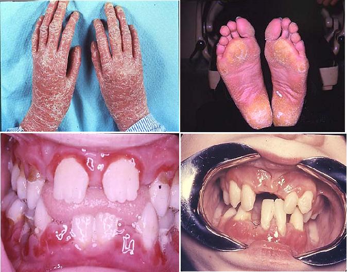
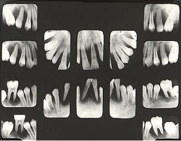
Papillon-Lefevre Syndrome
Papillon-Lefevre syndrome (PLS) belongs to a heterogenous group of 19 different skin diseases characterized by hyperkeratosis of the palms of the hands and soles of the feet (palmar-plantar hyperkeratosis). PLS is caused by mutations in the cathepsin C gene located on chromosome 11. Cathepsin C is a protease, normally found in high levels in epithelium and immune cells such as neutrophils, which acts to degrade proteins and activate proenzymes in immune cells. Patients with PLS have little or no cathepsin C activity.
PLS differs from other members of this group of hyperkeratoses in that patients with PLS universally have generalized rapid destruction of the periodontal attachment apparatus resulting in premature loss of primary and permanent teeth. The presence of neutrophil defects in PLS is commonly noted. Diminished chemotaxis, phagocytosis, and intracellular killing of certain bacteria have been reported in some but not all cases. It is possible that neutrophil defects are not entirely responsible for the findings in PLS. Some authors have hypothesized that the hereditary defect in PLS is located in the epithelial barrier, which in the gingival sulcus may lead to a reduced defense against pathogenic bacteria. Alterations in cementum, collagenolytic activity in the periodontal ligament, and osteoclastic activity have also been suggested in some patients with PLS. Taken together, these findings could explain the aggressive periodontal destruction seen in patients with PLS even in the absence of significant neutrophil abnormalities.
The periodontal condition in PLS is difficult to treat, and use of conventional mechanical debridement rarely has been successful. Systemic administration of synthetic retinoids, when combined with meticulous plaque control, debridement, topical antimicrobials such as chlorhexidine, and systemic antibiotic therapy, may give the best chance for preventing progression of periodontitis.
Treatment Retinoid therapy: Improves the skin condition but not the periodontal therapy. Periodontal condition: No effective treatment
Leukemias
Leukemia is a neoplastic disorder of the blood-forming tissues, primarily affecting leukocytes. This heterogenous group of diseases arises from a neoplastic proliferation in the bone marrow. The replacement of normal bone marrow elements by leukemic cells causes decreased production of erythrocytes, normal white blood cells, and platelets. The clinical result is anemia, with weakness, fatigue, pallor of skin, and mucous membranes; thrombocytopenia with associated bleeding tendencies; and leukopenias resulting in increased susceptibility to infection. Leukemias are classified as either acute or chronic, depending on the presentation of the disease. They are further classified relative to the predominant cell affected as either lymphocytic or myelocytic. Monocytic leukemias form a subgroup of myelocytic leukemia.
Oral involvement is common in leukemia and may represent the first sign of the disease. Dental professionals were responsible for initiating the diagnosis of leukemia in 25% to 33% of cases. Overall, 15% to 80% of patients with leukemia have oral manifestations, with the acute forms presenting oral signs in approximately 65% of cases, compared with only 30% in chronic leukemias. Oral petechiae or bleeding, mucosal ulceration, and gingival enlargement are the most common signs. Acute periodontal infection, pain, pharyngitis, and lymphadenopathy also may be seen.
Gingival enlargement may be localized or generalized and represents an infiltration of leukemic cells into the gingiva, and less frequently into bone (Fig. 3). Gingival enlargement is most common in acute monocytic leukemia (67% of cases), followed by acute myelomonocytic leukemia (18.5%), and acute myelocytic leukemia (4%).— The enlarged gingiva tends to be relatively firm in texture and most prominent in the interdental regions. The marginal tissues may be bluish-red or cyanotic. Gingival enlargement creates pseudopockets where plaque accumulates, stimulating a host response that may further exacerbate the swelling. Gingival bleeding is also common, and may be an early indicator of leukemia. Oral mucosal ulcers are a frequent finding in patients with leukemia. These lesions may result from bacterial invasion caused by severe leukopenia or from mucosal atrophy caused by a direct effect on epithelial cells of the chemotherapeutic drugs used to treat leukemia. Trauma from a dental prosthesis or teeth may result in large secondarily infected ulcers progressing to facial cellulitis and septicemia.
Biopsy of gingiva revealed large leukemic infiltrate.
Treatment for leukemia may include chemotherapy, radiation therapy, and BMT, each of which has the potential to produce a wide range of oral complications. Mucositis, xerostomia, and secondary infection with a variety of bacterial, viral, and fungal agents may occur. Candidiasis is almost universally seen in hospitalized patients with leukemia undergoing chemotherapy. Infections with unusual organisms (e.g., Pseudomonas and Klebsiella species) are common in this group of patients. Many drugs used for chemotherapy are neurotoxic and may cause intense oral pain, which is usually transient. These symptoms must be distinguished from pain of odontogenic origin. Patients undergoing BMT require special consideration because they receive very high-dose chemotherapy, often in combination with total body irradiation. The extreme immunosuppression experienced by patients with BMT predisposes to systemic spread of even mild infections. A large percentage of patients with BMT develop graft-versus-host disease, a condition where transplanted immunocompetent marrow cells recognize the host tissues as foreign and react against them, resulting in fever, mucosal ulcerations, skin erythema, and systemic involvement .
It is critical that dental needs be assessed as soon as a definitive diagnosis of leukemia has been rendered and a decision is made to initiate a radiation, chemotherapy, or BMT protocol. Unfortunately, oral care has been overlooked in the past, but aggressive promotion of dental intervention as a part of leukemia treatment protocols in recent years has dramatically decreased the incidence of oral complications.
During the acute phase of the disease only those procedures that are necessary to alleviate the discomfort and hemorrhaging should be performed. Conversely, during a period of remission every attempt should be made to achieve a state of periodontal health. The treatment should be conservative, consisting of the removal of all local irritants and instruction in good plaque control techniques. The distinct benefits of strict plaque control in severely granulocytopenic leukemia patients have been demonstrated: obtaining excellent gingival health
Severe gingival bleeding resulting from thrombocytopenia often can be managed successfully with localized treatment. The use of an absorbable gelatin sponge with topical thrombin or placement of microfibrillar collagen is often sufficient. Some authors report successful management of gingival bleeding with oral rinses of antifibrinolytic agents. If these measures are not successful in stopping blood flow from an oral site, platelet transfusions may be necessary.
Management of oral ulcers in patients with leukemia should be directed toward preventing the spread of localized infection and bacteremia, promoting healing of the lesion, and decreasing pain. Oral ulcers or extensive tissue sloughing may serve as the source of life-threatening septicemia in patients with leukemia . Topical antibacterial and antifungal medication should be used. Chlorhexidine mouth rinses are effective in reducing the severity of oral ulcerations, primarily by minimizing secondary infection of these lesions. Severe ulcers showing clinical signs of infection should be treated with a combination of topical medication and systemic antibiotics.
Patients with myelosuppressed leukemia are at risk for a variety of viral infections, most commonly herpes simplex, varicella zoster, and cytomegalovirus (CMV; Fig.5,6). These infections may become severe and must be recognized early. Herpes simplex virus and varicella zoster virus respond well to systemic acyclovir or other antiviral agents, and many patients with leukemia undergoing chemotherapy are treated prophylactically to prevent infection.
The predominant inherited coagulation disorders are hemophilia A, hemophilia B, and von Willebrand disease. Coagulopathies may also be acquired. Liver disease affects coagulation because most of the clotting factors are synthesized in the liver; thus the clinician should be wary of coagulation disorders in alcohol abusers and patients with hepatitis. Vitamin K deficiency, usually associated with long-term antibiotic usage or with malabsorption syndromes, can result in coagulation problems. Several of the clotting factors are dependent on vitamin K for their synthesis.
Rubeola (nine-day or red measles)
Measles, a disease recognised for over two thousand years, is a highly contagious, acute infection caused by the rubella virus. It usually occurs in children. It is seen in every country of the world. Before the use of vaccines, epidemics of measles occurred every two to five years. Cough, cold, fever and a skin rash that begins several days before the initial symptoms characterise the illness. Recovery from measles is usual, but serious complications of the respiratory and central nervous system may occur.
· Prodromal symptoms – fever, malaise, dry (occasional croupy) cough, coryza, conjunctivitis c clear d/c, marked photophobia.
· 1-2 days p prodromal symptoms – Koplik spots on the buccal mucosa.
· Koplik spots – tiny, bluish-white dots surrounded by red halos.
· Day 3 or 4 – blotchy, erythematous, blanching, maculopapular exanthem appears.
· Rash begins at the hairline and spreads cephalocaudally and involves palms and soles.
· Rash typically lasts 5 – 6 days.
· Can see desquimation in severe cases.
· Patients can be systemically ill
· Incubation period 9-10 days
· Patients contagious from 4 days prior to the rash until 4 days after the resolution of the rash
· Highly contagious – 90% for susceptible people
· High morbidity and mortality common in children in underdeveloped countries
· Peak season is late winter to early spring
· Potential complications – OM, PNA, obstructive laryngotracheitis, acute encephalitis
· Vaccination is highly effective in preventing disease
Measles virus belongs to the Morbillivirus group of the Paramyxovirus family. Humans are the only natural host for wild measles virus. The virus is easily destroyed but remains in the droplet form in air for several hours, especially under conditions of low relative humidity. It is spread by direct contact with droplets from respiratory secretions of infected persons. It is one of the most communicable of infectious diseases and is most infectious when cough and cold is at its peak. The virus invades the respiratory lining membrane and then enters the blood stream. It causes inflammation of the respiratory tract and may predispose to secondary bacterial pneumonia.
The incubation period is one to two weeks and is often longer in adults. The illness begins with symptoms of malaise, fever, loss of appetite, conjunctivitis, cough and cold lasting several days. This is followed by bluish-grey spots in the oral cavity (Koplik’s spots) and then a diffuse skin rash beginning on the face and proceeding down the body to involve the extremities. The rash lasts for five days and then peeling of the skin occurs. Several days after the appearance of the rash, the fever abates. The most common complications of measles involve the respiratory tract and the nervous systems. Bacterial super-infection can also cause middle ear infection or pneumonia in severe cases. Encephalitis may be acute or chronic, after measles infection. Transient hepatitis can also occur.
Severe measles can occur in persons who are immunocompromised such as those, being treated for malignancy or those with AIDS. Malnourished children in developing countries may also develop severe measles. In pregnant women, however, measles (rubeola) unlike German measles (rubella) does not cause any congenital anomalies.
Classic measles is diagnosed when a child develops along with cough, cold, conjunctivitis, Koplik’s spots and a skin rash. Leucopenia (a low white blood cell count) is common. Virus isolation in the laboratory is technically difficult. A four-fold increase in the measles antibody titre in acute and convalescent serum samples is considered diagnostic.
The disease is usually self-limited, and supportive therapy such as antipyretics and fluids are indicated. Bacterial super-infection should be promptly treated with appropriate antimicrobials. Prophylactic antibiotics are not known to be of value and are not recommended.
Measles can be prevented by administrating a live vaccine long before an anticipated exposure. It is now recommended that all healthy children be administered live measles vaccines at fifteen months of age. A second dose given in childhood, usually as a measles-mumps-rubella (MMR) is now routine. The first vaccine can be given between six and nine months of age in situations where the incidence of measles is high before the age of one year. Transient fever and rash develop about one week after vaccination in 5 – 15 percent of children. Live measles vaccine is contra-indicated in persons with defects in the cell-mediated immunity and in pregnant women.
Passive immunisation with antibodies is recommended for those at high risk of developing severe measles and for those who have been exposed to the infection. For example, children with malignant disease and those with defects in cell-mediated immunity. To be effective, passive immunisation must be given within six days after an exposure.
Rubella (german measles)
Rubella — commonly known as German measles or 3-day measles — is an infection that primarily affects the skin and lymph nodes. It is caused by the rubella virus (not the same virus that causes measles), which is usually transmitted by droplets from the nose or throat that others breathe in. It can also pass through a pregnant woman’s bloodstream to infect her unborn child.
It’s a generally mild disease in children; the primary medical danger of rubella is the infection of pregnant women because it can cause congenital rubella syndrome in developing babies.
Before a vaccine against rubella became available in 1969, rubella epidemics occurred every 6-9 years, most often among kids 5 to 9 years old. Many cases of congenital rubella occurred as well. Thanks to immunization, there are far fewer cases of rubella and congenital rubella.
Most rubella infections today appear in young, non-immunized adults rather than in kids. In fact, experts estimate that 10% of young adults are currently susceptible to rubella, which could pose a danger to children they might have someday.
· Little or no prodrome in children
· In adolescents – 1-5 days of low-grade fever, malaise, headache, adenopathy, sore throat, coryza
· Exanthem – discrete, pinkish red, fine maculopapular eruption – begins on the face and spreads cephalocaudally
· Rash becomes generalized in 24 hours and clears by 72 hours
· Forchheimer spots – small reddish spots on the soft palate – can sometimes be seen on day 1 of the rash
· Arthritis and arthralgias – frequent in adolescents and young women – beginning on day 2 or 3 lasting 5-10 days
· Up to 25% of patients are asymptomatic – serology testing may be necessary to establish the diagnosis
· Important in establishing the diagnosis if the patient is pregnant or has been in contact c a pregnant woman
· Peaks in late winter to early spring
· Contagious from a few days before the rash to a few days after the rash
· Incubation period 14-21 days
· Complications – rare in childhood – arthritis, purpura c or s thrombocytopenia, mild encephalitis
Signs and Symptoms
Rubella infection may begin with 1-2 days of mild fever (99-
The rubella rash can look like many other viral rashes. It appears as either pink or light red spots, which may merge to form evenly colored patches. The rash can itch and lasts up to 3 days. As the rash clears, the affected skin occasionally sheds in very fine flakes.
Other symptoms of rubella (these are more common in teens and adults) can include headache, loss of appetite, mild conjunctivitis(inflammation of the lining of the eyelids and eyeballs), a stuffy or runny nose, swollen lymph nodes in other parts of the body, and pain and swelling in the joints (especially in young women). Many people with rubella have few or no symptoms.
Rubella in a pregnant woman can cause congenital rubella syndrome, with potentially devastating consequences for the developing fetus. Children who are infected with rubella before birth are at risk for growth retardation; mental retardation; malformations of the heart and eyes; deafness; and liver, spleen, and bone marrow problems.
Contagiousness
The rubella virus passes from person to person through tiny drops of fluid from the nose and throat. People who have rubella are most contagious from 1 week before to 1 week after the rash appears. Someone who is infected but has no symptoms can still spread the virus.
Infants who have congenital rubella syndrome can shed the virus in urine and fluid from the nose and throat for a year or more and may pass the virus to people who have not been immunized.
Prevention
Rubella can be prevented by the rubella vaccine. Widespread immunization against rubella is critical to controlling the spread of the disease, thereby preventing birth defects caused by congenital rubella syndrome.
The vaccine is usually given to children at 12-15 months of age as part of the scheduled measles–mumps-rubella (MMR) immunization. A second dose of MMR is generally given at 4-6 years of age. As is the case with all immunization schedules, there are important exceptions and special circumstances. For example, if your child will be traveling outside the United States, the vaccine can be given as early as 6 months of age. Talk to your child’s doctor to see when the vaccine is needed.
The rubella vaccine should not be given to pregnant women or to a woman who may become pregnant within 1 month of receiving the vaccine. If you are thinking about becoming pregnant, make sure that you’re immune to rubella through a blood test or proof of immunization. If you’re not immune, you should receive the vaccine at least 1 month before you become pregnant.
Pregnant women who are not immune should avoid anyone who has the illness and should be vaccinated after delivery so that they will be immune during any future pregnancies.
Incubation
The incubation period for rubella is 14-23 days, with an average incubation period of 16-18 days. This means that it can take 2-3 weeks for a child to get rubella after they are exposed to someone with the disease.
Duration
The rubella rash usually lasts 3 days. Lymph nodes may remain swollen for a week or more, and joint pain can last for more than 2 weeks. Children who have rubella usually recover within 1 week, but adults may take longer.
Treatment
Rubella cannot be treated with antibiotics because they do not work against viral infections. Unless there are complications, rubella will resolve on its own.
Any pregnant woman who has been exposed to rubella should contact her obstetrician immediately.
Rubella is typically mild in kids, who often can be cared for at home. Monitor your child’s temperature and call the doctor if the fever climbs too high.
To relieve minor discomfort, you can give your child acetaminophen or ibuprofen. Do not give aspirin to a child with a viral illness because such use has been associated with the development of Reye syndrome, which can lead to liver failure and death.
Varicella (chickenpox)
Caused by the varicella-zoster virus (VZV), chickenpox used to be a common illness among kids in the United States (particularly among those under age 12). An itchy rash of spots that look like blisters can appear all over the body and be accompanied by flu-like symptoms. Chickenpox is very contagious, so an infected child should stay home and rest until the rash is gone.
Kids can be protected from VZV by getting the chickenpox (varicella) vaccine. The vaccine significantly reduces the chances of getting chickenpox. Vaccinated kids who do get chickenpox tend to have milder cases and quicker recoveries compared to those who contract the virus and aren’t immunized.
· Caused by varicella-zoster virus
· Highly contagious
· Brief prodrome of low-grade fever, URI symptoms, and mild malaise may occur
· Rapid appearance of puritic exanthem
· Lesions appear in crops – typically have 3 crops
· Crops begin in trunk and scalp, then spread peripherally
· Lesions begin as tiny erythematous papules, then become vesicles surrounded by red halos
· Lesions began to dry – umbilicated appearance, then surrounding erythema fades and a scab forms
· Hallmark – lesions in all stages of evolution
· All scabs slough off 10-14 days
· Scarring not typical unless superinfected
· Cluster in areas of previous skin irritation
· Puritic lesions on the skin
· Painful lesions along the oral, rectal, and vaginal mucosa, external auditory canal, tympanic membrane
· Occurs year-round, peaks in late autumn and late winter through early spring
· Incubation period ranges from 10-20 days
· Contagious 1-2 days prior to rash until all lesions are crusted over
· Complications – secondary bacterial skin infections (GAS), pneumonia, hepatitis, encephalitis, Reye syndrome
· Severe in the immunocompromised host – can be fatal
· Can have severe CNS, pulmonary, generalized visceral involvement (often hemorrhagic)
· Need to get varicella-zoster immunogloblin 96 hours post-exposure to possible varicella
Symptoms
Chickenpox often starts with a fever, headache, sore throat, or stomachache. These symptoms may last for a few days, with fever in the 101°-
Chickenpox causes a red, itchy skin rash that usually appears first on the abdomen or back and face, and then spreads to almost everywhere else on the body, including the scalp, mouth, arms, legs, and genitals.
The rash begins as multiple small red bumps that look like pimples or insect bites, usually less than a quarter of an inch wide. They appear in crops over 2 to 4 days and develop into thin-walled blisters filled with fluid. The blister walls break, leaving open sores, which finally crust over to become dry, brown scabs. The rash is very itchy, and cool baths or calamine lotion may help to manage the itching.
A hallmark of chickenpox is that all stages (red bumps, blisters, and scabs) can appear on the body at the same time. The rash may be more extensive or severe in kids who have skin disorders like eczema, or weak immune systems. Young kids tend to have a mild illness with fewer blisters than older children or adults. In rare cases, serious bacterial infections involving the skin, lungs, bones, joints, and the brain can occur.
Risk of Shingles
Anyone who has had chickenpox is at risk for developing a skin condition called shingles (herpes zoster) later in life. That’s because after an infection, VZV remains inactive ierve cells near the spinal cord and reactivates later as shingles, which can cause tingling, itching, or pain in one area of the body, followed by a rash with red bumps and blisters. Fortunately, this is a rare occurrence in kids and teens who have healthy immune systems.
It’s also uncommon for someone who’s been vaccinated against chickenpox to develop singles later in life. When it does happen, the case of shingles is usually milder and less likely to cause complications than in a person who wasn’t immunized.
Contagiousness
The chickenpox virus spreads both through the air (by coughing and sneezing), and by direct contact with mucus, saliva, or fluid from blisters. Chickenpox is contagious from about 2 days before the rash appears until all the blisters are crusted over. A child with chickenpox should be kept out of school until all blisters have dried, usually about 1 week. If you’re unsure about whether your child is ready to return to school, ask your doctor.
Chickenpox is very contagious — most kids with a sibling who’s been infected will get it as well (if they haven’t already had the disease or the vaccine), showing symptoms about 2 weeks after the first child does. To help keep it from spreading, make sure your kids wash their hands frequently, particularly before eating and after using the bathroom. And keep a child with chickenpox away from unvaccinated siblings as much as possible.
People who haven’t had chickenpox or the vaccine also can catch it from someone with shingles, but they cannot catch shingles itself. That’s because shingles can only develop from a reactivation of VZV in someone who has previously had chickenpox.
High-Risk Groups
Certain groups of people are more at risk for complications from chickenpox, including pregnant women and anyone with immune system problems. These groups should avoid others who have chickenpox.
If a pregnant woman who hasn’t had chickenpox in the past contracts it (especially in the first 20 weeks of pregnancy), the fetus is at risk for birth defects and the mother is at risk for more health complications than if she’d been infected when she wasn’t pregnant. If she develops chickenpox just before or after the child is born, the newborn is at risk for serious health complications. There is no risk to a developing baby if the mother develops shingles during pregnancy.
If a pregnant woman has had chickenpox before the pregnancy, the baby will be protected from infection for the first few months of life, since the mother’s immunity gets passed on to the baby through the placenta and breast milk.
Those at risk for severe disease or serious complications — such as newborns whose mothers had chickenpox at the time of delivery, patients with leukemia or immune deficiencies, and kids receiving drugs that suppress the immune system — may be given a medication after exposure to chickenpox to reduce its severity.
Prevention
The chickenpox vaccine is 99% effective at preventing the VZV infection in kids. Doctors recommend that kids receive the chickenpox vaccine twice — when they’re 12 to 15 months old, with a booster shot at 4 to 6 years old.
People 13 years of age and older who have never had chickenpox or haven’t gotten the vaccine should receive two doses of the vaccine at least 28 days apart to be protected. While few people who’ve been vaccinated actually develop chickenpox, those who do tend to develop very mild cases of the condition and recover quickly.
Healthy kids who have had chickenpox do not need the vaccine — they usually have lifelong protection against the illness.
Treatment
Since a virus causes chickenpox, doctors won’t prescribe antibiotics to treat it. However, antibiotics may be required if the sores become infected by bacteria. This is pretty common among kids because they often scratch and pick at the blisters.
An antiviral medicine might be prescribed for people with chickenpox who are at risk for complications. The decision to use this will depend on a child’s age and health, the extent of the infection, and the timing of the treatment. Your doctor can tell you if the medication is right for your child.
Dealing With Discomfort
To help relieve the itchiness, fever, and discomfort of chickenpox:
· Use cool wet compresses or give baths in cool or lukewarm water every 3 to 4 hours for the first few days. Oatmeal bath products, available at supermarkets and drugstores, can help to relieve itching. (Baths do not spread the rash.)
· Pat (don’t rub) the body dry.
· Put calamine lotion on itchy areas (but don’t use it on the face, especially near the eyes).
· Serve foods that are cold, soft, and bland because chickenpox in the mouth can make drinking or eating difficult. Avoid feeding your child anything highly acidic or especially salty, like orange juice or pretzels.
· Ask your doctor or pharmacist about pain-relieving creams to apply to sores in the genital area.
· Give your child acetaminophen regularly to help relieve pain if your child has mouth blisters.
· Ask the doctor about using over-the-counter medication for itching.
Never use aspirin to reduce pain or fever in kids with chickenpox because aspirin has been associated with the serious disease Reye syndrome, which can lead to liver failure and even death.
As much as possible, discourage kids from scratching. This can be difficult for them, so consider putting mittens or socks on your child’s hands to prevent scratching during sleep. In addition, trim fingernails and keep them clean to help lessen the effects of scratching, including broken blisters and infection.
Adenovirus
Adenoviral infections affect babies and young children much more often than adults. Childcare centers and schools sometimes have multiple cases of respiratory infections and diarrhea caused by adenovirus.
Adenoviral infections can occur at any time of the year, but:
· respiratory tract problems caused by adenovirus are more common in late winter, spring, and early summer
· conjunctivitis (pinkeye) and pharyngoconjunctival fever caused by adenovirus tend to affect older kids, mostly in the summer
Adenoviral infections can affect children of any age, but most occur in the first years of life — and most kids have had at least one before age 10. There are many different types of adenoviruses, so some kids can have repeated adenoviral infections.
· 30 distinct types
· Variety of infections including conjunctivitis, URIs, pharyngitis, croup, bronchitis, bronchiolitis, pneumonia (occ fulminant), gastroenteritis, myocarditis, cystitis, encephalitis
· Can be accompanied by a rash – variable iature
· Typically can see – conjunctivitis, rhinitis, pharyngitis c or s exudate, discrete, blanching, maculopapular rash
· Can see anterior cervical and preauricular LAD, low-grade fever, malaise
· Peak season is late winter through early summer
· Contagious during first few days
· Incubation period 6-9 days
Coxsackie
Coxsackieviruses are part of the enterovirus family of viruses (which also includes polioviruses and hepatitis A virus) that live in the human digestive tract. They can spread from person to person, usually on unwashed hands and surfaces contaminated by feces, where they can live for several days.
In cooler climates, outbreaks of coxsackievirus infections most often occur in the summer and fall, though they cause infections year-round in tropical parts of the world.
In most cases, coxsackieviruses cause mild flu-like symptoms and go away without treatment. But in some cases, they can lead to more serious infections.
o Brief prodome – low-grade fever, malaise, sore mouth, anorexia
o 1-2 days later, rash appears
o Oral lesions – shallow, yellow ulcers surrounded by red halos
o Cutaneous lesions – begin as erythematous macules then evolve to small, thick-walled, grey vesicles on an erythematous base
o Highly contagious
o Incubation period 2-6 days
o Lasts 2-7 days
o Peak season summer through early fall
o If no cutaneous lesions – herpangina
o less painful and less intense than herpes gingivostomatitis
Signs and Symptoms
Coxsackievirus can produce a wide variety of symptoms. About half of all kids infected with coxsackievirus have no symptoms. Others suddenly develop high fever, headache, and muscle aches, and some also develop a sore throat, abdominal discomfort, or nausea. A child with a coxsackievirus infection may simply feel hot but have no other symptoms. In most kids, the fever lasts about 3 days, then disappears.
Coxsackieviruses can also cause several different symptoms that affect different body parts, including:
· Hand, foot, and mouth disease, a type of coxsackievirus syndrome, causes painful red blisters in the throat and on the tongue, gums, hard palate, inside of the cheeks, and the palms of hands and soles of the feet.
· Herpangina, an infection of the throat which causes red-ringed blisters and ulcers on the tonsils and soft palate, the fleshy back portion of the roof of the mouth.
· Hemorrhagic conjunctivitis, an infection that affects the whites of the eyes. Hemorrhagic conjunctivitis usually begins as eye pain, followed quickly by red, watery eyes with swelling, light sensitivity, and blurred vision.
Occasionally, coxsackieviruses can cause more serious infections that may need to be treated in a hospital, including:
· viral meningitis, an infection of the meninges (the three membranes that envelop the brain and spinal cord)
· encephalitis, a brain infection
· myocarditis, an infection of the heart muscle
Newborns can be infected from their mothers during or shortly after birth and are more at risk for developing serious infection, including myocarditis, hepatitis, and meningoencephalitis (an inflammation of the brain and meninges). Iewborns, symptoms can develop within 2 weeks after birth.
Contagiousness
Coxsackieviruses are very contagious. They can be passed from person to person on unwashed hands and surfaces contaminated by feces. They also can be spread through droplets of fluid sprayed into the air when someone sneezes or coughs.
When an outbreak affects a community, risk for coxsackievirus infection is highest among infants and kids younger than 5. The virus spreads easily in group settings like schools, childcare centers, and summer camps. People who are infected with a coxsackievirus are most contagious the first week they’re sick.
Prevention
There is no vaccine to prevent coxsackievirus infection. Hand washing is the best protection. Remind everyone in your family to wash their hands frequently, particularly after using the toilet (especially those in public places), after changing a diaper, before meals, and before preparing food. Shared toys in childcare centers should be routinely cleaned with a disinfectant because the virus can live on these objects for days.
Kids who are sick with a coxsackievirus infection should be kept out of school or childcare for a few days to avoid spreading the infection.
The duration of an infection varies widely. For fever without other symptoms, a child’s temperature may return to normal within 24 hours, although the average fever lasts 3 to 4 days. Hand, foot, and mouth disease usually lasts for 2 or 3 days; viral meningitis can take 3 to 7 days to clear up.
Treating Coxsackievirus Infections
Depending on the type of infection and symptoms, the doctor may prescribe medications to make your child feel more comfortable. However, because antibiotics only work against bacteria, they can’t be used to fight a coxsackievirus infection.
Acetaminophen may be given to relieve any minor aches and pains. If the fever lasts for more than 24 hours or if your child has any symptoms of a more serious coxsackievirus infection, call your doctor.
Most kids with a simple coxsackievirus infection recover completely after a few days without needing any treatment. A child who has a fever without any other symptoms should rest in bed or play quietly indoors. Offer plenty of fluids to prevent dehydration.
Addison’s disease
Addison’s disease is characterized by chronic adrenal cortical insufficiency and may occur at all ages and a roughly equal gender distribution.
In the etiology of this disorder may be incriminated the prolonged use of corticosteroids (cortisone, hydrocortisone, prednisone) for various affections.
Due to the multitude of their action, cortical hormones are involved in the most important functions of the body, the body caot survive in their absence.
Addison Disease Symptoms vary in intensity depending by the degree of adrenal insufficiency.
Addison disease onset is insidious. State period is characterized by the following signs and symptoms:
· hyperpigmentation of the skin and mucous membranes, breast areola, large labia, scrotum, palm interlining, elbow, knee;
· hairy skin of the head, palms and plants are not pigmenting;
· hypotension – is increasing while standing and can occur faintness states, the systolic falls below 80 mmHg and minimum below 50 mmHg;
· fatigue and adynamic – are even more pronounced as the disease is more advanced; initially emphasized in the evening and subsequently become permanent;
· digestive symptoms – are multiple and variable in intensity, so during the “crisis” (acute adrenal insufficiency) patient may experience abdominal pain simulating a violent acute surgical abdomen, nausea, vomiting, diarrhea;
· other digestive symptoms are: loss of appetite, weight loss, “salt hunger”;
· neurological disorders – muscle atrophy, mixed sensory deficits;
· genital disorders – females: menstrual cycle disorders, frigidity, men – impaired spermatogenesis, decreased sexual appetite;
· urinary disorders – decreased amount of urine passed in 24 hours;
· intellectual activity is diminished.
Regarding laboratory investigations: intermediate metabolisms, blood count, hormones dosing, serum and urinary ionogram, radiological examinations. As follows:
– intermediate metabolisms: hypoglycemia (low blood sugar);
– low blood lipids;
– Hypocholesterolemia (lower blood cholesterol);
– anemia, eosinophilia, lymphocytosis;
– Hormone – ACTH (adrenocorticotropic pituitary hormone) is increased;
– ADH (antidiuretic hormone) is increased;
– blood cortisol value is low;
– Plasma aldosterone is low;
– 17 OH CS and 17 CS are low;
– Na (sodium), Cl (chlorine) are low;
– K (potassium) is increased;
– Urine – Na is high and K is low;
– Radiological examinations (abdominal radiography) may reveal adrenal tuberculosis, calcification in the adrenal glands, in this case the tuberculin skin test may be positive.
Cushing Syndrome
Adrenals are two glands located at the upper pole of the kidney. The corticoadrenal gland is the peripheral one and consists of three areas:
· glomerular area – at this level mineralocorticoid hormones are secreted (aldosterone, dezoxicorticosteron);
· fasciculated zone – the area which produces glucocorticoid hormones (corticosterone, cortisol, cortisone);
· reticulated area – produces androgens (testosterone, androstenlion, dehydroepiandrosterone) and a small amount of estrogen (estrone, estradiol).
Adrenal activity is under hypothalamic control (through secretion of corticoliberin) and pituitary control (through the secretion of adrenocorticotropic hormone).
Cushing syndrome is a condition that has as clinical expression the hyperfunction of adrenal gland. This syndrome occurs with predilection for women, with onset predominantly in the second decade of life (20 years).
Cushing’s Syndrome Causes
In terms of etiology, the syndrome may be caused by a benign tumor (adenoma of fasciculated area) that secrete glucocorticoids, or a malignanttumor (carcinoma). This malignancy (cancer) may present anarchic secretion of steroid hormones (mineralocorticoids, glucocorticoids), resulting a complex clinical picture.
Itenko-Cushing’s syndrome is made by hypothalamic dysfunction with excess secretion of corticoliberine (CRH) which stimulates the pituitary causing excessive secretion of adrenocorticotropic hormone which in turn will stimulate the adrenal gland, resulting in an hormonal excess of this gland.
If adrenocortical hypersecretion is due to an adenoma (benign tumor) located at the pituitary gland, which secretes adrenocorticotropic hormone and which in turn stimulates the adrenal gland secretion, then the condition is called Cushing’s disease.
There can be seen a paraneoplastic Cushing (a series of bronchial malignancies, of thymus, of pancreas can secrete adrenocorticotropic substances acting hormone-like).
Cushing’s symptoms
If the tumor, localized at adrenal level, predominantly secretes cortisol then appears the clinical picture of hypercortizolism. It is characterized by an insidious onset, with weight gain, appearance of hyperpilosity (overgrowth of hair), blood pressure fluctuations, menstrual disorders in women, sexual dynamic disturbances both in men and women.
The state period of the disease is characterized by the following signs and symptoms:
– Obesity – affects the face and trunk, but respects the members;
– Skin – are harsh, dry, purple stretch marks (located on the abdomen, thighs and armpit), ecchymosis (bruising), acne, virilism in women;
– Facies – is red, vultuos;
– Muscle – at this level may occur severe muscle atrophy with physical fatigue;
– Bones – bone demineralization occurs with predominant involvement of the vertebrae, ribs, pelvic bones. This demineralization process can lead to severe pain or fractures;
– Cardio-vascular system – hypertension appears;
– Nervous system, mental disorders such as emotional lability, anxiety, depression, impaired memory and concentration;
– Uro-genital apparatus – in women can occur virilizing syndrome (hair extends typically like male distribution areas – face, anterior chest, the lower abdomen, the front of the thighs; voice thickens) if the tumor secrete an excess of androgens, also in this context, can install the defeminization phenomena (menstrual disorders, sterility, frigidity);
In men – hair growth intensifies, testicles atrophy, impotence occurs, gynecomastia (growth of mammary glands in men);
– Associated endocrine disorders – endocrine secondary hypothyroidism and diabetes.

Cushing syndrome analysis
For diagnosis of Cushing’s sydrome laboratory determinations are necessary: metabolic, hormonal and imaging exams.
The blood picture is characterized by: neutrophilia (increased neutrophils), lymphopenia, eozinopeniaand increased platelet count.
Metabolic analysises highlights:
– Glucose – increased;
– Lipids – high;
– Cholesterol – increased;
– Plasma Na – increased;
– K from plasma – decreased;
Hormonal determinations consists of dosing plasma cortisol (which is increased) and disappears the normal rhythm of secretion.
Imaging consist in CT (may reveal an adrenal tumor), turkish saddle ray (may reveal a pituitary tumor), and skeletal X-rays (for diagnosis of osteoporosis).
Cushing syndrome is a chronic condition in which evolution can occur a number of complications. The most important complications that may occur are hypertension, heart failure, steroid diabetes, low resistance to infections, severe mental disorders with depressive phenomena with a tendency to suicide.
Cushing syndrome treatment
Treatment of Cushing’s syndrome depends on the etiology of this disorder. If the substrate of this disease is a adrenocortical tumor (benign or malignant) then will be practiced the unilateral removal of the adrenal gland. Surgery is followed by hormone replacement therapy with glucocorticoids.
If the substrate of the condition is represented by an inoperable carcinoma (cancer) then is used chemotherapy.
In Cushing’s disease the treatment addresses both to adrenal gland and pituitary gland (pituitary radiotherapy – to prevent tumor growth at this level).
In Cushing’s paraneoplastic syndrome the treatment is addressed primarily to tumor.
Diabetes mellitus
Diabetes mellitus (or diabetes) is a chronic, lifelong condition that affects your body’s ability to use the energy found in food. There are three major types of diabetes: type 1 diabetes, type 2 diabetes, and gestational diabetes.
All types of diabetes mellitus have something in common. Normally, your body breaks down the sugars and carbohydrates you eat into a special sugar called glucose. Glucose fuels the cells in your body. But the cells need insulin, a hormone, in your bloodstream in order to take in the glucose and use it for energy. With diabetes mellitus, either your body doesn’t make enough insulin, it can’t use the insulin it does produce, or a combination of both.
The cells can’t take in the glucose, it builds up in your blood. High levels of blood glucose can damage the tiny blood vessels in your kidneys, heart, eyes, or nervous system. That’s why diabetes — especially if left untreated can eventually cause heart disease, stroke, kidney disease, blindness, and nerve damage to nerves in the feet.
Type 1 Diabetes
Type 1 diabetes is also called insulin-dependent diabetes. It used to be called juvenile-onset diabetes, because it often begins in childhood.
Type 1 diabetes is an autoimmune condition. It’s caused by the body attacking its own pancreas with antibodies. In people with type 1 diabetes, the damaged pancreas doesn’t make insulin.
This type of diabetes may be caused by a genetic predisposition. It could also be the result of faulty beta cells in the pancreas that normally produce insulin.
A number of medical risks are associated with type 1 diabetes. Many of them stem from damage to the tiny blood vessels in your eyes (called diabetic retinopathy), nerves (diabetic neuropathy), and kidneys (diabetic nephropathy). Even more serious is the increased risk of heart disease and stroke.
Treatment for type 1 diabetes involves taking insulin, which needs to be injected through the skin into the fatty tissue below. The methods of injecting insulin include:
· Syringes
· Insulin pens that use pre-filled cartridges and a fine needle
· Jet injectors that use high pressure air to send a spray of insulin through the skin
· Insulin pumps that dispense insulin through flexible tubing to a catheter under the skin of the abdomen
A periodic test called the A1C blood test estimates glucose levels in your blood over the previous three months. It’s used to help identify overall glucose level control and the risk of complications from diabetes, including organ damage.
Having type 1 diabetes does require significant lifestyle changes that include:
· Frequent testing of your blood sugar levels
· Careful meal planning
· Daily exercise
· Taking insulin and other medications as needed
People with type 1 diabetes can lead long, active lives if they carefully monitor their glucose, make the needed lifestyle changes, and adhere to the treatment plan.
Type 2 Diabetes
By far, the most common form of diabetes is type 2 diabetes, accounting for 95% of diabetes cases in adults. Some 26 million American adults have been diagnosed with the disease.
Type 2 diabetes used to be called adult-onset diabetes, but with the epidemic of obese and overweight kids, more teenagers are now developing type 2 diabetes. Type 2 diabetes was also called non-insulin-dependent diabetes.
Type 2 diabetes is often a milder form of diabetes than type 1. Nevertheless, type 2 diabetes can still cause major health complications, particularly in the smallest blood vessels in the body that nourish the kidneys, nerves, and eyes. Type 2 diabetes also increases your risk of heart disease and stroke.
With Type 2 diabetes, the pancreas usually produces some insulin. But either the amount produced is not enough for the body’s needs, or the body’s cells are resistant to it. Insulin resistance, or lack of sensitivity to insulin, happens primarily in fat, liver, and muscle cells.
People who are obese — more than 20% over their ideal body weight for their height — are at particularly high risk of developing type 2 diabetes and its related medical problems. Obese people have insulin resistance. With insulin resistance, the pancreas has to work overly hard to produce more insulin. But even then, there is not enough insulin to keep sugars normal.
There is no cure for diabetes. Type 2 diabetes can, however, be controlled withweight management, nutrition, and exercise. Unfortunately, type 2 diabetes tends to progress, and diabetes medications are ofteeeded.
An A1C test is a blood test that estimates average glucose levels in your blood over the previous three months. Periodic A1C testing may be advised to see how well diet, exercise, and medications are working to control blood sugar and prevent organ damage. The A1C test is typically done a few times a year.
Gestational Diabetes
Diabetes that’s triggered by pregnancy is called gestational diabetes (pregnancy, to some degree, leads to insulin resistance). It is often diagnosed in middle or late pregnancy. Because high blood sugar levels in a mother are circulated through the placenta to the baby, gestational diabetes must be controlled to protect the baby’s growth and development.
According to the National Institutes of Health, the reported rate of gestational diabetes is between 2% to 10% of pregnancies. Gestational diabetes usually resolves itself after pregnancy. Having gestational diabetes does, however, put mothers at risk for developing type 2 diabetes later in life. Up to 10% of women with gestational diabetes develop type 2 diabetes. It can occur anywhere from a few weeks after delivery to months or years later.
With gestational diabetes, risks to the unborn baby are even greater than risks to the mother. Risks to the baby include abnormal weight gain before birth, breathing problems at birth, and higher obesity and diabetes risk later in life. Risks to the mother include needing a cesarean section due to an overly large baby, as well as damage to heart, kidney, nerves, and eye.
Treatment during pregnancy includes working closely with your health care team and:
· Careful meal planning to ensure adequate pregnancy nutrients without excess fat and calories
· Daily exercise
· Controlling pregnancy weight gain
· Taking diabetes insulin to control blood sugar levels if needed
A few rare kinds of diabetes can result from specific conditions. For example, diseases of the pancreas, certain surgeries and medications, or infections can cause diabetes. These types of diabetes account for only 1% to 5% of all cases of diabetes.

