Investigation of physico-chemical properties and chemical composition of
normal urine.
Investigation of pathological components of urine.

Kidney – the couple organ, which is responsible for
excriting of final products of metabolism and for homeostasis. They regulate
water and mineral metabolism, acid-base balance, excriting of nitrogenous
slags, osmotic pressure. Also they regulate arterial pressure and
erhythropoesis.
Understanding how the urinary system helps maintain homeostasis by removing
harmful substances from the blood and regulating water balance in the body is
an important part of physiology. Your kidneys, which are the main part of the urinary
system, are made up of millions of nephrons that act as individual filtering
units and are complex structures themselves. The ureters, urethra, and urinary
bladder complete this intricate system.
Kidney functions in organism:
a) excretion of final metabolic products;
b) maintaining of acid-base balance;
c) water-salts balance regulation;
d) endocrine function.(The kidneys secrete a variety of hormones,
including erythropoietin,
and the enzyme renin. Erythropoietin is released in response to hypoxia (low levels of oxygen at tissue level)
in the renal circulation. It stimulates erythropoiesis (production of red blood cells) in the bone marrow. Calcitriol,
the activated form of vitamin D,
promotes intestinal absorption of calcium and the renalreabsorption of phosphate.
Part of the renin-angiotensin-aldosterone system,
renin is an enzyme involved in the regulation of aldosterone levels.)
The
kidney participates in whole-body homeostasis,
regulating acid-base
balance, electrolyte concentrations, extracellular fluid volume, and regulation
of blood
pressure. The kidney accomplishes these homeostatic functions both
independently and in concert with other organs, particularly those of the endocrine
system. Various endocrine hormones coordinate these endocrine
functions; these include renin, angiotensin
II, aldosterone, antidiuretic hormone, and atrial natriuretic peptide, among others.
Many of
the kidney's functions are accomplished by relatively simple mechanisms of
filtration, reabsorption, and secretion, which take place in the nephron.
Filtration, which takes place at therenal
corpuscle, is the process by which cells and large proteins are
filtered from the blood to make an ultrafiltrate that eventually becomes urine.
The kidney generates
http://www.youtube.com/watch?v=6x5pVoMb_vI&feature=related
Physical and chemical characteristics and
components of urine:
a) volume, physical and chemical properties of urine;
b) inorganic components of urine;
c) organic components of urine.
Key words and phrases:
http://www.youtube.com/watch?v=glu0dzK4dbU
Nephron – is the structural and functional unit of
kidney.


Urine – fluid with different organic and inorganic
compounds, which must be excreted (excess of water, final products of nitrogen
metabolism, xenobiotics, products of protein’s decay, hormones, vitamins and
their derivates). Most of them present in urine in a bigger amount than in
blood plasma. So, urine formation – is not passive process (filtration and
diffusion only).
In basis of urine formation lay 3 processes:
filtration, reabsorbtion and secretion.

Glomerulal filtration. Water and low
weight molecules go to the urine with help of following powers: blood
hydrostatic pressure in glomerulas (near 70 mm Hg), oncotic press ure of blood plasma proteins (near 30 mm Hg) and
hydrostatic pressure of plasma ultrafiltrate in glomerulal capsule (near 20 mm Hg). In normal
conditions, as You see, effective filtration pressure is about 20 mm Hg.


Hydrostatic pressure depends from correlation between
opening of a. afference and a. efference.
Primary urine formed in result of filtration (about 200 L per day). Between
all blood plasma substances only proteins don’t present in primary urine. Most
of these substances are undergone to the following reabsorbtion. Only urea,
uric acid, creatinin, and other final products of different metabolic pathways
aren’t undergone to the reabsorbtion.
For evaluate of filtration used clearance (clearance for
some substance – it is an amount of blood plasma in ml, which is cleaned from
this substance after 1 minute passing through kidney).
Drugs, which stimulate blood circulation in kidney
(theophyllin), also stimulate filtration. Inflammatory processes of renal
tissue (nephritis) reduce filtration, and azotaemia occurred (accumulation of
urea, uric acid, creatinin, and other metabolic final products).
Glomerulal
filtration. Water and low weight molecules go to
the urine with help of following powers: blood hydrostatic pressure in
glomerulas (near
Hydrostatic pressure depends from correlation between opening of a.
afference and a. efference.

Primary urine formed in result of filtration (about
For evaluate of filtration used clearance (clearance for some substance – it is a amount of blood plasma
in ml, which is cleaned from this substance after 1 minute passing through
kidney).
Drugs which stimulate blood circulation in kidney (theophyllin), also
stimulate filtration. Inflammatory processes of renal tissue (nephritis) reduce
filtration, and azotaemia occurred (accumulation of urea, uric acid, creatinin,
and other metabolic final products).
The
development of the clearance concept
ABSTRACT:
The clearance concept is central to three critical areas of nephrology; it was
a key feature to early conceptual analysis of the nature of urine formation, it
was utilized as a measure of kidney function in advancing renal diseases, and
it was a pivotal concept to elucidate the physiology of the kidneys. This paper
describes the clearance concept as currently understood and then it examines
how clearance was utilized to understand these various aspects of kidney
function.

Introduction
The concept of clearance is central to
three major areas of nephrology. First, the nature of urine for mation was
explored to a great extent using clearance techniques. Second, the early search
for measures of kidney function with advancing disease resorted to clearance
procedures, particularly involving urea and creatinine. Third, the physiology
of the kidney was examined and developed with great power and sophistication by
the deepening theoretical understanding of the concept of clearance accompanied
by ingenious analytical techniques and procedures. There is an additional
domain which hovered over the studies of urine formation. This had to do with
the per vasive resort to vitalism as an explanation of physiologic regulation.
My task was to examine the birth and evolution of the clearance concept. For
purposes of exposition, it may be helpful to describe first the clearance
concept as it is currently understood and then recount how the concept emerged
and developed during the 19th and early 20th centuries, as it was repeatedly
invoked to analyze the process of urine formation, the failure of renal
function, and the nature of physiologic regulation.
THE CONCEPT OF CLEARANCE
Figure 1 gives the definition of clearance in currently
conventional units of time and concentration. It is evident from the formula
that the numerator is a rate of excretion (mg/mm); the denominator is a plasma
concentration (mg/mL). Therefore, the clearance of any substance is expressed
as mL/min (mg/min x mL/mg=mL/min). Clearance, therefore, has the dimensions of
a volume per unit time. This simultaneous measurement of the excretion rate of
a solute and a flow rate of fluid from which the solute is derived has resulted
in some confusion. Fig 2, modified from Cassin and Vogh, emphasizes that the
kidney removes (clears) a small fraction of a substance from each mL of total
flow. The clearance, therefore, of any substance is the virtual volume of
plasma flow required to supply the amount of the substance excreted in any one
minute.
NATURE OF URINE
FORMATION
The historical evolution of the clearance concept is
intimately connected with studies examining the nature of urine formation.
Particularly noteworthy reviews have been published by Smith (2), Bradley (3),
Thurau, Davis and Haberle (4), Gottschalk (5), Schuster and Seldin (14). In the
early 19th centur y, Johannes Muller (18011858) advanced a theor y of urine
formation that rested on two prevalent concepts current at the time: 1) fluid
movement was a secretory process mediated by glands; 2) the activity of the
secretor y system required vitalistic forces that could not be reduced to physical
processes. Despite enormous contributions to microscopic anatomy, he denied
that the glomerulus was directly connected with the renal tubules, and ascribed
urine formation to the secretory activity of the tubules, regarding the kidney
as a gland. Notwithstanding the powerful currents of vitalism at the time, Carl
Ludwig (1816-1895) came to the study of kidney function with an uncompromising
physicochemical orientation. He appreciated the role of the afferent and
efferent arteriole in elevating the hydrostatic pressure in the intervening
glomerulus, thereby facilitating the movement of a protein-free ultrafiltrate,
containing all the elements to be found in the urine, and restraining the
passage of protein and formed elements. To account for the different composition
of blood and urine, Ludwig proposed that some unspecified chemical force
promoted active sodium chloride reabsorption while some property of the tubular
wall restrained urea back-diffusion. No vital force was postulated, although
the nature of the “chemical force” promoting reabsorption was unspecified.
Simultaneously and independently, William Bowman (1816-1892) also postulated
that water was separated from blood at the glomerulus, but he assumed that
solutes remained in the blood and were subsequently secreted into the urine by
the tubules. This was an expression of the prevalent view of glandular
secretion mediating solute movement. The central feature of Ludwig’s theory
that urine formation was critically linked with glomerular pressure, was challenged
by Rudolph Heidenhain (1834-1897). On the basis of calculations of a clearance
type, he concluded that to attribute urea excretion to filtration alone would
require

His
conception of a trifold process of ultrafiltration at the glomerulus by
physical forces, reabsorption of most of the filtrate in part by active tubular
processes, and active secretion of certain solutes by the tubular epithelia is
remarkably close to modern views. His summary is worth quoting . It
should be emphasized that the use of clearance calculations by Heidenhain and
their reinterpretation by Cushny served to make creditable the conceptual model
of the comparatively modest magnitude of urine flow in a setting of huge
volumes of glomerular filtration.
DIGRESSION ON VITALISM
Although Cushny may have over-emphasized
the commitment of Heidenhain and others to vitalism, there is no question that
the concept of vital activity, not reducible to physical forces, was a powerful
conceptual factor that infected the theories of renal function. For most of the
19th centur y, a basic problem in biology was conceived to be the distinction
between living and non-living matter. A mechanistic explanation assumed that
organic and non-organic matter were not irreducibly different. A vitalistic
explanation assumed that a reduction of living to non-living phenomena is in
principle impossible. Embryology was a dominating biologic discipline. To
provide a flavor of the intellectual climate surrounding the study of renal
function, it may be helpful to review briefly the prevailing embryologic
studies. Landmark studies exemplified by the work of Hans Driesch are
summarized in a comprehensive publication in 1914 (7). In a series of studies
on embr yonic sea-urchins, he demonstrated that rearrangement of cells at the
blastomere stage had no effect on normal development. Moreover, a single
blastomere, isolated from the rest at the two - or fourcell stage, can develop
into a normal sea-urchin embr yo.

The conclusion was drawn that spatio
temporal location is irrelevant to development, and that non-physical forces
“entelechies” are “wholemaking” factors which have no quantitative
characteristics. It was only the gradual advancement of physical and biologic
science that could meet the vitalistic arguments. Organic chemistr y was shown
to be a misnomer. The synthesis of urea, heretofore found only in living
organisms, from CO2 and NHby3 Wöhler in 1828 (7) led to the
view that organic chemistr y was simply the chemistr y of carbon compounds.
Purpose and purposiveness were explained by reference to integrated and
adjustible feed-back systems. The ability of blastomere cells to develop
differently in different transplant locations in ontogenesis, unlike a machine
where each part fulfills a designed function, is explainable in principle by
genetic theory. And finally “energy” input required to impart selectivity is
not confined to hydrostatic or oncotic forces. On a conceptual level, it was
pointed out by the logical positivist philosopher C.I. Hengel that vitalism has
no predictive power, offering neither verifiable predictions nor providing
models of coherent mechanisms. It was the increasing power of the physical
sciences that gradually undermined the recourse to postulated entities which
could not be identified, characterized, or worst of all, refuted. It was these
reasons which led Ludwig and Cushny to vigorously reject vitalistic
explanations.
UREA AND UREA CLEARANCE
Bright in 1836 recognized that the
concentration of blood urea rose in patients with chronic renal disease (3).
Ambard (8) showed that the blood level of urea was related to urea excretion
and formulated an equation which was designed to register impairment of renal
function.
However, the equation involved a square
root function which obscured the physiologic significance of the relationship
between urinar y excretion and blood urea concentration. Addis (9), in 1917,
showed that at maximal urine flows the ratio of the excretion of urea per hour
and the blood urea concentration was constant in any one individual. This
expression represented the urea clearance per hour, an approximation of
glomerular filtration rate. Austin, Stillman and Van Slyke (10) showed that the
rate of urine flow influenced urea excretion independently of the blood level
and renal excretory capacity. In a later study (11), it was demonstrated that
above urine flows of 2ml/min (augmentation limit), the relationship between
urea excretion and plasma concentration in any one individual was constant, and
expressed by a simple formu1a:
Curea = Urea / Purea
The term,
clearance, was introduced with this analysis. Since blood urea concentration is
frequently used as an index of filtration rate, it is worthwhile examining the
factors which influence it independently of intrinsic renal function. It has
alredy been pointed out that urine flow influences urea clearance. Urea
undergoes a complex intrarenal recycling process, the fractional reabsorption
increasing from 35% of the filtered load in hydrated states to 60% in
dehydration. The blood urea concentration is influenced by a variety of factors
independent of renal function. Changes in urine flow affect blood urea in a
manner which depends on the nephron segment where fluid is being reabsorbed.

Fig. 4

Fig. 5
The proximal tubule is highly permeable
to urea, and is the principal segment of passive reabsorption. The distal
nephron is less permeable to urea, even in the presence of antidiuretic
hormone. If ever ything else is left constant, salt depletion will produce more
azotemia at the same low rate of urine flow than will water restriction,
because salt depletion accelerates proximal reabsorption while water
restriction accelerates principally distal reabsorption (12). Protein loads
also influence blood urea concentration independent of renal function. Figure 4
lists the sources of protein loads. Factors 1-5 serve to increase protein loads
while factor 6 reduces it. Figure 5 (13) illustrates the effect of protein
intake at various levels of renal function.

Fig. 6
It should be emphasized that at low filtration rates, the
BUN is very sensitive to protein loads, as is illustrated by Figure 6 (13).
Figure 7 summarizes the various factors which influence both the BUN and urea
clearance. It is evident that the interpretation of the BUN as a rough index of
GFR requires correction for the numerous factors which influence its
concentration independent of renal function. Urea clearance circumvents some of
the distorting effects of protein loads. Nevertheless, it is always less than
inulin clearance, but tends to rise toward inulin clearance with advanced renal
failure. These various factors are discussed in detail in ref. 14.
PHYSIOLOGY OF THE KIDNEY
Although Addis and Van Slyke had
published landmark studies on urea clearance as a measure of renal function,
the precise relation between urea clearance and glomerular filtration rate was
not appreciated. Rehberg (15, 16) introduced creatinine as a marker of
glomerular filtration rate, but was unaware that it was secreted by the
tubules, and therefore would give falsely high values, especially if its plasma
concentration was raised by infusions. Smith was skeptical that creatinine
would be an ideal marker for glomerular filtration, since it underwent
secretion in aglomerular fish, and might do the same in mammals, a supposition
that proved correct. He then went on to elaborate the criteria for an ideal
marker of GFR (Fig. 8).

Fig. 7

Fig. 8
The
failure of sugars to be secreted in aglomerular fish led Smith ultimately to
identify inulin as an ideal marker (17, 18). Simultaneously and independently,
Richards and his associates also demonstrated in micropuncture studies that
inulin fulfilled the requirements for an ideal marker of GFR (19). In Figure 9,
inulin excretion is shown to increase in direct proportion to its plasma
concentration when GFR is constant (a); inulin clearance is constant over a
wide range of plasma inulin concentrations (b); inulin clearance is constant
over a wide range of urine flows (c). Findings such as these established inulin
as a kind of gold standard for GFR (14). Smith went on to develop methods for
measuring renal blood flow, utilizing diodrast as a marker and the Fick
principle to calculate total renal blood flow. Since the Fick principle required
renal vein catheterization, it was unsuitable for routine use. Para-amino
hippurate (PAH) was identified as a substance which, at low plasma
concentrations, was almost completely secreted into the tubular urine. This
eliminated the need for renal catherization, since renal venous PAH could be
assumed to be close to zero. Subsequent studies of tubular maximum transport
capacity using glucose and many other substances provided a measure of
functioning tubular mass.

Fig. 9

Fig. 10
These various measures allowed Smith to portray the
various functional aspects of normal and diseased kidneys in remarkable detail,
as summarized in his Porter lectures (20). From a clinical and physiologic
point of view, the various measures of renal function that Smith and others
explored over the years are summarized in Figure 10. Smith has remarked how
fruitful the clearance concept has proved to be. From rather tentative
beginnings it has stimulated the search for novel analytic procedures, allowed
for the assessment of renal function, and most of all provided a conceptual
rallying point for insight and understanding of renal physiology.
Reabsorption. Lenght of renal tubules is about
All substances can be divided into 3 group:
1. Actively reabsorbed substances.
2. Substances, which are reabsorbed in a little amount.
3. Non-reabsorbed substances.
To the first group belong Na+, Cl-, Mg2+,
Ca2+, H2O, glucose and other monosaccharides, amino
acids, inorganic phosphates, hydrocarbonates, low-weight proteins, etc.


Na+ reabsorbed by active transport to the epitelium cell, then – into
the extracellular matrix. Cl- and HCO3-
following Na+ according to the electroneutrality principle, water –
according to the osmotic gradient. From extracellular matrix substances go to
the blood vessels. Mg2+ and Ca2+ are reabsorbed with help
of special transport ATPases. Glucose and amino acids use the energy of Na+
gradient and special carriers. Proteins are reabsorbed by endocytosis.
Urea and uric acid are little reabsorbable substances.
Creatinin, mannitol, inulin and some other substances are
non-reabsorbable.
Henle’s loop play important role in the reabsobtion process. Its
descendent and ascendent parts create anti-stream system, which has big
capacity for urine concentration and dilution. Fluid which passes from proximal
part of renal tubule to the descendent part of Henle’s loop, where
concentration of osmotic active substances higher than in kidney cortex. This
concentration is due to activity of thick ascendent part of Henle’s loop, which
is non-penetrated for water and which cells transport Na+ and Cl-
into the interstitium. Wall of descendent part is penetrated for water and here
water pass into the interstitium by osmotic gradient but osmotic active
substances stay in the tubule. Ascendent part continue to reabsorb salt
hypertonically, even in the absence of aldosteron, so that fluid entering the
distal tubule still has a much lower osmolality than does interstitial fluid.
Extracellular
homeostasis
The kidney is responsible for
maintaining a balance of the following substances:
|
Substance |
Description |
||||
|
If glucose is not reabsorbed by the kidney, it
appears in the urine, in a condition known as glycosuria. This is associated with diabetes mellitus. |
reabsorption (almost 100%) viasodium-glucose transport proteins(apical) and GLUT (basolateral). |
– |
– |
– |
|
|
All are reabsorbed nearly completely. |
reabsorption |
– |
– |
– |
|
|
Regulation of osmolality. Varies
with ADH |
reabsorption
(50%) via passive transport |
secretion |
– |
reabsorption in medullary collecting ducts |
|
|
Uses Na-H antiport, Na-glucose
symport, sodium ion channels (minor) |
reabsorption
(65%, isosmotic) |
reabsorption (25%, thick ascending, Na-K-2Cl symporter) |
reabsorption (5%, sodium-chloride
symporter |
reabsorption (5%, principal cells), stimulated by aldosterone via ENaC |
|
|
Usually follows sodium. Active
(transcellular) and passive (paracellular) |
reabsorption |
reabsorption (thin ascending, thick ascending, Na-K-2Cl symporter) |
reabsorption (sodium-chloride
symporter) |
– |
|
|
absorbed osmotically along with solutes |
reabsorption
(descending) |
– |
reabsorption (regulated by ADH, via arginine vasopressin receptor 2) |
||
|
Helps maintain acid-base balance. |
reabsorption
(80–90%) |
reabsorption
(thick ascending) |
– |
||
|
Uses vacuolar H+ATPase |
– |
– |
– |
secretion
(intercalated cells) |
|
|
Varies upon
dietary needs. |
reabsorption
(65%) |
reabsorption (20%, thick ascending, Na-K-2Cl symporter) |
– |
secretion (common, via Na+/K+-ATPase, increased by aldosterone), or
reabsorption (rare, hydrogen potassium ATPase) |
|
|
reabsorption |
reabsorption (thick ascending) via passive transport |
– |
– |
||
|
Calcium and magnesium compete, and an excess of one
can lead to excretion of the other. |
reabsorption |
reabsorption
(thick ascending) |
reabsorption |
– |
|
|
Excreted as titratable acid. |
reabsorption (85%) viasodium/phosphate cotransporter. inhibited by parathyroid hormone. |
– |
– |
– |
|
|
|
reabsorption
(100%) viacarboxylate
transporters. |
– |
– |
– |
The body is very
sensitive to its pH.
Outside the range of pH that is compatible with life, proteins are denatured and
digested, enzymes lose their ability to function, and the body is unable to
sustain itself. The kidneys maintain acid-base homeostasis by regulating the pH of the blood plasma.
Gains and losses of acid and base must be balanced. Acids are divided into
"volatile acids" and
"nonvolatile acids".[14] See also titratable
acid.
The major homeostatic control point for maintaining this
stable balance is renal excretion. The kidney is directed to excrete or retain
sodium via the action of aldosterone, antidiuretic hormone(ADH, or vasopressin), atrial natriuretic peptide (ANP), and other hormones. Abnormal
ranges of the fractional excretion of sodium can imply acute tubular necrosis or glomerular dysfunction.
Some substances (K+, ammonia and other) are secreted into urine in the distal part
of tubules. K+ is changed to Na+ by the activity of Na+-K+ATPase.

WATER REGULATION BY THE KIDNEYS
The water content of the body can vary depending on
various factors. Hot weather and physical activity such as exercise make us
sweat and so lose body fluids. Drinking tends to be at irregular intervals when
socially convenient. This means that sometimes the body has too little water
and needs to conserve it and sometimes too much water and needs to get rid of
it. Most of the control of water conservation takes place in the distal and
collecting tubules of the nephrons under control of anti-diuretic hormone,
(ADH), sometimes called vasopressin. This hormone is released by the posterior
pituitary under control of the hypothalamus in the mid-brain area. The
hypothalamus monitors the water content of the blood. If the blood contains too
little water (indicating dehydration) then more ADH is released. If the blood
stream contains too much water (indicating over-hydration)
then less ADH is released into the blood 
Release of ADH from the posterior pituitary into the blood stream
ADH released from
the pituitary travels in the blood stream to the peritubular capillaries of the
nephron. ADH binds to receptors on the distal and collecting tubules of the
nephrons which causes water channels to open in the tubule walls. This allows
water to diffuse through the tubule walls into the interstitial fluid where it
is collected by the peritubular capillaries. The more ADH present, the more
water channels are open and the more water is reabsorbed,

Reabsorption of
water from the filtrate under the influence of ADH
Over 99% of the
filtrate produced each day can be reabsorbed. The amount of water reabsorbed
from the filtrate back into the blood depends on the water situation in the
body. When the body is dehydrated, most of the filtrate is reabsorbed but note
that even in cases of extreme of water shortage, the kidneys will continue to
produce around 500 ml of urine each day in order to perform their excretory function.
Peculiarities of biochemical processes in kidney.
Kidney have a very high level of metabolic processes.
They use about 10 % of all O2, which used in organism. During
24 hours through kidney pass 700-900 L of blood. Carbohydrates are the main fuel for kidney. Glycolysis,
ketolysis, aerobic oxidation and phosphorylation are very intensive in kidney.
A lot of ATP formed in result.
Metabolism of proteins also presents in kidney in high
level. Especially, glutamine deaminase is very active and a lot of free ammonia
formed. In kidney take place the first reaction of creatin synthesis.
Kidney have plenty of different enzymes: LDG (1, 2, 3,
5), AsAT, AlAT. Specific for kidney is alanine amino peptidase, 3rd isoform.
Utilization of glucose in cortex and medulla is
differs one from another. Dominative type of glycolysis in cortex is aerobic
way and CO2 formed in result. In
medulla dominative type is anaerobic and glucose converted to lactate.
Two sources contribute to the renal ammonia: blood
ammonia (is about one-third of excreted ammonia), and ammonia formed in the
kidney. The predominant source for ammonia production within the kidney is
glutamine, the most abundant amino acid in plasma, but a small amount may
originate from the metabolism of other amino acids such as asparagine, alanine,
and histidine. Ammonia is secreted into the tubular lumen throughout the entire
length of the nephron. Secretion occurs both during normal acid-base balance
and in chronic acidosis. Metabolic acidosis is accompanied by an adaptive
increase in renal ammonia production with a corresponding increase in urinary
ammonium excretion.
Kidney cortex like liver appear to be unique in that
it possess the enzymatic potential for both glucose synthesis from
noncarbohydrate precursors (gluconeogenesis) and glucose degradation via the
glycolytic pathway. Gluconeogenesis is important when the dietary supply of
glucose does not satisfy the metabolic demands. Under these conditions, glucose
is required by the central nervous system, the red blood cells, and, possibly,
other tissues which cannot obtain all their energy requirements from fatty
acids or ketone body oxidation. Also, gluconeogenesis may be important in the
removal of excessive quantities of glucose precursors from the blood (lactate
acid after severe exercise for example). Although the biosynthetic pathways are
similar, there are several important differences in the factors, which regulate
gluconeogenesis in the two organs. 1) The liver utilizes predominately pyruvate,
lactate and alanine. The kidney cortex utilizes pyruvate, lactate, citrate,
α-ketoglutarate, glycine and glutamine. 2) Hydrogen ion activity has
little effect upon hepatic gluconeogenesis, but it has marked effects upon
renal gluconeogenesis. Thus, when intracellular fluid pH is reduced (metabolic
acidosis, respiratory acidosis or potassium depletion), the rates of
gluconeogenesis in slices of renal cortex are markedly increased. The ability
of the kidney to convert certain organic acids to glucose, a neutral substance,
is an example of a nonexcretory mechanism in the kidney for pH regulation.
Renin-Angiotensin mechanism

The renin–angiotensin
system (RAS) or the renin–angiotensin–aldosterone
system (RAAS) is a hormone system that
regulates blood pressure and water (fluid) balance.
When blood volume is low, juxtaglomerular cells in the kidneys
secrete renin directly
into circulation. Plasma renin then
carries out the conversion of angiotensinogen released
by the liver to angiotensin I. Angiotensin I is subsequently converted
to angiotensin II by the enzyme angiotensin converting enzyme found in
the lungs. Angiotensin II is a potent vaso-active peptide that causes blood
vessels to constrict, resulting in increased blood pressure. Angiotensin II
also stimulates the secretion of the hormone aldosterone from
the adrenal cortex. Aldosterone causes the tubules
of the kidneys to increase the reabsorption of sodium and water into the blood.
This increases the volume of fluid in the body, which also increases blood
pressure.

If the renin–angiotensin–aldosterone system is abnormally
active, blood pressure will be too high. There are many drugs that interrupt
different steps in this system to lower blood pressure. These drugs are one of
the main ways to control high blood pressure (hypertension), heart failure,kidney failure,
and harmful effects of diabetes.
The system can be
activated when there is a loss of blood volume or a drop in blood
pressure (such as in hemorrhage).
This loss of pressure is interpreted by baroreceptors in
the carotid sinus. In alternative fashion, a decrease
in the filtrate NaCl concentration and/or decreased filtrate flow rate will
stimulate the macula densa to signal the juxtaglomerular cells to release
renin.
·
If the perfusion of
the juxtaglomerular apparatus in the
kidney's macula densa decreases, then the
juxtaglomerular cells (granular cells, modified pericytes in the glomerular
capillary) release the enzyme renin.
·
Renin cleaves
a zymogen,
an inactive peptide,
called angiotensinogen, converting it into angiotensin I.
·
Angiotensin I is
then converted to angiotensin II by angiotensin-converting enzyme (ACE),[5] which
is thought to be found mainly in lung capillaries.
One study in 1992 found ACE in all blood vessel endothelial cells.[6]
·
Angiotensin II is the major bioactive
product of the renin-angiotensin system, binding to receptors on intraglomerular mesangial cells, causing
these cells to contract along with the blood vessels surrounding them and
causing the release of aldosterone from the zona glomerulosa in
the adrenal cortex. Angiotensin II acts as an endocrine, autocrine/paracrine,and intracrinehormo.

Cardiovascular effects

It is believed that
angiotensin I may have some minor activity, but angiotensin II is the major
bio-active product. Angiotensin II has a variety of effects on the body:
·
Throughout the body, it is a potent vasoconstrictor of arterioles.
·
In the kidneys, it constricts glomerular arterioles,
having a greater effect on efferent arterioles than afferent. As with most other
capillary beds in the body, the constriction of afferent arterioles increases the arteriolar resistance,
raising systemic arterial blood pressure and decreasing the blood flow.
However, the kidneys must continue to filter enough blood despite this drop in
blood flow, necessitating mechanisms to keep glomerular blood pressure up. To
do this, angiotensin II constricts efferent arterioles, which forces blood to
build up in the glomerulus, increasing glomerular pressure. The glomerular
filtration rate(GFR) is thus maintained, and blood filtration can
continue despite lowered overall kidney blood flow. Because the filtration
fraction has increased, there is less plasma fluid in the downstream
peritubular capillaries. This in turn leads to a decreased hydrostatic pressure
and increased oncotic pressure (due to unfiltered plasma proteins) in the
peritubular capillaries. The effect of decreased hydrostatic pressure and
increased oncotic pressure in the peritubular capillaries will facilitate
increased reabsorption of tubular fluid.
·
Angiotensin II decreases medullary blood
flow through the vasa recta. This decreases the washout of NaCl and urea in the
kidney medullary space. Thus, higher concentrations of NaCl and urea in the
medulla facilitate increased absorption of tubular fluid. Furthermore,
increased reabsorption of fluid into the medulla will increase passive
reabsorption of sodium along the thick ascending limb of the loop of Henle.
·
Angiotensin II stimulates Na+/H+ exchangers located on the apical membranes
(faces the tubular lumen) of cells in the proximal tubule and thick ascending
limb of the loop of Henle in addition to Na+ channels in the collecting ducts. This
will ultimately lead to increased sodium reabsorption
·
Angiotensin II stimulates the hypertrophy
of renal tubule cells, leading to further sodium reabsorption.
·
In the adrenal
cortex, it acts to cause the release of aldosterone.
Aldosterone acts on the tubules (e.g., the distal convoluted tubules and the cortical collecting
ducts) in the kidneys, causing them to reabsorb more sodium and water from the urine. This
increases blood volume and, therefore, increases blood pressure. In exchange
for the reabsorbing of sodium to blood, potassiumis secreted into the tubules,
becomes part of urine and is excreted.
·
Release of anti-diuretic hormone (ADH),
also called vasopressin – ADH is made in the hypothalamus and
released from the posterior pituitary
gland. As its name suggests, it also exhibits vaso-constrictive
properties, but its main course of action is to stimulate reabsorption of water
in the kidneys. ADH also acts on the central nervous system to increase an individual's appetite
for salt, and to stimulate the sensation of thirst.
These effects directly
act in concert to increase blood pressure.
Local
renin-angiotensin systems
Locally expressed
renin-angiotensin systems have been found in a number of tissues, including the kidneys, adrenal
glands, the heart, vasculature and nervous
system, and have a variety of functions, including local cardiovascular
regulation, in association or independently of the systemic renin-angiotensin
system, as well as non-cardiovascular functions. Outside the kidneys, renin is
predominantly picked up from the circulation but may be secreted locally in
some tissues; its precursor prorenin is highly expressed in tissues and more
than half of circulating prorenin is of extrarenal origin, but its
physiological role besides serving as precursor to renin is still
unclear.Outside the liver, angiotensinogen is picked up from the circulation or
expressed locally in some tissues; with renin they form angiotensin I, and
locally expressed angiotensin converting enzyme, chymase or other enzymes can transform it into
angiotensin II. This process can
be intracellular or interstitial.
In the adrenal glands,
it is likely involved in the paracrine regulation of aldosterone secretion, in the heart and
vasculature, it may be involved in remodeling or vascular tone, and in the brain where it is largely independent of the
circulatory RAS, it may be involved in local blood pressure regulation. In addition, both the central and peripheral nervous systems can use angiotensin
for sympathetic neurotransmision. Other
places of expression include the reproductive system, the skin and digestive
organs. Medications aimed at the systemic system may affect the expression of
those local systems, beneficially or adversely.
|
|

Peculiarities of biochemical processes in
kidney.
Kidney have a very high level of metabolic processes. They use about 10
% of all O2, which used in organism. During 24 hours through kidney
pass 700-
Metabolism of proteins also present in kidney in high level. Especially,
glutamine deaminase is very active and a lot of free ammonia formed. In kidney
take place the first reaction of creatin synthesis.
Kidney have plenty of different enzymes: LDG (1, 2, 3,
5), AsAT, AlAT. Specific for kidney is alanine amino peptidase, 3rd isoform.
Utilization of glucose in cortex and medulla is differ one from another.
Dominative type of glycolysis in cortex is aerobic way and CO2
formed in result. In medulla dominative type is anaerobic and glucose converted
to lactate.
Two sources contribute to the renal ammonia: blood ammonia (is about
one-third of excreted ammonia), and ammonia formed in the kidney. The
predominant source for ammonia production within the kidney is glutamine, the
most abundant amino acid in plasma, but
a small amount may originate from the metabolism of other amino acids
such as asparagine, alanine, and histidine. Ammonia is secreted into the
tubular lumen throughout the entire length of the nephron. Secretion occurs
both during normal acid-base balance and in chronic acidosis.Metabolic acidosis
is accompanied by an adaptive increase in renal ammonia production with a
corresponding increase in urinary ammonium excretion.
Kidney cortex like liver appear to be unique in that it possess the
enzymatic potential for both glucose synthesis from noncarbohydrate precursors
(gluconeogenesis) and glucose degradation via the glycolytic pathway.
Gluconeogenesis is important when the dietary supply of glucose does not
satisfy the metabolic demands. Under these conditions, glucose is required by
the central nervous system, the red blood cells, and possibly other tissues
which cannot obtain all their energy requirements from fatty acids or ketone
body oxidation. Also, gluconeogenesis may be important in the removal of
excessive quantities of glucose precursors from the blood (lactate acid after
severe exercise for example). Although the biosynthetic pathways are similar,
there are several important differences in the factors which regulate
gluconeogenesis in the two organs. 1) The liver utilizes predominately
pyruvate, lactate and alanine. The kidney cortex utilizes pyruvate, lactate,
citrate, α-ketoglutarate, glycine and glutamine. 2) Hydrogen ion
activity has little effect upon hepatic gluconeogenesis, but it has marked
effects upon renal gluconeogenesis. Thus, when intracellular fluid pH is
reduced (metabolic acidosis, respiratory acidosis or potassium depletion), the
rates of gluconeogenesis in slices of renal cortex are markedly increased. The
ability of the kidney to convert certain organic acids to glucose, a neutral
substance, is an example of a nonexcretory mechanism in the kidney for pH
regulation.
Regulation of urine formation.
Na-uretic hormone (produced in heart)
decrease reabsorbtion of Na+, and quantity of urine increased.
Aldosteron and some other hormones (vasopressin, renin, angiotensin II) increase Na-reabsorption
and decrease quantity of urine.
Role of kidney in acid-base balance
regulation.
The kidneys have two important roles in the maintaining
of the acid-base balance: to reabsorb bicarbonate from and to excrete hydrogen
ions into urine. 4500 mmol of bicarbonate are filtered into the primary
filtrate of urine daily, but only 2 mmol of it are finally excreted. 70-80% of
bicarbonate is reabsorbed in the first part of proximal tubule, 10-20% in the
loop of Henle and 5-10% in the distal tubule and collecting ducts. (Jalanko
& Holmberg 1998)
Carbonic anhydrase plays an important role in the
reabsorption in the proximal tubule. Disturbance in the reabsorption of
bicarbonate in the proximal tubule leads to metabolic acidosis, hyperchloremia
and alkalotic urine. This disease is named as "type II renal tubular
acidosis" (N25.8). (Jalanko & Holmberg 1998)
Renal tubules actively secrete hydrogen ions. Most of
this takes place in the distal part of the nephron, but active transport of
hydrogen ions occurs in the proximal tubule, too. The H-ATPase of the apical
cell membrane secretes hydrogen ions into urine. For each hydrogen ion
secreted, one bicarbonate molecule is transported to the interstitial fluid,
from there it diffuses into the bloodstream. Fifty mmol of hydrogen ions are
normally excreted daily. (Jalanko & Holmberg 1998)
If the hydrogen ions are not properly secreted into the
collecting ducts, the result is metabolic acidosis, hypokalemia, hypocalcemia,
nephrocalcinosis and an alkalotic urine. This disease is called "type I
renal tubular acidosis" (N25.8). (Jalanko & Holmberg 1998)
The maximal hydrogen ion gradient, against which the
transport mechanism can secrete H+ ions,
corresponds to a urine pH of
The source of the hydrogen ions secreted by the tubular
cells is not completely certain. It is probably produced by dissociation of H2CO3.
The acid-secreting cells contain carbonic anhydrase, which facilitates the
rapid formation of H2CO3 from CO2 and water. The renal acid secretion is
mainly regulated by the changes in the intracellular pCO2, potassium
concentration, carbonic anhydrase activity and adrenocortical hormone
concentration. (Ganong 1991)
Kidney have some mechanisms for maintaining acid-base balance. Na+
reabsorbtion and H+ secretion play very important role.
1. Primary urine has a lot of Na2HPO4 (in
dissociated form). When Na+ reabsorbed, H+ secreted into
urine and NaH2PO4 formed.
2. Formation of hydrocarbonates. Inside renal cells carboanhydrase forms
from CO2 and H2O H2CO3, which
dissociated to H+ and HCO3-. H+ excreted from
cell into urine (antiport with Na+) and leaded with urine. Na+
connect with HCO3-, NaHCO3 formed and go to
the blood, thereupon acidity decreased.
3. Formation of free ammonia. NH3 used for formation of NH4+
(H+ ion associted), and different acid metabolites excreted as
ammonia salts.
The organs involved in
regulation of external acid-base balance are the lungs are the kidneys.
The lungs are important
for excretion of carbon dioxide (the respiratory acid) and there is a huge
amount of this to be excreted: at least 12,000 to 13,000 mmols/day.
In contrast the kidneys
are responsible for excretion of the fixed acids and this is also a critical
role even though the amounts involved (70-100 mmols/day) are much smaller. The
main reason for this renal importance is because there is no other way to
excrete these acids and it should be appreciated that the amounts involved are
still very large when compared to the plasma [H+] of only 40 nanomoles/litre.
There is a second
extremely important role that the kidneys play in acid-base balance, namely the
reabsorption of the filtered bicarbonate. Bicarbonate is the predominant
extracellular buffer against the fixed acids and it important that its plasma
concentration should be defended against renal loss.
In acid-base balance,
the kidney is responsible for 2 major activities:
·
Reabsorption of filtered bicarbonate:
4,000 to 5,000 mmol/day
·
Excretion of the fixed acids (acid anion
and associated H+): about 1 mmol/kg/day.
Both these processes
involve secretion of H+ into
the lumen by the renal tubule cells but only the second leads to excretion of H+ from the body.
The renal mechanisms
involved in acid-base balance can be difficult to understand so as a simplification we will consider the processes
occurring in the kidney as involving 2 aspects:
·
Proximal tubular mechanism
·
Distal tubular mechanism
The contributions of the proximal tubules to acid-base balance are:
·
firstly, reabsorption of bicarbonate
which is filtered at the glomerulus
·
secondly, the production of ammonium
The next 2 sections explain these roles in more detail.
Daily filtered bicarbonate equals the product of the daily glomerular
filtration rate (180 l/day) and the plasma bicarbonate concentration (24
mmol/l). This is 180 x 24 = 4320 mmols/day (or usually quoted as between 4000
to 5000 mmols/day).
About 85 to 90% of the filtered bicarbonate is reabsorbed in the proximal
tubule and the rest is reabsorbed by theintercalated cells of the distal tubule and collecting
ducts.
The reactions that occur are outlined in the diagram. Effectively, H+ and HCO3- are formed from CO2 and H2O in a reaction catalysed
by carbonic anhydrase. The actual reaction involved is probably formation of H+ and OH- from water, then reaction of OH- with CO2 (catalysed by carbonic anhydrase) to
produce HCO3-. Either way, the end result is the same.
The H+ leaves the
proximal tubule cell and enters the PCT lumen by 2 mechanisms:
·
Via a Na+-H+ antiporter (major route)
·
Via H+-ATPase (proton pump)
Filtered HCO3- cannot
cross the apical membrane of the PCT cell. Instead it combines with the
secreted H+ (under the
influence of brush border carbonic anhydrase) to produce CO2 and H2O. The CO2 is lipid soluble and easily crosses
into the cytoplasm of the PCT cell. In the cell, it combines with OH- to produce bicarbonate. The HCO3-crosses
the basolateral membrane via a Na+-HCO3- symporter. This symporter is
electrogenic as it transfers three HCO3- for every one Na+. In
comparison, the Na+-H+ antiporter
in the apical membrane is not electrogenic because an equal amount of charge is
transferred in both directions.
The basolateral membrane also has an active Na+-K+ ATPase (sodium pump) which transports
3 Na+ out per 2 K+ in. This pump is electrogenic in a
direction opposite to that of the Na+-HCO3- symporter. Also the sodium pump keeps
intracellular Na+ low which
sets up the Na+ concentration
gradient required for the H+-Na+ antiport at the apical membrane. The H+-Na+ antiport is an example of secondary active transport.
The net effect is the reabsorption of one molecule of HCO3 and one molecule of Na+ from the tubular lumen into the blood
stream for each molecule of H+ secreted.
This mechanism does not lead to the net excretion of any H+ from the body as the H+ is consumed in the reaction with the
filtered bicarbonate in the tubular lumen.
[Note: The differences in functional properties of the apical membrane from
that of the basolateral membranes should be noted. This difference is
maintained by the tight junctions which link adjacent proximal tubule cells.
These tight junctions have two extremely important functions:
Gate function: They limit access of luminal
solutes to the intercellular space. This resistance can be altered and this
paracellular pathway can be more open under some circumstances (ie the ‘gate’
can be opened a little).
Fence function: The junctions maintain different
distributions of some of the integral membrane proteins. For example they act
as a ‘fence’ to keep the Na+-H+ antiporter limited to the apical
membrane, and keep the Na+-K+ ATPase limited to the basolateral
membrane. The different distribution of such proteins is absolutely essential
for cell function.]
The 4 major factors which control bicarbonate reabsorption are:
·
Luminal HCO3- concentration
·
Luminal flow rate
·
Arterial pCO2
·
Angiotensin II (via decrease in cyclic AMP)

An increase in any of these four factors causes an increase in bicarbonate
reabsorption. Parathyroid hormone also has an effect: an increase in hormone
level increases cAMP and decreases bicarbonate reabsorption.
The mechanism for H+ secretion
in the proximal tubule is described as a high capacity, low gradient system:
The high capacity refers to the large amount (4000 to 5000 mmols) of H+ that is secreted per day. (The actual
amount of H+ secretion
is 85% of the filtered load of HCO3-).
The low gradient refers to the low pH gradient as tubular pH can be
decreased from 7.4 down to 6.7-7.0 only.
Though no net excretion of H+ from
the body occurs, this proximal mechanism is extremely important in acid-base
balance. Loss of bicarbonate is equivalent to an acidifying effect and the
potential amounts of bicarbonate lost if this mechanism fails are very large.
Ammonium (NH4) is produced predominantly within the proximal
tubular cells. The major source is from glutamine which enters the cell from
the peritubular capillaries (80%) and the filtrate (20%). Ammonium is produced
from glutamine by the action of the enzyme glutaminase. Further ammonium is
produced when the glutamate is metabolised to produce alpha-ketoglutarate. This
molecule contains 2 negatively-charged carboxylate groups so further metabolism
of it in the cell results in the production of 2 HCO3- anions. This occurs if it is oxidised
to CO2 or if it is
metabolised to glucose.
The pKa for ammonium is so high that
both at extracellular and at intracellular pH, it is present entirely in the
acid form NH4+. The previous idea that lipid soluble NH3 is produced in the tubular cell,
diffuses into the tubular fluid where it is converted to water soluble NH4+ which is now trapped in the tubule
fluid is incorrect.
The subsequent situation with ammonium is complex. Most of the ammonium is
involved in cycling within the medulla. About 75% of the proximally produced
ammonium is removed from the tubular fluid in the medulla so that the amount of
ammonium entering the distal tubule is small. The thick ascending limb of the
loop of Henle is the important segment for removing ammonium. Some of the
interstitial ammonium returns to the late proximal tubule and enters the
medulla again (ie recycling occurs).
An overview of the situation so far is that:
·
The ammonium level in the DCT fluid is
low because of removal in the loop of Henle
·
Ammonium levels in the medullary
interstitium are high (and are kept high by the recycling process via the thick
ascending limb and the late PCT)
·
Tubule fluid entering the medullary
collecting duct will have a low pH if there is an acid load to be excreted (and
the phosphate buffer has been titrated down.
If H+ secretion
continues into the medullary collecting duct this would reduce the pH of the
luminal fluid further. A low pH greatly augments transfer of ammonium from the
medullary interstitium into the luminal fluid as it passes through the medulla.
The lower the urine pH, the higher the ammonium excretion and this ammonium
excretion is augmented further if an acidosis is present. This augmentation
with acidosis is 'regulatory' as the increased ammonium excretion by the kidney
tends to increase extracellular pH towards normal.
If the ammonium returns to the blood stream it is metabolised in the liver
to urea (Krebs-Henseleit cycle) with net production of one hydrogen ion per
ammonium molecule.
Regulation of Renal H+ Excretion
The discussion above has described the mechanisms involved in renal acid
excretion and mentioned some factors which regulate acid excretion.
The major factors which regulate renal bicarbonate reabsorption and acid
excretion are:
1. Extracellular volume
Volume depletion is associated with Na+ retention and this also enhances HCO3
reabsorption. Conversely, ECF volume expansion results in renal Na+ excretion and secondary decrease in
HCO3 reabsorption.
2. Arterial pCO2
An increase in arterial pCO2 results in increased renal H+ secretion and increased bicarbonate
reabsorption. The converse also applies. Hypercapnia results in an
intracellular acidosis and this results in enhanced H+ secretion. The cellular processes
involved have not been clearly delineated. This renal bicarbonate retention is
the renal compensation for a chronic respiratory acidosis.
3. Potassium & Chloride Deficiency
Potassium has a role in bicarbonate reabsorption. Low intracellular K+ levels result in increased HCO3 reabsorption in the kidney. Chloride
deficiency is extremely important in the maintenance of a metabolic alkalosis
because it prevents excretion of the excess HCO3 (ie now the bicarbonate
instead of chloride is reabsorbed with Na+ to maintain electroneutrality).
4. Aldosterone & cortisol (hydrocortisone)
Aldosterone at normal levels has no role in renal regulation of acid-base
balance. Aldosterone delpetion or excess does have indirect effects. High
aldosterone levels result in increased Na+ reabsorption and increased urinary
excretion of H+ and K+ resulting in a metabolic alkalosis.
Conversely, it might be thought that hypoaldosteronism would be associated with
a metabolic acidosis but this is very uncommon but may occur if there is
coexistent significant interstitial renal disease.
5. Phosphate Excretion
Phosphate is the major component of titratable acidity. The amount of
phosphate present in the distal tubule does not vary greatly. Consequently,
changes in phosphate excretion do not have a significant regulatory role in
response to an acid load.
6. Reduction in GFR
It has recently been established that a reduction in GFR is a very
important mechanism responsible for the maintenance of a metabolic alkalosis.
The filtered load of bicarbonate is reduced proportionately with a reduction in
GFR.
7. Ammonium
The kidney responds to an acid load by increasing tubular production and
urinary excretion of NH4+. The mechanism involves an
acidosis-stimulated enhancement of glutamine utilisation by the kidney
resulting in increased production of NH4+ and HCO3- by the tubule cells. This is very
important in increasing renal acid excretion during a chronic metabolic
acidosis. There is a lag period: the increase in ammonium excretion takes
several days to reach its maximum following an acute acid load. Ammonium
excretion can increase up to about 300 mmol/day in a chronic metabolic acidosis
so this is important in renal acid-base regulation in this situation. Ammonium
excretion increases with decreases in urine pH and this relationship is
markedly enhanced with acidosis.
![]()
Role of kidney in water balance regulation.
Excessive entrance of
water leads to dilution of extracellular fluid. Decreasing of osmolality
inhibits secretion of antidiuretic hormone. Walls of collective tubules stay
non-penetrated to water and dilutive urine formed.
If volume of blood circulation increases, circulation in kidney
increases also and hyperosmotic medium of kidney medulla removed. Some
substances in these conditions return into blood. So, excess of water carried
with urine and a lot of soluble substances are reabsorbed into blood. After
water loading stopped, hyperosmolality in kidney medulla returns for previous
stage during some days.
Physical and chemical characteristics of urine.

Urine amount (diures) in healthy people is 1000-2000 ml per day.
Day-time diures is in 3-4 times more than night-time.
Normal colour of urine is yellow (like hay or amber), what is due to
presence of urochrom (derivate of urobilin or urobilinogen). Some another
colour substances are uroerythrin (derivate of melanine), uroporphyrines,
rybophlavine and other. Colour depends from urine concentration.
Urine varies in appearance, depending principally upon a body's level of hydration,
as well as other factors. Normal urine is a transparent solution ranging from
colorless to amber but is usually a pale yellow. In the urine of a healthy
individual the color comes primarily from the presence of urobilin.
Urobilin in turn is a final waste product resulting from the breakdown of heme from hemoglobin during the destruction of aging blood
cells.
Colorless urine indicates over-hydration, generally preferable to
dehydration (though it can remove essential salts from the body). Colorless
urine in drug tests can suggest an attempt to avoid detection of illicit drugs
in the bloodstream through over-hydration.
·
Dark yellow urine is often indicative of
dehydration.
·
Yellowing/light orange may be caused by
removal of excess B vitamins from the bloodstream.
·
Certain medications such as rifampin and phenazopyridine can cause orange urine.
·
Bloody urine is termed hematuria,
a symptom of a wide variety of medical conditions
·
Dark orange to brown urine can be a
symptom of jaundice, rhabdomyolysis,
or Gilbert's syndrome.
·
Black or dark-colored urine is referred
to as melanuria and may be caused by a melanoma.
·
Pinkish urine can result from the
consumption of beets.
·
Greenish urine can result from the
consumption of asparagus.
·
Reddish or brown urine may be caused by porphyria (not to be confused with the harmless,
temporary pink or reddish tint caused by beeturia).
·
Blue urine can be caused by the
ingestion of methylene
blue, e.g. in medications
·
Blue urine stains can be caused by blue diaper syndrome
Urine is transparent. This characteristic depends from amount of
different salts (oxalates, urates, phosphates), amount of present epitelium
cells and leucocytes.
Density of urine depends from concentration of soluble substances.
Borders of variation are from 1002 to 1035 g/l. Near 60-
The pH of urine can vary between 4.6 and 8,
with neutral (7) being norm. In persons with hyperuricosuria,
acidic urine can contribute to the formation of stones of uric acid in the kidneys, ureters, or bladder. Urine pH can be monitored by a physician or at home.
A diet high in citrus, vegetables, or dairy can increase urine pH (more
basic). Some drugs also can increase urine pH, including acetazolamide,
potassium citrate, and sodium bicarbonate.
A diet high in meat can decrease urine pH (more acidic). Cranberries, popularly thought to
decrease the pH of urine, have actually been shown not to acidify urine. Drugs that can decrease urine pH
include ammonium
chloride, chlorothiazide diuretics, and methenamine mandelate.[14][15]
In normal conditions urine has acid or weak acid reaction (pH=5,3-6,8).
This depends from presence of NaH2PO4 and KH2PO4.
Fresh urine has a specific smell, which is due to presence of flying
acids. But a lot of microorganisms, which are present in urine, split urea and
free ammonia formed.
Exhaustive detailed description of the composition of human urine can be
found in NASA Contractor Report No. NASA CR-1802, D. F. Putnam, July 1971. That report provided detailed chemical
analyses for inorganic and organic constituents, methods of analysis, chemical
and physical properties and its behavior during concentrative processes such as
evaporation, distillation and other physiochemical operations. Urine is an
aqueous solution of greater than 95% water, with the remaining constituents, in
order of decreasing concentration urea 9.3 g/L, chloride 1.87 g/L, sodium 1.17 g/L, potassium 0.750 g/L, creatinine 0.670 g/L and other dissolved ions, inorganic
and organic compounds.
Urine is sterile until
it reaches the urethra, where epithelial
cells lining the
urethra are colonized by facultatively anaerobic Gram negative rods and cocci. Subsequent
to elimination from the body, urine can acquire strong odors due to bacterial
action, and in particular the release of ammonia from the breakdown of urea.
Some
diseases alter the quantity and consistency of urine, such as diabetes introducing sugar. Consuming beets can result in beeturia (pink/red urine containing betanin)
for some 10–14% of the popu Urine is a liquid produced by the kidneys to remove
waste products from the bloodstream. Human urine is yellowish in color and
variable in chemical composition, but here is a list of its primary components.
Human urine consists primarily of water, with
organic solutes including urea, creatinine, uric acid, and trace amounts of
enzymes, carbohydrates, hormones, fatty acids, pigments, and mucins, and
inorganic ions such as sodium (Na+), potassium (K+),
chloride (Cl-), magnesium (Mg2+), calcium (Ca2+),
ammonium (NH4+), sulfates (SO42-),
and phosphates (e.g., PO43-). A representative chemical
composition would be:
water 95%
urea 9.3 g/l
chloride 1.87 g/l
sodium 1.17 g/l
potassium 0.750 g/l
creatinine 0.670 g/l
Organic compounds of urine.
Proteins. Healthy people excretes 30 mg of proteins per day. As a rule these are
low weight proteins.
Urea. This is main part of organic compounds in urine. Urea nitrogen is about
80-90 % of all urine nitrogen. 20-
Uric acid. Approximately 0,6-1,0 g of uric acid is excreted per day in form
of different salts (urates), mainly in form of sodium salt. Its amount depends
from food.
Creatinin and
creatin. Near 1-
Amino acids. Per day healthy person excretes 2-
Couple substances. Hypuric acid (benzoyl glycine) is excreted in amount 0,6-
Indican (potassium
salt of indoxylsulfuric acid). Per day excrition of indican
is about 10-
Organic acids. Formic, acetic, butyric, β-oxybutyric, acetoacetic and some other
organic acids are present in urine in a little amount.
Vitamines. Almost all vitamines can be excreted via kidney, especially, water-soluble.
Approximately 20-30 mg of vit C, 0.1-0.3 mg of vit B1, 0.5-0.8
mg of vit B2 and some products of vitamine’s metabolism. These data
can be used for evaluating of supplying our organism by vitamines.
Hormones. Hormones and their derivates are always present in urine. Their amount
depends from functional state of endocrinal glands and liver. There is a very
wide used test – determination of 17-ketosteroids in urine. For healthy man
this index is 15-
Urobilin. Present in a little amount, gives to urine yellow colour.
Bilirubin. In normal conditions present in so little amount that cannot be found
by routine methods of investigations.
Glucose. In normal conditions present in so little amount that cannot be found
by routine methods of investigations.
Galactose. Present in the newborn’s urine, when digestion of milk or
transformation of glalactose into glucose in the liver are violated.
Fructose. It is present in urine very seldom, after eating a lot of fruits,
berries and honey. In all other cases it indicates about liver’s disorders, diabetes mellitus.
Pentoses. Pentoses are excreted after eating a lot of fruits, fruit juices, in
case of diabetes mellitus and steroid diabetes, some intoxications.
Ketone bodies. In normal conditions urine contains 20-50 mg of ketone bodies and this
amount cannot be found by routine methods of clinical investigations.
Porphyrines. Urine of healthy people contains a few I type porphyrines (up to 300
mkg per day).

Inorganic compounds of urine.
Urine of healthy people contains 15-
NaCl. Per day near 8-
Potassium. Twenty-four hours urine contains 2-
Different drugs can change excretion of Na and K. For example,
salicylates and cortikosteroids keep Na and amplify excretion of K.
Calcium. Twenty-four hours urine contains 0.1-
Magnesium. Content of magnesium in urine is 0.03-
Iron. Amount of iron in urine is about 1 mg per day.
Phosphorus. In urine are present one-substituted phosphates of potassium and
sodium. Their amount depends from blood pH. In case of acidosis two-substituted
phosphates (Na2HPO4) react with H+ and
one-substituted phosphates (NaH2PO4) formed. In case of
alkalosis one-substituted phosphates react with bases and two-substituted
phosphates formed. So, in both cases amount of phosphates in urine increases.
Sulfur. Amount of sulfur in twenty-four hours urine is 2-
Ammonia. Ammonia is excreted in ammonium sulfates and couple substances.
Ammonium salts make up 3-6 % of all nitrogen in urine. Their amount depends
from character of food and blood pH.

Urine analysis infers valuable information in a variety of ailments. Physical
characteristics of urine have been used as diagnostic and prognostic
tool from the time immemorial by the health physicians. We know that the
major functions of kidneys are:

·
Removal of water not needed by the body
fluids, the amount depending on the balance between glomerular filtrate and he
degree of tubular reabsorption;
·
The excretion of certain substances
normally present in the plasma when their concentration rises above a certain
level;
·
The selective reabsorption of substances
such as glucose which are of value to the body;
·
The excretion of useless substances; and
·
Regulation of acid base balance.
Disordered renal function may lead to a change in the volume of the urine excreted per day
along with remarkable changes in its physical and chemical properties and
microscopic contents. Urine analysis is the very first
investigation of diagnostic importance not only in renal disorders but also in
other diseases like diabetes, liver disease, jaundice etc. In diagnostic
pathology the extent of abnormalities could only be understood in comparison
with the reference values obtained from similar investigations in normal
individuals. Hence, it is important to have an understanding of normal
parameters of physical and chemical
characteristics of urine.
Characteristics of normal urine:
·
Quantity: The
quantity averages 1500 to 2000 ml in an adult man daily. It may vary with the
amount of fluid taken. In fact it is linked with the protein metabolism; higher
is the protein intake higher will be the urinary output since the urea produced
from the protein needs to be flushed out from the body. Higher is the urea
production in the body, the higher is the volume of urine to excrete it.
·
Color: The
color should be clear pale amber without any deposits. However, a light
flocculent cloud of mucus may sometimes be seen floating in the normal urine.

·
Specific gravity: It varies from 1.010 to 1.025. Specific gravity is determined
with urinometer.

·
Odor: The
odor is aromatic.
·
Reaction: The
reaction of normal urine is slightly acidic with an average pH of 6.0.
Composition of normal
urine: Urine is mainly composed of water, urea
and sodium chloride. I an adult taking about
·
Water: Near
about 96%
·
Solids: About
4% (urea 2% and other metabolic products 2%. Other metabolic products include:
uric acid, creatinine, electrolytes or salts such as sodium chloride, potassium
chloride and bicarbonate).
§
Urea is
one of the end products of protein metabolism. It is prepared from the deaminated
amino-acid in the liver and reach the kidneys through blood circulation
(The normal blood urea level is 20-40 mg/dl). About
§
Uric Acid: The
normal level of uric acid in blood is 2 to 6 mg/dl and about 1.5 to
§
Creatinine: Creatinine
is the metabolic waste of creatin in muscle. Purine bodies, oxalates,
phosphates, sulphates and urates are the other metabolic products.
§
Electrolytes or salts such as sodium chloride and potassium chloride are also excreted in
the urine to maintain the normal level in blood. These are the salts which are
the part of our daily diet and are always taken in excess and need to be
excreted to maintain normal physiological balance.
Indeces of renal functions disorders.
Proteinuria. Proteinuria can be kidney
and extralidney origin. Kidney proteinuria is due to damages of nephrons, when
blood plasma proteins can pass through glomerular membranes. In this case
albumins and globulins are present in urine. Extra kidney proteinuria is due to
damages of urinary tracts and prostate.
Hematuria. This pathological
component is due to damages of kidney or urinary tracts. In most cases
nephrolytiasis is accompanied by hematuria. Some times hematuria is indicator
of traumatical kidney damage.
Glucosuria. In most cases glucosuria
is a symptom of diabetes mellitus, when level of glucose in blood more than
kidney threshold (8-10 mmol/L). But sometimes glucose can be present in urine
even its level in blood is normal. This is so called “renal glucosuria” which
is due to disorders of glucose reabsorbtion in tubules.
Pyuria. In normal urine
leucocytes are present in a very little amount. Due to different inflammatory
processes of urinary tracts, urine bladder, prostate, also due to
nephrolytiasis amount of leucocytes in urine increases and this situation is
called “pyuria”.
Creatin. Decreasing of creatin in
urine is index of kidney insufficiency.
Proteinuria (PU)
• Means pathological
presence of protein in definitive urine (i.e. > 0.15 g/24 hr.)
• Is common sign of
glomerular impairment:
• Mechanical:
non-selective proteinuria
• Charge loss: selective
PU - albuminuria
• Tubular proteinuria:
the presence of proteins normally filtered, which are not
resorbed in tubules
• Extrarenal causes:
myoglobine, Hb e
Proteinuria (from protein and urine)
means the presence of an
excess of serum proteins in the urine. The excess protein in the urine often causes the
urine to become foamy, although foamy urine may also be caused by bilirubin in the urine (bilirubinuria), retrograde ejaculation, pneumaturia
(air bubbles in the urine) due to a fistula, or drugs such as pyridium.
Proteinuria may be a
sign of renal (kidney)
damage. Since serum proteins are readily reabsorbed from urine, the presence of
excess protein indicates either an insufficiency of absorption or impaired
filtration. Diabetics may suffer from damaged nephrons and develop proteinuria. The most
common cause of proteinuria is diabetes, and in any person with proteinuria and
diabetes, the etiology of the underlying proteinuria should be separated into
two categories: diabetic proteinuria versus the field.
With severe proteinuria, general hypoproteinemia can develop
which results in diminished oncotic pressure. Symptoms of diminished
oncotic pressure may include ascites, edema andhydrothorax.
Conditions with proteinuria as a sign
Proteinuria may be a feature of the
following conditions:
·
Nephrotic syndromes (i.e. intrinsic renal failure)
·
Toxic lesions of kidneys
·
Amyloidosis
·
Collagen vascular diseases (e.g.
systemic lupus erythematosus)
·
Dehydration
·
Glomerular diseases, such as membranous glomerulonephritis, focal
segmental glomerulonephritis, minimal change disease (lipoid
nephrosis)
·
Strenuous exercise
·
Stress
·
Benign orthostatic (postural)
proteinuria
·
Focal segmental glomerulosclerosis (FSGS)
·
IgA
nephropathy (i.e.
Berger's disease)
·
Membranoproliferative
glomerulonephritis
·
Diabetes
mellitus (diabetic nephropathy)
·
Drugs (e.g. NSAIDs, nicotine, penicillamine, lithium
carbonate, gold and other heavy metals, ACE
inhibitors, antibiotics,
or opiates (especially heroin)[16]
·
Infections (e.g. HIV, syphilis, hepatitis,
poststreptococcal infection, urinary schistosomiasis)
·
Hypertensive nephrosclerosis
·
Organ
rejection: Kidney transplant patients may have gamma-globulins in
their urine if the kidneys start to reject.[17]
·
Familial Mediterranean fever
·
Systemic lupus erythematosus
·
Rheumatoid arthritis
·
Glycogen storage disease type 1
·
A urinary tract infection which has
spread to the kidney(s)
This list is incomplete; you can help by expanding it.
Conditions with proteinuria consisting mainly of
Bence-Jones proteins as a sign
·
Waldenstrom's macroglobulinemia
·
Chronic lymphocytic leukemia
·
Malignancies (e.g., lymphoma, other
cancers)
There are three main
mechanisms to cause proteinuria:
·
Due to disease in glomerulus
·
Because of increased quantity of
proteins in serum (overflow proteinuria)
·
Due to low reabsorption at proximal
tubule (Fanconi
syndrome)
Proteinuria can also be
caused by certain biological agents, such as bevacizumab (Avastin) used in cancer treatment, or
by excessive fluid intake (drinking in excess of
Glycosuria or glucosuria is the excretion of glucose into the urine. Ordinarily, urine contains no glucose
because the kidneys are able to reclaim all of the filtered glucose back into
the bloodstream. Glycosuria is nearly always caused by elevated blood glucose
levels, most commonly due to untreated diabetes
mellitus. Rarely, glycosuria is due to an intrinsic problem with
glucose reabsorption within the kidneys themselves, a condition termed renal glycosuria.
Glycosuria leads to excessive water loss into the urine with resultant
dehydration, a process called osmotic
diuresis.
Glycosuria refers to sugar in the urine. Less than 0.1% of glucose normally filtered
by the glomeruli appears in the urine, and less than 130 mg should appear in
the urine over a 24-hour period. Glucose is present in glomerular filtrate but
is reabsorbed by the kidney's proximal tubule. If the blood glucose level
exceeds the capacity of the tubules to reabsorb all the glucose present in the
glomerular filtrate, the renal threshold is reached and glucose spills into the
urine. A finding of glycosuria indicates that the person is hyperglycemic or
has a lowered renal threshold for glucose. The renal threshold for glucose is
approximately 160 to 190mg/dl of blood; glucose does not appear in the urine
until the blood glucose rises above this level.
Occasionally, glycosuria may be a normal finding,
such as after eating a heavy meal or during times of emotional stress. Some
individuals have a benign condition in which they have a lower than usual renal
threshold for glucose, but have normal blood glucose levels. In pregnancy, the
renal threshold for glucose may be lowered so that small amounts of glycosuria
may be present. Patients on hyperalimentation may have glycosuria if the
carbohydrate solution is being infused faster than the pancreas can produce
insulin. The most common reason for glycosuria is diabetes mellitus. Urine
glucose tests are used to screen for diabetes, to confirm a diagnosis of
diabetes, or to monitor diabetic control.
Renal glycosuria, also known as renal glucosuria, is a rare condition in which the
simple sugar
glucose
is excreted in the urine despite normal or low blood glucose levels. With
normal kidney (renal) function, glucose is excreted in the urine only when
there are abnormally elevated levels of glucose in the blood. However, in those
with renal glycosuria, glucose is abnormally elevated in the urine due to
improper functioning of the renal tubules, which are primary components of nephrons,
the filtering units of the kidneys.
Hematuria, or haematuria, is the presence of red blood
cells (erythrocytes) in the urine. It may be idiopathic
and/or benign,
or it can be a sign that there is a kidney stone
or a tumor
in the urinary tract (kidneys, ureters, urinary
bladder, prostate, and urethra), ranging from trivial to lethal. If white blood
cells are found in addition to red blood cells, then it is a signal
of urinary tract infection.
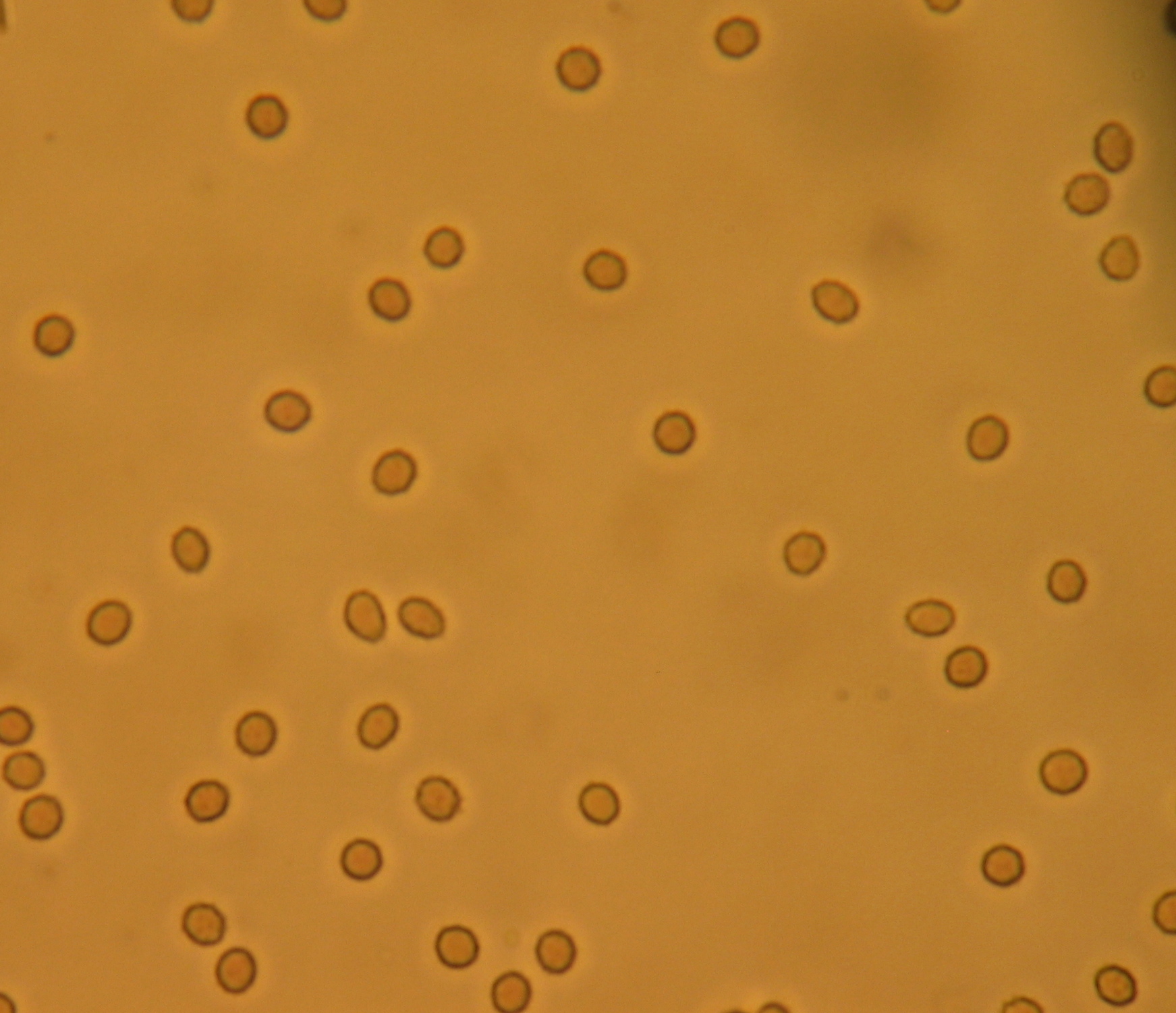
Microscopic hematuria
Occasionally "hemoglobinuria"
is used synonymously, although more precisely it refers only to hemoglobin
in the urine.
Red discoloration of the urine can
have various causes:
- Red
blood cells
- Microscopic hematuria (small amounts
of blood, can be seen only on urinalysis
or light microscopy)
- Macroscopic hematuria (or
"frank" or "gross") hematuria
- Hemoglobin
(only the red pigment, not the red
blood cells)
- Other pigments
- Myoglobin
in myoglobinuria
- Porphyrins
in porphyria
- Betanin,
after eating beets
- Drugs such as Rifampicin
and Phenazopyridine
Diagnosis

Acute hematuria due to trauma.
Often, the diagnosis is made on the basis of the medical history and some blood tests—especially
in young people in whom the risk of malignancy is negligible and the symptoms
are generally self-limiting.
Ultrasound
investigation of the renal tract is often used to distinguish between various
sources of bleeding. X-rays
can be used to identify kidney stones, although CT scanning is more precise.
In older patients, cystoscopy with biopsy of suspected lesions is often employed to investigate
for bladder
cancer.
If combined with pain, it may be loin pain hematuria syndrome.

The dipstick method of chemical
analysis of urine


Pathological components of urine, which are occur due to different
metabolic disorders in organism.
Ketonuria. Due to some diseases and pathological conditions (diabetes mellitus,
starvation, severe heart weakness, when amount of fat in the food more than
amount of carbohydrates) level of ketone bodies increases in a big amount (up
to 20-50 g per day). This is the index of deep metabolic
disorders, especially in carbohydrates metabolism.
Bilirubinuria. It occurs in case of hepatic parenhimatous inflammatory processes or in
case of obstruction of gall bladder ductus. Urine has a colour as dark bear.
After some times it stays yellow-green (bilirubin oxidized to biliverdin).
Urobilinuria. Increasing of amount of urobilin is due to haemolytic or parenhimatous
hepatitis, when decomposition of mesobilinogen in liver is depressed.
Creatinuria. Amount of creatin in urine increases due to different pathological processes
in muscles like myopathy and myodistrophy, starvation, hypovitaminosis E,
radiation sickness, hyperthyreosis. Also this is present in small children and
in women after delivery.
Indicanuria. Increasing of indican’s level in urine is due to intensification of
decay proteins in the intestines, weaken of intestine peristaltic (atony,
constipation), and chronic diseases, which are accompanied by intensive
decomposition of proteins (tuberculosis, for example).
Phenylketonuria. Innate deficiency of phenylalanine hydroxilase in liver makes
transformation of phenylalalnine in tyrosine impossible.