Nutrition for Disorders of the Liver,
Gallbladder, and Pancreas
Each year, millions of Americans are diagnosed with digestive disorders,
ranging from the occasional upset stomach to the more life-threatening colorectal
cancer. They encompass disorders of the gastrointestinal tract, as well as the
liver, gallbladder, and pancreas.
The
Liver: Anatomy and Functions
Anatomy of the liver:
The liver is located in the
upper right-hand portion of the abdominal cavity, beneath the diaphragm, and on
top of the stomach, right kidney, and intestines. Shaped like a cone, the liver
is a dark reddish-brown organ that weighs about
There are two distinct sources
that supply blood to the liver, including the following:
oxygenated
blood flows in from the hepatic artery
nutrient-rich
blood flows in from the hepatic portal vein
The liver holds about one pint
(13 percent) of the body's blood supply at any given moment. The liver consists
of two main lobes, both of which are made up of thousands of lobules. These
lobules are connected to small ducts that connect with larger ducts to
ultimately form the hepatic duct. The hepatic duct transports the bile produced
by the liver cells to the gallbladder and duodenum (the first part of the small
intestine).
Did you know?
The liver can lose
three-quarters of its cells before it stops functioning.
In addition, the liver is the
only organ in the body that can regenerate itself.
Functions of the liver:
The liver regulates most chemical
levels in the blood and excretes a product called bile, which helps carry away
waste products from the liver. All the blood leaving the stomach and intestines
passes through the liver. The liver processes this blood and breaks down the
nutrients and drugs into forms that are easier to use for the rest of the body.
More than 500 vital functions have been identified with the liver. Some of the
more well-known functions include the following:
production
of bile, which helps carry away waste and break down fats in the small
intestine during digestion
production
of certain proteins for blood plasma
production
of cholesterol and special proteins to help carry fats through the body
conversion
of excess glucose into glycogen for storage (glycogen can later be converted
back to glucose for energy)
regulation
of blood levels of amino acids, which form the building blocks of proteins
processing
of hemoglobin for use of its iron content (the liver stores iron)
conversion
of poisonous ammonia to urea (urea is an end product of protein metabolism and
is excreted in the urine)
clearing
the blood of drugs and other poisonous substances
regulating blood clotting
resisting
infections by producing immune factors and removing bacteria from the bloodstream
When the liver has broken down
harmful substances, its by-products are excreted into the bile or blood. Bile
by-products enter the intestine and ultimately leave the body in the form of
feces. Blood by-products are filtered out by the kidneys, and leave the body in
the form of urine.
Disorders of the Liver
There are many disorders of
the liver that require clinical care by a physician or other healthcare
professional. Listed in the directory below are some, for which we have provided
a brief overview.
If you cannot find the
information in which you are interested, please visit the Liver
Disorders Online Resources page in this Web site for an Internet/World Wide Web
address that may contain additional information on that topic.
About 95 million persons are
affected by all digestive problems. Digestive disorders account for 35 million
physician office visits per year.
Most digestive diseases are
very complex, with subtle symptoms, and the causes of many remain unknown. They
may be inherited, or develop from multiple factors such as stress, fatigue,
diet, or smoking. Abusing alcohol imposes the greatest risk for digestive
diseases.
Reaching a diagnosis requires
a thorough and accurate medical history and physical examination. Some patients
may need to undergo more extensive diagnostic evaluations, including lab tests,
endoscopic procedures, and imaging techniques. Physicians who specialize in the
treatment of digestive problems are called gastroenterologists.
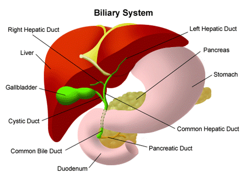
Liver,
Biliary, and Pancreatic Disorders
More than 25 million
The liver is the largest organ
in the human body. It is also one of the most important ones. The biliary
system consists of the bile ducts, gallbladder, and pancreas - all closely
associated with the functioning of the liver.
Some liver, biliary, and
pancreatic diseases are congenital (present at birth). Others can be prevented.
In any case, whether these diseases are congenital, injury-related,
viral-induced, or alcohol-induced, they can be devastating to a person's health
and require medical care.
Alcohol-Induced
Liver Disease
Did you know?
Women are more prone to liver damage from drinking alcohol than men.
What is alcohol-induced liver disease?
Alcohol-induced liver disease,
as the name implies, is caused by excessive consumption of alcohol and is a common,
but preventable, disease.
There are three primary types
of alcohol-induced liver disease, including the following:
fatty
liver Fatty liver is excessive accumulation of fat inside the liver cells.
Fatty liver is the most common alcohol-induced liver disorder. The liver is
enlarged, causing upper abdominal discomfort on the right side.
alcoholic
hepatitis Alcoholic hepatitis is an acute inflammation of the liver,
accompanied by the destruction of individual liver cells and scarring. Symptoms
may include fever, jaundice, an increased white blood cell count, an enlarged,
tender liver, and spider-like veins in the skin.
alcoholic
cirrhosis Alcoholic cirrhosis is the destruction of normal liver tissue,
leaving non-functioning scar tissue. Symptoms may include those of alcoholic
hepatitis, in addition to portal hypertension, enlarged spleen, ascites, kidney
failure, confusion, or liver cancer.
What are the symptoms of alcohol-induced liver disease?
Symptoms of alcohol-induced
liver disease depend on how much and how long a person has been drinking
alcohol. The following are the most common symptoms of alcohol-induced liver disease.
However, each individual may experience symptoms differently. Symptoms may
include:
enlarged liver
fever
jaundice
- yellowing of the skin and eyes.
increased white blood cell count
spider-like
veins in the skin
portal hypertension
enlarged spleen
ascites
- fluid build-up in the abdominal cavity.
kidney failure
confusion
The symptoms of
alcohol-induced liver disease may resemble other medical conditions or
problems. Always consult your physician for a diagnosis.
How is alcohol-induced liver disease diagnosed?
In addition to a complete
medical history and physical examination, diagnostic procedures for
alcohol-induced liver disease may include the following:
laboratory tests
liver
function tests - a series of special blood tests that can determine if the
liver is functioning properly.
liver
biopsy - a procedure in which tissue samples from the liver are removed (with a
needle or during surgery) from the body for examination under a microscope.
Treatment for alcohol-induced liver disease:
Specific treatment for
alcohol-induced liver disease will be determined by your physician based on:
your
age, overall health, and medical history
extent of the disease
your
tolerance for specific medications, procedures, or therapies
expectations
for the course of the disease
your opinion or preference
The goal of treatment is to
restore some or all normal functioning to the liver. Treatment usually begins
with abstinence from alcohol. The liver has great restorative power and is
often able to repair some of the damage caused by alcohol. In most cases, the
only damage it cannot reverse is scarring from cirrhosis.
Chronic
Liver Disease /Cirrhosis
What is chronic liver disease?
Chronic liver disease is marked by the gradual
destruction of liver tissue over time. Several liver diseases fall under this
category, including the following:
cirrhosis of the liver
fibrosis of the liver
What is cirrhosis of the
liver?
Cirrhosis is the 12th
leading cause of death in the United States, according to the National
Institute of Diabetes and Digestive and Kidney Diseases (NIDDK). Because of
chronic damage to the liver, scar tissue slowly replaces normal functioning
liver tissue, progressively diminishing blood flow through the liver. As the
normal liver tissue is lost, nutrients, hormones, drugs, and poisons are not
processed effectively by the liver. In addition, protein production and other
substances produced by the liver are inhibited.
What are the symptoms of
cirrhosis?
Symptoms of cirrhosis vary, depending on severity
of the condition. Mild cirrhosis may not exhibit any symptoms at all. The
following are the most common symptoms of cirrhosis. However, each individual
may experience symptoms differently. Symptoms may include:
abnormal nerve function
ascites
- fluid build-up in the abdominal cavity.
breast enlargement in men
coughing up or vomiting blood
curling
of fingers (Dupuytren's contracture of the palms)
gallstones
hair loss
itching
jaundice
- yellowing of the skin and eyes.
kidney failure
liver encephalopathy
muscle loss
poor appetite
portal hypertension
redness of palms
salivary gland enlargement in cheeks
shrinking of testes
spider-like
veins in the skin
weakness
weight loss
The symptoms of cirrhosis may resemble other
medical conditions or problems. Always consult your physician for a diagnosis.
What causes cirrhosis?
The most common cause of cirrhosis
is alcohol abuse. Other causes include the following:
hepatitis and other viruses
use of certain drugs
chemical exposure
bile duct obstruction
autoimmune diseases
obstruction
of outflow of blood from the liver (i.e., Budd-Chiari syndrome)
heart and blood vessel disturbances
alpha1-antitrypsin deficiency
high blood galactose levels
high
blood tyrosine levels at birth
glycogen storage disease
cystic fibrosis
diabetes
malnutrition
hereditary
accumulation of too much copper (Wilson's Disease) or iron (hemochromatosis)
How is cirrhosis diagnosed?
In addition to a complete medical history and
physical examination, diagnostic procedures for cirrhosis may include the
following:
laboratory tests
liver
function tests - a series of special blood tests that can determine if the
liver is functioning properly.
liver
biopsy - a procedure in which tissue samples from the liver are removed (with a
needle or during surgery) from the body for examination under a microscope.
cholangiography
- x-ray examination of the bile ducts using an intravenous (IV) dye (contrast).
computed
tomography scan (CT or CAT scan) - a diagnostic imaging procedure using a
combination of x-rays and computer technology to produce cross-sectional images
(often called slices), both horizontally and vertically, of the body. A CT scan
shows detailed images of any part of the body, including the bones, muscles,
fat, and organs. CT scans are
more detailed than general x-rays.
ultrasound
(Also called sonography.) - a diagnostic imaging technique, which uses
high-frequency sound waves and a computer to create images of blood vessels,
tissues, and organs. Ultrasounds are used to view internal organs of the
abdomen such as the liver, spleen, and kidneys and to assess blood flow through
various vessels.
Treatment for cirrhosis:
Specific treatment for
cirrhosis will be determined by your physician based on:
your
age, overall health, and medical history
extent of the disease
your
tolerance for specific medications, procedures, or therapies
expectations
for the course of the disease
your opinion or preference
Cirrhosis is a progressive
liver disease, and damage sustained to the liver is irreversible. However, with
proper nutrition, avoidance of certain toxins (such as alcohol), vitamin
supplementation, and management of cirrhosis complications, further liver
damage can often be delayed or stopped. In severe cases of cirrhosis, liver
transplantation may be considered.
What is fibrosis?
Fibrosis is the growth of scar
tissue due to infection, inflammation, injury, or even healing. The overgrowth
of scar tissue can occur in almost any organ. Fibrosis in the liver can inhibit
the organ's proper functioning. Liver fibrosis is usually the result of
cirrhosis.
Congenital
Liver Defects
What are congenital liver defects?
Defects of the liver at birth
usually affect the bile ducts. Though rare, some congenital liver defects
include the following:
biliary
atresia - a condition in which the bile ducts are absent or have developed
abnormally.
choledochal
cyst - a malformation of the hepatic duct that can obstruct flow of bile in
infants.
What are the indicators that a congenital liver defect may be present?
Congenital liver defects that
affect the flow of bile share some common symptoms. The following are the most
common symptoms of congenital liver defect. However, each individual may
experience symptoms differently. Symptoms may include:
jaundice
- yellowing of the skin and eyes.
dark urine
pale stool
The symptoms of congenital
liver defects may resemble other medical conditions or problems. Always consult
your child's physician for a diagnosis.
How are congenital liver defects
diagnosed?
Congenital liver defects that affect the flow of
bile are usually diagnosed at birth or shortly afterward. In addition to a
complete medical history and physical examination, diagnostic procedures for a
congenital liver defect may include the following:
laboratory tests
liver
function tests - a series of special blood tests that can determine if the
liver is functioning properly.
liver
biopsy - a procedure in which tissue samples from the liver are removed (with a
needle or during surgery) from the body for examination under a microscope.
Treatment for congenital liver
defects:
Specific treatment for congenital liver defects
will be determined by your child's physician based on:
your
child's age, overall health, and medical history
extent of the disease
your
child's tolerance for specific medications, procedures, or therapies
expectations
for the course of the disease
your opinion or preference
Treatment may include surgery to reconstruct or
bypass the bile ducts. Sometimes, a liver transplant may be necessary.
Hepatitis
There are several types of hepatitis that
require clinical care by a physician or other healthcare professional. Listed
in the directory below are some, for which we have provided a brief overview.
If you cannot find the information in which you
are interested, please visit the Liver
Disorders Online Resources page in this Web site for an Internet/World Wide Web
address that may contain additional information on that topic.
Hepatitis
In-Depth Report
In-Depth From A.D.A.M.
Background
Hepatitis is a disorder in
which viruses or other mechanisms produce inflammation in liver cells,
resulting in their injury or destruction. The liver is the largest organ in the
body, occupying the entire upper right quadrant of the abdomen. It performs over 500 vital functions including:
· The liver processes all of the nutrients the
body requires, including proteins, glucose, vitamins, and fats.
· The liver manufactures bile, the greenish fluid
stored in the gallbladder that helps digest fats.
· One of the liver's major contributions is to
render harmless potentially toxic substances, including alcohol, ammonia,
nicotine, drugs, and harmful by-products of digestion.
· Old red blood cells are removed from the blood
by the liver and spleen, and the iron contained in them is recycled to the bone
marrow to make new red blood cells.

The esophagus, stomach, large
and small intestine -- aided by the liver, gallbladder, and pancreas -- convert
the nutritive components of food into energy and break down the non-nutritive
components into waste to be excreted.
Damage to the liver can impair
these and many other processes. Hepatitis varies in severity from a
self-limited condition with total recovery to a life-threatening or life-long
disease. It can occur from many
different causes:
· In the most common hepatitis cases (viral
hepatitis), specific viruses incite the immune system to fight off infections. Specific immune factors become over-produced that
cause injury.
· Hepatitis can also result from an autoimmune
condition, in which abnormal immune factors attack the body's own liver cells.
· Inflammation of the liver can also occur from
medical problems, drugs, alcoholism, chemicals, and environmental toxins.
No matter what the cause of
hepatitis, it can take either an acute (short term) or chronic (long term)
form. In some cases, acute hepatitis develops into a chronic condition, but
chronic hepatitis can also occur on its own. Although chronic hepatitis is
generally the more serious condition, patients having either condition can
experience varying degrees of severity.
Acute Hepatitis. Acute hepatitis can begin
suddenly or gradually, but it has a limited course and rarely lasts beyond 1 or
2 months. Usually, there is only spotty liver cell damage and evidence of immune
system activity. Rarely, acute hepatitis can cause severe, even
life-threatening, liver damage.
Chronic Hepatitis. The chronic forms of
hepatitis last for prolonged periods. Doctors usually categorize chronic hepatitis by indications of severity:
· Chronic persistent hepatitis is usually mild and
nonprogressive or slowly progressive, causing limited damage to the liver.
· Chronic active hepatitis involves extensive
liver damage and cell injury beyond the portal tract.
Viral
Hepatitis
Most cases of hepatitis are
caused by viruses that infect liver cells and begin replicating. They are defined by the letters A through G:
· Hepatitis A, B, and C are the most common viral
forms of hepatitis. Investigators are still looking for additional viruses that
may be implicated in hepatitis unexplained by the current known viruses.
· Other hepatitis viruses include hepatitis E and
hepatitis G. Like hepatitis A, hepatitis E is caused by contact with
contaminated food or water. It is not serious except in pregnant women, when it
can be life threatening. Hepatitis G is always chronic and most likely has the
same modes of transmission as hepatitis C. It does not appear to have serious
effects.
Scientists do not know exactly
how these viruses actually cause hepatitis (inflammation in the liver). As the
virus reproduces in the liver, several proteins and enzymes, including many
that attach to the surface of the viral protein, are also produced. Some of
these may be directly responsible for liver damage. Researchers are
investigating elevated levels of specific immune factors, including T cell
sub-types in the liver of hepatitis C and B patients. T cells are important
infection fighters in the immune system that in some cases release powerful
inflammatory substances (tumor necrosis factor and interferon gamma) that can
cause considerable damage leading to hepatitis B or C.
Autoimmune
Chronic Hepatitis
Autoimmune chronic hepatitis
accounts for about 20% of all chronic hepatitis cases. Like other autoimmune
disorders, this condition develops because a genetically defective immune
system attacks the body's own cells and organs (in this case the liver). The
attack is triggered by an environmental factor, probably a virus. Suspects
include the measles virus, a hepatitis virus, or the Epstein-Barr virus, which
causes mononucleosis. It is also possible that a reaction to a drug or other
toxin that affects the liver also triggers an autoimmune response in
susceptible individuals. In about 30% of cases, autoimmune hepatitis is
associated with other disorders that involve autoimmune attacks on other parts
of the body.
Hepatitis
Caused by Alcohol and Drugs
Alcohol. About 10 - 35% of heavy
drinkers develop alcoholic hepatitis. In the body, alcohol breaks down into
various chemicals, some of which are very toxic to the liver. After years of
drinking, liver damage can be very severe, leading to cirrhosis in about 10 -
20% of cases. Although heavy drinking itself is the major risk factor for
alcoholic hepatitis, genetic factors may play a role in increasing a person's
risk for alcoholic hepatitis. Women who abuse alcohol are at higher risk for
alcoholic hepatitis and cirrhosis than are men who drink heavily. High-fat
diets may also increase the risk in heavy drinkers.
Drugs. Because the liver plays such
a major role in metabolizing drugs, hundreds of medications can cause reactions
that are similar to those of acute viral hepatitis. Symptoms can appear
anywhere from 2 weeks to 6 months after starting drug treatment. In most cases,
they disappear when the drug is withdrawn, but in rare circumstances they may
progress to serious liver disease. Drugs most noted for liver interactions
include halothane, isoniazid, methyldopa, phenytoin, valproic acid, and the
sulfonamide drugs. Very high doses of acetaminophen (Tylenol) have been known
to cause severe liver damage and even death, particularly when used with
alcohol.
Nonalcoholic
Fatty Liver Disease (NAFLD)
Nonalcoholic fatty liver
disease (NAFLD) affects between 10 - 24% of the population. It covers several
conditions, including nonalcoholic steatohepatitis (NASH). NAFLD has features
similar to alcoholic hepatitis, particularly a fatty liver, but it occurs in
individuals who drink little or no alcohol. Severe obesity and diabetes are the
major risk factors for NAFLD as well as complications from NAFLD. NAFLD is
usually benign and very slowly progressive. In certain patients, however, it
can lead to cirrhosis, liver failure, or liver cancer. <!--[For more
information, see In-Depth Report #75: Cirrhosis.]-->
In-Depth From A.D.A.M. Diagnosis
In people suspected of having
or carrying viral hepatitis, doctors will measure certain substances in the
blood.
· Bilirubin. Bilirubin is one of the most important
factors indicative of hepatitis. It is a red-yellow pigment that is normally
metabolized in the liver and then excreted in the urine. In patients with
hepatitis, the liver cannot process bilirubin, and blood levels of this
substance rise. (High levels of bilirubin cause the yellowish skin tone, known
as jaundice.)
· Liver Enzymes
(Aminotransferases). Enzymes known as aminotransferases, including
aspartate (AST) and alanine (ALT), are released when the liver is damaged.
Measurements of these enzymes, particularly ALT, are the least expensive and
most noninvasive tests for determining severity of the underlying liver disease
and monitoring treatment effectiveness. Enzyme levels vary, however, and are
not always an accurate indicator of disease activity. (For example, they are
not useful in detecting progression to cirrhosis.)

Blood is drawn from a vein
(venipuncture), usually from the inside of the elbow or the back of the hand. A
needle is inserted into the vein, and the blood is collected in an air-tight
vial or a syringe. Preparation may vary depending on the specific test.
General Tests to Determine Causes of Viral
Hepatitis
Radioimmunoassays. To identify the particular virus
causing hepatitis, blood tests called radioimmunoassays are performed.
Typically, radioimmunoassays identify particular antibodies, which are
molecules in the immune system that attack specific antigens. (Antigens
are any molecules that the body considers threatening or dangerous and which
can be targeted by antibodies.) Some of these tests can pinpoint hepatitis
antigens directly. These tests, however, have limitations:
· There may not be sufficient numbers of
antibodies to be detectable by blood tests for up to weeks or months after
hepatitis develops. Blood tests that are taken too early may miss these signs
of infection.
· Antibodies also linger after patients recover,
so a positive antibody test can indicate a previous infection but does not necessarily
determine if the infection is active.
The assays for individual
hepatitis viruses may differ.
Polymerase Chain Reaction. In some cases of hepatitis C,
a polymerase chain reaction (PCR), may be performed. PCR is able to make
multiple copies of the virus’ genetic material to the point where it is
detectable.
Liver Biopsies
A liver biopsy may be
performed for acute viral hepatitis caught in a late stage or for severe cases
of chronic hepatitis. No laboratory tests for enzyme or viral levels can truly
determine the actual damage to the liver. A biopsy helps determine treatment
possibilities, the extent of damage, and the long-term outlook.
The biopsy requires abdominal
surgery, most often laparoscopy. This procedure takes about an hour. It requires general anesthesia and involves the
following steps:
· The surgeon makes one or more small incisions
(about 0.5 - 1.0 inch) in the abdomen.
· Carbon dioxide or nitrous oxide is delivered
through the incision to inflate the abdomen so that the involved area is
visible.
· The surgeon inserts a thin tube, called a
laparoscope, which contains a tiny camera. Surgical instruments are also
inserted through the incision to remove the liver tissue for biopsy.

A liver biopsy is
not a routine procedure, but is performed when it is necessary to determine the
presence of liver disease and to look for malignancy, cysts, parasites, or other
pathology. The actual procedure is only slightly uncomfortable. Most of the
discomfort arises from being required to lie still for several hours afterwards
to prevent bleeding from the biopsy site.
A less invasive procedure,
called a minilaparoscopy, uses a smaller scope and may prove to reduce the time
of the procedure.
Screening
for Liver Cancer
Patients with cirrhosis are
usually screened for liver cancer using tests for a substance called
alpha-fetoprotein (AFP) and ultrasound. It is not known, however, if such
screening has much impact on survival, since it is not very sensitive and has a
high rate of false positives (suggesting the presence of cancer when it is not
actually present). Screening is not necessary in patients without cirrhosis.
In-Depth From A.D.A.M. Hepatitis A
About a third of the U.S.
population has antibodies to hepatitis A, indicating previous infection by the
virus. The hepatitis A virus infects up to 200,000 Americans every year and
causes symptoms in about 134,000 of them. Almost 30% are children under age 15.
Hepatitis A (formerly called
infectious hepatitis) is excreted in feces and transmitted by contaminated food
and water. Eating shellfish taken from sewage-contaminated water is a common
means of contracting hepatitis A. Infected people can transmit it to others if
they do not take strict sanitary precautions. Hepatitis A is infectious for 2 -
4 weeks before symptoms develop and for a few days afterward.
People at risk for passing the
infection along or being infected include:
· International travelers. Hepatitis A is the
hepatitis strain people are most likely to encounter in the course of
international travel. In fact, in spite of the availability of a vaccine, the
increase in travel to underdeveloped countries has kept the incidence of
hepatitis A steady in Western nations. The incidence may even be increasing.
· Day care employees and children. It is estimated
that between 11 - 16% of hepatitis A cases occur among day care employees and
children who attend day care. The risk for children attending day care is very
low, however, if hygienic precautions are used, particularly when changing
babies and handling diapers.
· Sexually active homosexual men.
· Intravenous drug users.
· Health care, food industry, and sewage workers.

A fly may act as a mechanical
vector of diseases such as hepatitis A, which means the fly carries the
infective organism on its feet or mouth parts and contaminates food or water
which a person then consumes. A biological vector actually develops an
infective organism in its body and passes it along to its host, usually through
its saliva. A fly can be a biological vector, as in the transmission of
leishmaniasis by the sandfly.
Symptoms
of Acute Hepatitis
Symptoms of acute viral
hepatitis may begin suddenly or develop gradually. They may be so mild that
patients mistake the disease for the flu. They include:
· Nearly all patients experience some fatigue and
often have mild fever.
· Gastrointestinal problems are very common,
including nausea, vomiting, a general feeling of discomfort in the abdomen, or
a sharper pain that may occur in the upper right area of the abdomen. This pain
tends to increase during jerking movements, such as climbing stairs or riding
on a bumpy road.
· Gastrointestinal problems can also lead to loss
of appetite, weight loss, and dehydration.
· After about 2 weeks, dark urine and jaundice (a
yellowish color in the skin and whites of the eyes) develops in some, but not
all, patients. (Children tend
not to develop jaundice.)
· About half of all patients have light colored
stools, muscle pain, drowsiness, irritability, and itching, usually mild.
· Diarrhea and joint aches occur in about a
quarter of patients.
· The liver may be tender and enlarged, and most
people have mild anemia.
· In about 10% of patients, the spleen is
enlarged.
Preventing
Hepatitis A Infections When Traveling to High-Risk Countries
Travelers should take the
following precautions:
· Get vaccinated against hepatitis A and possibly
B if traveling for long periods of time to countries where epidemics occur.
· Use only carbonated bottled water for brushing
teeth and drinking. (Remember that ice cubes can carry infection.) Boiling
water is the best method for eliminating infectious organisms. Bringing the
water to a good boil for at least a minute generally renders it safe to drink.
· Heated food should be hot to the touch and eaten
promptly.
· Don’t buy food from street vendors.
· Beware of sliced fruit that may have been washed
in contaminated water. Travelers
themselves should peel all fresh fruits and vegetables.
· Avoid dairy products.
· Avoid raw or undercooked meat and fish.
Vaccinations for Hepatitis A
Two vaccines (Havrix, Vaqta)
are now available, both very safe and effective for preventing hepatitis A
(HAV). They can be given along with immune globulin and other vaccines. A
combination Hep A - Hep B vaccine (Twinrix) that contains both Havrix and
Engerix-B (a hepatitis B vaccine) is also available.

Immunization is a process
to initiate or augment resistance to an infectious disease. The goal of
immunization is to prevent, and in some cases eradicate, potentially serious,
life-threatening diseases. Candidates for HAV
Vaccinations. Vaccinations for hepatitis A are recommended for:
· Children age 12 - 23 months
(the U.S. Centers for Disease Control and Prevention recommends that children
receive the first dose of the hepatitis A vaccine when they are 12 months old,
and a second dose 6 months later). Hepatitis A used to affect mostly children,
but now occurs mostly in adults.
· Travelers to developing
countries. (Travelers should also receive immune globulin if they are visiting
high-risk areas within 4 weeks of the vaccination.)
· Sexually active homosexual men
· Illegal drug users, especially
those who inject drugs
· Health care workers
· People with chronic liver disease
· People with hemophilia or
other blood-clotting disorders
Side Effects. Although there are few side effects,
allergic responses from the vaccination can occur. Hair loss has been reported
in very few people after a second administration. There may be pain at the
injection site. (Havrix causes more pain at the injection site than Vaqta.)
Symptoms of Hepatitis A
Symptoms are usually mild,
especially in children, and generally appear between 2 - 6 weeks after exposure
to the virus. Adult patients are more likely to have fever, jaundice, and
itching that can last up to several months.
General Outlook for People Infected with
Hepatitis A
Hepatitis A is the least
serious of the common hepatitis viruses. It does not directly kill liver cells,
and there is no risk for a chronic form. Severe (fulminant) hepatitis is the
only major concern, but even if it develops, it is almost always less dangerous
than with other viral types. Only 1 in a 1,000 patients is at risk for death
from this complication. If hepatitis A infection occurs in patients with
hepatitis C, however, superinfections can occur, even without cirrhosis,
leading to a life-threatening form of fulminant hepatitis. (Infection of
patients with hepatitis B who do not have cirrhosis does not appear to be as
dangerous.)
Specific Tests for Hepatitis A
Radioimmunoassays are
generally used to identify IgM antibodies, first produced to fight hepatitis A.
They appear early in the course of the disease and usually can be identified as
soon as symptoms appear. IgM antibodies disappear during recovery, but those
known as IgG antibodies persist, and their presence can be used to indicate a
previous infection.
Treatments and Measures to Prevent Transmission
of Hepatitis A
The primary goals for managing
acute viral hepatitis are to provide adequate nutrition, to prevent additional damage
to the liver, and to prevent transmission to others.
Precautions for Preventing
Transmission of Hepatitis A. Because hepatitis A and hepatitis E are usually
passed through contaminated food, people with these viruses should not prepare
food for others. Unfortunately,
these viruses are most contagious before symptoms appear.
· Using hot water when cleaning utensils or
clothing is essential. Heating a contaminated article for 1 minute kills the
virus. Simple household bleach is effective for disinfecting hard surfaces.
Sterilizing is not necessary. Still, even with strong precautions, utensils
used by the patient for eating and cooking should be kept separate from those
used by others.
· Abstain from sexual activity or take strict
precautions.
· Abstain from alcohol. Moderate drinking after
recovery is not harmful for most people.
In-Depth From A.D.A.M.
Hepatitis B and D
Hepatitis B and D were
formerly called serum hepatitis. Hepatitis B is mainly transmitted through
blood transfusions, contaminated needles, and sexual contact. Blood screening
has reduced the risk from transfusions. It can also be passed from cuts,
scrapes, and other breaks in the skin. Hepatitis D virus can replicate only by
attaching to hepatitis B and therefore cannot exist without the B virus being
present.
Risk Factors for Hepatitis B. About 1.2 million Americans
are chronically infected with hepatitits B and between 20 - 30% acquired the
infection when they were children. Men are at higher risk than women. Among
ethnic groups living in the United States, Asians are at highest risk, due to
the high rate of hepatitits B in Asian countries. Fortunately, in the US the
number of new infections has declined dramatically -- by 67% between 1990 and
2002. In 2003, 7,526 cases were reported compared to over 20,000 in 1990. The
greatest decrease has occurred in children. Among young adults and people
living in the Northeast, however, the incidence has increased since 1999. This
may indicate that sexual activity is an important route for viral transmission
and that the protective effect of the vaccine has not yet reached older,
high-risk groups. Also, as with hepatitis A, the increase in travelers to
underdeveloped nations may be responsible for the steady rate.
Hepatitits B is far more common
overseas and about 600,000 people die each year from conditions, such as liver
cancer or cirrhosis, that are related to chronic hepatitis B. Nearly 70% of
these infections were acquired during infancy or early childhood.
People at risk include:
· Drug users who share needles.
· Children of infected mothers.
Pregnant women with hepatitis B can transmit the virus to their babies. Even if
they are not infected at birth, unvaccinated children of infected mothers run a
60% risk of developing hepatitits B before age 5. Children are more likely than
adults to become chronic carriers, although between 6 - 12% of children
spontaneously recover each year.
· People with multiple sex
partners or other high-risk sexual behavior.
· Hospital workers and others
exposed to blood products. Contaminated medical instruments, including
fingerstick devices used for more than one individual, have been known to
transmit the virus.
· Staff members and clients of
institutions for the developmentally disabled.
· Prisoners.
· Immigrants from areas where
the disease rate is high. (International travelers who spend long periods in
such areas may also be at risk.)
People at highest risk for
becoming chronic carriers of the virus include:
· Children infected before age
5, including newborns, most of whom become carriers.
· Infected people with damaged
immune systems, such as AIDS patients.
Risk Factors for Hepatitis D. Hepatitis D occurs only in
people with hepatitis B. It is not common in the U.S. and the incidence of this
hepatitis is declining rapidly overseas. Experts anticipate that it will be
extremely rare in the near future. Those who recover from hepatitis B are
immune to further infection from both hepatitis B and D viruses.
Lifestyle
Precautions for Preventing Hepatitis B and Hepatitis C Virus Transmission
The following are some
precautions for preventing the transmission of hepatitits B or hepatitits C:
· All objects contaminated by blood from patients
with hepatitis B or C must be handled with special care. (Restrictions on food
preparation are not necessary for these hepatitis viruses.)
· Patients with viral hepatitis should abstain
from sexual activity or take strict precautions. Infected patients should use
condoms and contraceptives that prevent passage of the virus, possibly even in
relationships that last for years. Women partners or infected women should
abstain from sexual activity during menstruation. Either partner with
infections that cause bleeding in the genital or urinary areas should avoid
sexual activity until the infection is no longer active.
· Couples with an infected partner or people
sharing household with an infected person should avoid sharing personal items,
such as razors or toothbrushes.
Note: There is no evidence
that the viruses can be passed through casual contact, or other contact without
exposure to blood, including kissing, hugging, sneezing, or coughing or by
sharing eating utensils or drinking glasses. People infected with chronic hepatitis
B or C should not be excluded from work, school, play, childcare or any social
or work settings on the basis of their infection.
Symptoms of Hepatitis B
Symptoms appear long after the
initial infection, usually 4 - 24 weeks. Many patients may not even experience
them or they may be mild and flu-like. About 10 - 20% of patients have a fever
and rash. Nausea is not common. Sometimes there is general aching in the
joints. The pain can resemble arthritis, affecting specific joints and
accompanied by redness and swelling.
Outlook
for Patients with Hepatitis B
Most people with hepatitis B
recover from the virus. The risk of progressing to the chronic form of
hepatitis B is age dependent. Only 2 - 6% of people who are older than 5 years
old when they acquire the virus will develop chronic hepatitis B. The risk for
chronic hepatitis in children age 1 - 5 years is 30%, and the risk for infants
under the age of 1 is up to 90%. In the U.S., about 1.25 million people are
chronically infected with hepatitis B. Worldwide, about 400 million people are
chronically infected.
Chronic hepatitis B infection
significantly increases the risk for liver damage, including cirrhosis and
liver cancer. In fact, hepatitis B is the leading cause of liver cancer
worldwide. According to a 2006 Lancet study, liver disease, especially liver
cancer, is the main cause of death in people with chronic hepatitis B. Because
of these high risks, it is very important that patients with chronic hepatitis
B receive regular screenings for liver cancer.
Patients with hepatitis B who
are co-infected with hepatitis D may develop a more severe form of acute
infection than those who have only hepatitis B. Co-infection with hepatitis B
and D increases the risk of developing acute liver failure. Patients with
chronic hepatitis B who develop chronic hepatitis D also face high risk for
cirrhosis. Hepatitis D occurs only in people who are already infected with
hepatitis B.
Specific Tests for Identifying Hepatitis B
A diagnosis of hepatitis B
relies on measuring the liver enzymes aspartate (AST) and alanine (ALT) --
released when the liver is damaged -- assays to identify the viral DNA, and a
liver biopsy.
Doctors must then determine if
the condition is chronic but inactive or whether it is more aggressive. This is
done by identifying a specific antigen called HBsAg, which is a protein that is
found in the blood in early stages of hepatitis B and suggests the presence of
a viral replication. Most people develop antibodies to this antigen during
convalescence. Their condition is referred to as HBeAG negative, or anti-HBe,
and suggests that infection is on the wane. About 5 - 10% of people do not
clear the infection but become carriers of the antigen (called HBsAG-positive).
Evidence of its persistence for more than 6 months suggests that the condition
is chronic.
Tests can identify specific
genetic types of hepatitis B virus (designated A to G). It is not clear how
significant they are in treating patients with hepatitits B.
It is important to remember,
however, that viral levels are not an accurate measure of actual liver damage.
Only a biopsy can determine this.
To diagnose hepatitis D using an
antibody test, hepatitis B must already have been identified.
Preventing Hepatitis B and its Transmission
General precautions for
preventing hepatitis B when traveling are the same as those for hepatitis A. In
infected people, precautions for preventing transmission are similar to those
for hepatitis C.
Vaccinations for Prevention of
Hepatitis B. Several inactivated virus vaccines, including Recombivax HB, GenHevac B,
Hepagene, and Engerix-B, can prevent hepatitis B and are safe even for infants
and children. A triple-antigen hepatitis B vaccine (Hepacare) is proving to be
effective for people who do not respond to the standard vaccines. Vaccination
programs are also helping to reduce the risk for liver cancer. A combination
vaccine (Twinrix) that contains Engerix-B and Havrix, a hepatitis A vaccine, is
now approved for people with risk factors for both hepatitis A and B.

The hepatitis B
vaccine is recommended for people who are at higher risk, including people who
live with someone with hepatitis B and healthcare workers.
Until recently, the vaccine
contained a mercury-based preservative called thimerosal. In response to
concerns, professional organizations recommended suspending vaccinations in
infants with noninfected mothers. In 1999, a thimerosal-free vaccine became
available, and medical centers are now urged to continue vaccinations.
Unfortunately, even after the thimerosal-free vaccine became available, a
number of hospitals still have not restored vaccination of all infants. This is
a safe vaccine. Parents should be sure their children are immunized.
Candidates for Hepatitits B
Vaccinations. Experts now recommend that all infants and children not previously vaccinated
be immunized by the time they reach seventh grade.
Typical schedules for
hepatitis B vaccinations in childhood are as follows:
· All infants should receive the hepatitis B
vaccine soon after birth and before hospital discharge. (The first dose may be
given by age 2 months if the mother has no evidence of infection. Infants of
mothers infected with hepatitits B should be treated with immune globulin plus
the hepatitis vaccine within 12 hours of birth. Vaccinating the newborn
prevents infection from being transmitted from mother to child.)
· The second dose should be given at least 4 - 6
weeks after the first dose. The third dose is given at least 8 weeks after the
second dose (typically when the baby is 6 - 23 months old).
· Children who are 11 - 12 years old and who have
not been immunized should receive two or three doses of the vaccine (depending
on the brand) given over a few months.
Hepatitis B vaccine protection
lasts at least 10 years. Booster shots after that may be recommended, depending
on continuing risk such as sexual exposure.
The following adults are at
very high risk and should be vaccinated:
· Health care and public safety workers who may be
exposed to blood products. Such individuals have a risk for hepatitis B virus
that ranges from 15 - 30%.
· People in the same household as hepatitits B
infected individuals. (Unvaccinated people who have had intimate exposure to
people with hepatitits B may be protected with immune globulin, which is
sometimes administered with the vaccine.)
· Travelers to developing countries.
· Patients who require transfusions and have not
been infected with hepatitits B. (Those with blood clotting disorders should
have the vaccination administered under the skin, not injected in the muscle.)
· Sexually active homosexual or heterosexual
individuals with multiple partners or who engage in high-risk sexual behavior.
· People with any sexually transmitted diseases.
Other people at risk who may
benefit from vaccinations include:
· Patients and workers in mental institutions and
morticians.
· Patients on hemodialysis. (People on
hemodialysis may need larger doses or boosters. They also may need to be
re-vaccinated if blood tests indicate they are losing immunity.)
· People who use injected drugs.
· Pregnant women at risk for the virus should be
vaccinated. There is no evidence that the vaccine is dangerous to the fetus.
· People receiving treatments or who have
conditions that suppress the immune system may need the vaccination, although
its benefits for this group are unclear except for those at high risk, such as
people with HIV or spleen abnormalities.
The regimen in adults is
typically three doses given over 6 months. People with alcoholism may need high
doses.
Soreness at the injection site
is the most common side effect. There have been some reports of nerve
inflammation after vaccinations for hepatitis B, and there has been some
concern about three small studies associating the vaccine with an insignificant
increase in multiple sclerosis. Recent studies, however, have found no evidence
to support these concerns. Nonetheless, some groups oppose the vaccination in
children who are not in high-risk groups. It should be strongly stressed that
worldwide 65 million people with chronic hepatitis are expected to die from
liver disease. Vaccinations save lives. For example, in Taiwan, where infection
rates are high and infants are at risk for hepatitis B from infected mothers,
vaccination programs have significantly reduced the risk for liver cancer.
Treatments
for Chronic Hepatitis B
Six drugs are currently
approved in the United States for treatment of chronic hepatitis B:
· Peginterferon alfa-2a (Pegasys)
· Interferon-alfa-2b (Intron)
· Adefovir (Hepsera)
· Lamivudine (Epivir)
· Entecavir (Baraclude)
· Telbivudine (Tyzeka)
These drugs block the replication of hepatitits
B in the body. Some also help boost the immune system. A doctor will decide
which drug to prescribe based on a patient’s age, disease severity, and other
factors. Each drug has various advantages and disadvantages in terms of cost,
efficacy, side effects, and likelihood of drug resistance. A combination of
drugs may also be prescribed.
Peginterferon alfa-2a. Peginterferon alfa-2a
(Pegasys) was approved in 2005 for treatment of chronic hepatitis B.
(Peginterferon is also called pegylated interferon.) The drug was previously
approved in 2002 for treatment of chronic hepatitis C. Pegasys prevents the
hepatitis B virus from replicating and also helps boost the immune system. It
is given as a weekly injection. Peginterferon is sometimes prescribed in
combination with lamivudine (Epivir).
Interferon Alpha. For many years, interferon
alfa-2b (Intron) was the standard drug for hepatitis B. The drug is usually
taken by injection every day for 16 weeks. (It does not appear to help
hepatitis D.) Unfortunately, even in hepatitis B, the virus recurs in almost
all cases, although this recurring mutation may be weaker than the original
strain. Administering the drug for longer periods may produce sustained
remission in more patients while still being safe. Interferon is also effective
in eligible children, although long-term effects are unclear.
Lamivudine, Entecavir, and Telbivudine.
These drugs are classified as nucleoside analogs. Lamivudine (Epivir or 3TC) is
an antiretroviral drug that is used to treat human immunodeficiency virus (HIV)
as well as hepatitis B. Studies suggest that lamivudine reduces viral count in
over half of hepatitis B patients who take it as sole therapy for about a year.
It is less expensive than interferon-alfa and has fewer side effects, but may
not work as well as interferon-alfa for long-term therapy. A major problem with
lamivudine is the development of mutated viral strains that become resistant to
the drug, particularly in areas where the virus is common. About 20% of
patients who take lamivudine develop drug resistance.
In 2005, the FDA approved entecavir (Baraclude)
for treatment of adults with chronic hepatitis B. In clinical trials, entecavir
worked better than lamivudine for treating hepatitits B. Entecavir appears to
have less risk of drug resistance than lamivudine. Studies also suggest that it
may be a good alternative treatment for patients who have developed resistance
to lamivudine. Questions have been raised about the drug’s possible cancer
risks. Ongoing studies are evaluating this risk.
In 2006, the FDA approved telbivudine (Tyzeka),
the newest nucleoside analog drug, for treatment of chronic hepatitis B.
Adefovir. Adefovir (Hepsera) belongs
to a class of antiviral drugs called nucleotide analogs. (Nucleotides are
related to nucleosides but have a slightly different chemical structure.)
Nucleotide analogs block an enzyme involved in the replication of viruses.
Adefovir costs more than lamivudine, but may be effective against
lamivudine-resistant strains of hepatitits B. The drug must be taken on a
long-term basis. A 2006 study indicated that when patients stopped taking
adefovir after 48 weeks, the hepitatis B virus resumed replication. Patients
who took the drug for a longer period (144 weeks) continued to benefit from
treatment. Another 2006 study indicated that for some patients, adefovir
remains effective for up to 5 years, although resistance occurs in about 20% of
patients.
Drug Warnings. In 2004, the FDA issued two
drug warnings for patients with hepatitits B. The HIV drug tenofovir (Viread)
should not be used to treat patients with HIV who are co-infected with
hepatitits B as the drug may increase hepatitis severity. The lymphoma drug
rituximab (Rituxan) may reactivate hepatitits B. Patients with lymphoma should
be screened for hepatitits B. In 2007, the FDA revised the label for entecavir
(Baraclude); patients who are co-infected with hepatitits B and HIV should take
entecavir only if they are also taking antiretroviral HIV drugs.
Investigational Drugs.
· Emtricitabine is a nucleoside analog drug used
to treat HIV and AIDS. It is being
investigated for chronic hepatitits B.
· Pegylated interferon alfa-2b (Peg-Intron) and
alfa-2a (Pegasys) are approved for treatment of chronic hepatitis C. They are
being investigated alone and in combination with other drugs, such as ribavirin
(Copegus, Rebetol), for treatment of hepatitits B. The combination of pegylated
interferon and ribavirin is the standard treatment for hepatitis C.
· Thymosin Alpha 1 (Zadaxin), also called
thymalfasin, is a synthetic version of a substance derived from the thymus
gland (which is responsible for maturation of immune factors called T-cells).
It appears to be safe for hepatitis B patients when used alone or in
combination with interferon. It is approved in many countries, but not the
United States.
Liver Transplantation. If the disease progresses to
liver failure, liver transplantation may be an option. It is not foolproof,
however. Viral recurrence is high in patients with hepatitis B. However,
regular, lifelong injections of hepatitis B immune globulin (HepaGam B) can
reduce the risk for re-infection following liver transplantation.
In-Depth From A.D.A.M. Hepatitis C
Hepatitis C is spread by contact
with infected human blood. It is the most common blood-borne infection in the
country. Until blood screening began in 1990, the hepatitis C virus was
primarily transmitted through blood transfusions. Now, hepatitis C is
transmitted mainly through intravenous drug use and sharing needles. Nearly
half of people infected with hepatitis C have a history of injecting drugs.
People who received a blood transfusion before 1992 are also at high risk, as
are people who have had 20 or more sexual partners. Hepatitis C can also be
passed from an infected mother to her baby during birth. (Breast-feeding does
not increase the risk of transmission.)

Hepatitis C is a virus-caused
liver inflammation which may cause jaundice, fever and cirrhosis. Persons who
are most at risk for contracting and spreading hepatitis C are those who engage
in unprotected sex with multiple partners. About 4 million Americans
have had an initial hepatitis C infection and an estimated 3.2 million have
chronic hepatitis C. Hepatitis C affects about 170 million people worldwide.
Most people with chronic hepatitis C are unaware that they have it. It is not
possible to predict which patients will develop the chronic form of hepatitis
C.
Ethnic Groups. In general, hepatitis C
occurs most commonly in non-Caucasian men ages 30 - 49 years. Over 6% of
African-Americans are infected with hepatitis C, about two to three times the
risk for Caucasians.
Symptoms of Hepatitis C
Most patients with hepatitis C
do not experience symptoms. If they appear at all, symptoms develop about 1 – 2
months after a person is infected. Symptoms of progressive chronic viral
hepatitis may be very subtle. In some patients, itchy skin is the first
symptom. Overall, fatigue is the most common symptom. Many patients do not
experience any symptoms at all. Chronic hepatitis C can be present for 10 - 30
years, and cirrhosis or liver failure can sometimes develop before patients
experience any clear symptom.
Some evidence suggests,
however, that patients with chronic hepatitis C often experience an impaired
quality of life, mostly from fatigue. Fatigue can impair daily function,
vitality, and mood in ways that are similar to other chronic diseases. The
severity of the fatigue is not necessarily related to the degree of liver
injury. Some patients develop pain in small joints in the body (such as the
hand) that may be nearly indistinguishable from symptoms of rheumatoid
arthritis, fibromyalgia, or carpal tunnel syndrome. Recent research suggests
that sexual dysfunction may be common among men with chronic hepatitis C. Other
nonspecific symptoms include abdominal discomfort, loss of appetite,
depression, and difficulty concentrating.
Outlook for Patients with Hepatitis C
Acute Form. Acute hepatitis C is rarely
recognized, since there are no symptoms in up to 80% of patients. About 15 -
45% of acute cases clear up on their own without becoming chronic. Early treatment
with interferon drugs can significantly reduce the risk for progression to
chronic hepatitis.
Chronic Form. About 55 - 85% of infected
people develop chronic hepatitis. Chronic hepatitis C poses a risk for
cirrhosis, liver cancer, or both.
· Five - 20% of patients with chronic hepatitis C
develop cirrhosis over a period of 20 – 30 years. The longer the patient has
had the infection, the greater the risk. Patients who have had hepatitis C for
more than 60 years have a 70% chance of developing cirrhosis.
· Seventy percent of patients with chronic
hepatitis C eventually develop chronic liver disease.
· Of these patients, 4% eventually develop liver
cancer. (Liver cancer rarely develops without cirrhosis first being present.)
About 1 - 5% of people with
chronic hepatitis C eventually die from liver diseases (cirrhosis or liver
cancer). However, according to a 2006 Lancet study, intravenous
drug-related deaths are more common than liver-related deaths among younger
female patients (ages 15 - 24) infected with hepatitis C or hepatitis C and B.
Patients with chronic
hepatitis C may also be at higher risk for non-liver disorders, including the
following:
· Cryoglobulinemia (a disorder in which protein
clumps form in the blood). This can cause skin rash and ulcers, kidney
problems, arthritis, and sensations (such as tingling or pain) in the hands and
feet. People with such symptoms may have particular difficulties with
interferon, which can have similar side effects.
· Porphyria cutanea tarda (a disorder that causes
skin color and texture changes and sensitivity to light).
· Certain autoimmune disorders, particularly
hypothyroidism and rheumatoid arthritis.
· Type 2 diabetes, particularly among younger people
with hepatitis C who are overweight.
· Some experts believe that hepatitis C may infect
the central nervous system in certain patients, possibly accounting for the
fatigue, depression, or both experienced by patients who have even relatively
mild cases.
· Certain types of lymphomas (cancers of the
lymphatic system). According to a 2007 study in the Journal of the American
Medical Association, hepatitis C infection increases the risk of developing
non-Hodgkin’s lymphoma by 20 - 30%. The risk for a particular type of
non-Hodgkin’s lymphoma, Waldenstrom’s macroglobulinemia, increases by 300%.
However, this study only evaluated male Vietnam War veterans, so these risks
may not apply to the general public.
Specific Tests for Identifying Hepatitis C and
Determining its Severity
Tests for Liver Enzymes. Blood tests showing elevated
liver enzymes, particularly alanine aminotransferase (ALT), plus symptoms of
hepatitis (jaundice, fatigue) are often first signs of acute hepatitis. In
chronic hepatitis, however, liver enzymes may be normal or fluctuate. They also
can be elevated even after the virus has cleared.
Tests to Identify the Virus. The standard first test for
diagnosing hepatitis C is known as enzyme-linked immunosorbent assay (ELISA or
EIA). The antibody for hepatitis C is used to identify the virus. The antibody
may not show up for 6 weeks to 1 year after the onset of the disease, however,
so its absence is not necessarily an indication of a healthy liver. A test
called an immunoblot assay (called RIBA) may also be used to confirm the
presence of the virus. An accurate home test (Hepatitis C Check) is now
available. It supplies a lancet for obtaining a drop of blood, which is sent to
the laboratory for EIA and possibly RIBA analysis. Results take about a week.
Tests to Identify Genetic
Types and Viral Load. Additional tests called hepatitis C RNA assays may
be used to confirm the diagnosis. They use a polymerase chain reaction (PCR) to
detect the RNA (the genetic material) of the virus. Such tests may be performed
if there is some doubt about a diagnosis but the doctor still firmly believes
the virus is present.
hepatitis C RNA assays also
determine virus levels (called viral load). Such levels do not reflect the
severity of the condition or speed of progression, as they do for other
viruses, such as HIV. However, high viral loads suggest a poorer response to
treatment with interferons.
Such techniques may also help
determine the genotype of the virus, which can be helpful in determining a
treatment approach. There are six main genetic types of hepatitis C and more
than 50 subtypes. They do not appear to affect the rate of progression of the
disease itself, but they can differ significantly in their effects on response
to treatment. Genotype 1 is the most difficult to treat and is the cause of up
to 75% of the cases in the U.S. The other common genetic types are types 2
(15%) and 3 (7%), which are more responsive to treatment. People with hepatitis
C need to have their genotype tested so that doctors can make appropriate
treatment recommendations.
Researchers are working on
developing a genetic test to identify patients with chronic hepatitis C who are
most at risk of developing cirrhosis. In 2007, scientists announced they had
made progress on a test that measures variations in seven genes to calculate a
“Cirrhosis Risk Score.” The researchers hope that this experimental test may
eventually help doctors decide which patients should receive early treatment
with alpha-interferon and ribavirin.
Liver Biopsy. Only a biopsy can determine
the extent of injury in the liver. Some doctors now recommend biopsies for all
patients with chronic hepatitis C, regardless of severity, because of the risk
for liver damage even in patients without symptoms. If a biopsy does not show
any scarring and liver enzymes are normal, patients can be assured that the
outlook is very favorable.
Prevention of Hepatitis C
No vaccines are available, but
immune globulin helps protect against developing hepatitis C after
transfusions. Periodic doses of immune globulin in sexual partners of infected
people also appear to be protective. In infected people, preventing
transmission is similar to those for hepatitis B.
Treatments for Chronic Hepatitis C
Interferons. Interferons are natural
proteins that activate certain immune functions in the body and have anti-viral
properties. The natural interferons used for chronic hepatitis B and C are
called type I interferons. They are given by injection, need to be taken three
times a week, and include the following:
· Interferon alfa 2b (Intron A). Used for both
hepatitis B and C.
· Interferon alfa 2a (Roferon-A). Mostly used for
hepatitis C.
· Interferon alfa-n1 (Wellferon). Approved but
mostly used in Canada for hepatitis C.
Newer synthetic interferons
have been developed that are showing some advantages over the natural forms:
· Pegylated interferon (PegINF). Pegylated
interferons use a small molecule called polythelene glycol (PEG), which
attaches to a protein and extends the activity of the interferon. This action
allows the drug to be taken only once a week. Drugs available include pegylated
interferon alfa-2b (Peg-Intron) and alfa-2a (Pegasys).
· Interferon alfacon-1 (Infergen). This drug is
called a consensus interferon (CIFN) because it was genetically developed using
the most commonly occurring amino acid sequences from each of the natural type
1 alpha interferons. It is 5 - 10 times more biologically active than natural
type 1 interferons. CIFN is usually given three times a week when used as
initial treatment for hepatitis C.
Interferon Candidates. The best candidates for
interferon treatments are patients who are at greatest risk for cirrhosis. Factors suggesting a higher risk for cirrhosis
include:
· Detectable virus levels as determined by an
assay test.
· High levels of aminotransferase enzyme for more
than 6 months.
· Indication of liver scarring on biopsy.
Patients who are not good candidates for
interferon and are usually ineligible include:
· Women who are pregnant or planning to become
pregnant soon.
· Patients with advanced cirrhosis. (It is unclear
if the drug improves survival in patients with advanced cirrhosis and, in any
case, it may be dangerous for them.)
· Patients with fluid in the abdomen (ascites).
· Patients with anemia or risk factors for anemia
should not take the combination treatments, although they may be candidates for
interferon alone.
Several kinds of patients are ineligible for
treatment because of the high risk for noncompliance and the severe psychiatric
effects of the drugs. They include patients with psychiatric and medical
problems and substance abusers. Some doctors believe that these patients could
benefit from treatment.
Side Effects and Complications
of Treatment with Interferon. Common side effects of any interferon are flu-like
symptoms (fever, chills, muscle aches) that usually occur within 6 hours and
gradually decline over 1 - 2 weeks. (Pegylated interferon may pose a higher
risk for these symptoms than the natural interferons.)
Chronic or more serious effects include:
· Emotional and mental changes. Depression can be
very severe, and cases of suicidal thoughts have been reported. Other mental
and emotional symptoms include anxiety, amnesia, confusion, irritability,
impaired concentration, decreased alertness, memory problems, and mental
slowing.
· Changes in sensation.
· Weight loss.
· Skin rashes.
· Hair loss.
· Gastrointestinal problems, including nausea,
vomiting, and diarrhea, and, in severe cases intestinal bleeding and ulcers.
· Fatigue and general weakness.
· Back pain.
· Complications in the lungs, including worsening
of asthma. In severe cases, interferon can cause shortness of breath,
inflammation in the lungs, and pneumonia.
· Possible negative effects on cholesterol and
lipid levels.
· Heart rhythm disturbances, which, in rare cases,
can be serious.
· Mild anemia.
· Drop in platelet and white blood cell counts,
increasing susceptibility to bacterial infections.
· May trigger an autoimmune response, possibly
causing anemia, diabetes, lupus-like symptoms, hypothyroidism, or even
autoimmune hepatitis.
· Complications in the eye, including bleeding
that, in some cases, may lead to loss of vision if not detected promptly.
· Rare reports of acute pancreatitis.
· In children, interferon therapy temporarily
disrupts growth.
Patients have a difficult time with prolonged
therapy. Over 20% drop out if treatment lasts longer than 2 years. Depression
is the most common reason for stopping the treatment.
Several different methods of administering
interferons are under investigation to help reduce some of the problems
associated with injections. These methods include pills, pumps, and controlled
release implants.
Interferons in Combination
with Ribavirin. Ribavirin, a nucleoside analog drug, does not work alone, but it can double
sustained response rates when combined with an interferon.
Pegylated interferon combined with ribavirin is
the gold standard treatment for chronic hepatitis C in both adults and
children. It achieves response rates of up to 50% for patients infected with
hepatitis C genotype 1 (the most common genotype form in the U.S.) and up to
80% for patients infected with genotypes 2 or 3. Interferon alone is usually
reserved for patients who cannot tolerate ribavirin.
A 2005 study suggested that some patients with
hepatitis C genotypes 2 or 3 may be able to benefit from a shorter course of
combination treatment (12 weeks) than the standard 24-week treatment duration.
A shorter treatment time may reduce the risk of side effects. However, a 2007
study in the New England Journal of Medicine found that 16 weeks of
combination therapy in patients with these genotypes did not work as well as
the 24-week regimen. Given the significant side effects associated with
combination pegylated interferon and ribavirin treatment, particularly anemia,
researchers are actively investigating how to identify which patients may be
able to succeed with shorter treatment duration.
PegINF combinations may help slow progression of
scarring, and have even achieved improvement in some patients who already have
cirrhosis. Whether the combination treatment protects against future liver
cancer is still unclear. (A higher total dose, rather than a longer duration of
treatment, may be the critical factor for protection.)
Side Effects of Combination
Treatment. The side effects of the combination include those of both interferon and
ribavirin. Interferon side effects may occur more often in the combination
treatment. Combination treatment side
effects may include:
· Anemia occurs in about 22% of patients who take
combination treatment versus 1% who take interferon alone. This complication is
reversible and usually stabilizes after 1 - 2 months of treatment. However,
some patients may become so anemic that they have to stop the medication. Since
anemia can worsen heart disease, patients with a history of significant heart
problems should not be treated with ribavirin. Other nucleoside analogues are
being investigated that may have a lower risk for anemia than ribavirin.
· Flu-like symptoms such as fever, headaches, and
muscle aches are the most common side effect.
· Reduced white blood cell count.
· Skin disorders such as dry skin and rash.
· Coughing and shortness of breath.
· Gastrointestinal symptoms (nausea, indigestion,
lack of appetite).
· Emotional and psychological symptoms, such as
severe sleep disturbances, depression, irritability, and anxiety.
· Combination treatment in pregnant women poses a
very high risk for birth defects.
Determining Treatment Success. Doctors measure treatment
success and approaches based on the patient’s response to the treatments:
· Early Response. These are patients who respond
to the drug right away. This means that their viral count drops very rapidly
within the first few weeks of treatment and is still undetectable at 12 weeks.
(One difficulty in deciding when to stop treatment, even in responders, is the
inability to predict at 12 weeks which of these patients will relapse and which
ones will have a sustained response.)
· Sustained Response. Patients who are free of the
virus longer than 6 months are considered to be sustained responders. The
overall sustained response rates with the current standard combination of
pegylated interferon and ribavirin is over 50%, with certain factors predicting
higher or lower response rates.
· Relapse. In relapse, the virus comes back again
and requires retreatment. This is usually due to the development of mutant
strains that are resistant to the drugs or because the original dose was too low.
· Nonresponse. Patients are considered to be
nonresponders if the virus is still detectable 12 weeks after interferon alone
or after 24 weeks of combination therapy. Treating these patients again has achieved only a 15% response.
People at Risk for Poor
Response to Combination Treatment. The following patients have a greater risk for
not responding to combination treatment with interferon and ribavirin:
· People at high risk for aggressive hepatitis C.
· Having a high viral count.
· Having a specific genetic type of the virus.
Patients with genotype 1 do not respond as well to combination treatment as
patients with genotypes 2 or 3.
· Older age (especially older than 60 years).
· African-Americans are less responsive to
treatment than Caucasians or Asians. The reasons for this are unclear.
Failure can be due to other, modifiable factors,
which should be assessed before stopping treatment, particularly in patients
who had interferon alone. They
include:
· Interferon dose was too low.
· Patient did not comply fully with the treatment.
· Patient was consuming alcohol.
· Treatment time was too short. Some evidence
suggests that response can significantly improve for many patients with
genotype 1 if treatment time is extended to 48 weeks.
Even if viral levels linger, interferon
treatment may still have benefits. For example, patients with normal liver
enzyme levels appear to have almost no risk for liver damage, even if viral
levels persist after treatment. Evidence also suggests that interferon reduces
liver scarring and may reduce the risk for liver cancer in some patients, even
if the treatment does not eliminate the virus. More research is needed,
however, to confirm these findings.
Investigational Drugs for Hepatitis
C. The current drugs used for hepatitis C still do not meet the needs of all
patients. They are expensive, have significant side effects, do not work in
half the patients who take them, and are unsuitable in many others. Investigation
is ongoing to find better solutions. Drugs that may show promise include:
· Albinterferon alfa-2b (Albuferon). This
long-acting form of interferon-alfa may have fewer side effects and require
less dosing than pegylated interferons. It is currently being tested in
combination with ribavirin in Phase II trials for patients with genotype 1
chronic hepatitis C.
· Thymosin Alpha 1 (Zadaxin), also called
thymalfasin, is a synthetic version of a peptide derived from the thymus gland
(which is responsible for maturation of immune factors called T cells). It is
being used for hepatitis B and is under investigation for hepatitis C in
combinations interferon.
· Celgosivir. Celgosivir is a new type of
antiviral drug, which blocks alpha-glucosidase, an enzyme involved in viral
replication. Celgosivir is being studied in combination with pegylated
interferon alfa-2b and ribavirin. The drug is derived from the Australian chestnut tree.
· Eltrombopag (Revolade). Thrombocytopenia,
reduced production of blood platelets, is a condition that affects patients
with hepatitis C and cirrhosis. Patients with thrombocytopenia cannot tolerate
standard antiviral therapy. Researchers hope that eltrombopag, a drug that
stimulates platelet production, may help normalize platelet levels so that they
can start antiviral drug treatment.
· Statins. Statin drugs are used for the treatment
and management of cholesterol. Researchers are studying whether they may help
improve liver enzyme levels in patients with hepatitis C.
Other drugs under investigation include
vaccines, genetic therapies known as antisense oligonucleotides or monoclonal
antibodies, and drugs that will help prevent or reduce progression of liver
scarring or progression to liver cancer. Even if successful, none of these
drugs will be available for many years.
Liver Transplantation for
Hepatitis C. If the disease progresses to the point where it becomes life-threatening,
liver transplantation may be an option. Nearly 40% of liver transplant patients
are infected with hepatitis C. However, liver transplantation is not a cure for
hepatitis C. The virus nearly always returns. One study of patients with
hepatitis C reported 5-year risks for viral recurrence of 80% and for cirrhosis
of 10%. A 2004 study found that the hepatitis C virus comes back with more
severity in livers from living donors than livers taken from cadavers.
Researchers are investigating retreatment with antiviral drugs.
Disease
Recurrence
In both hepatitis B and C, the disease often
persists or returns despite treatment. The virus continually generates many
“mutant viruses” that differ just slightly from the parent virus. These mutated
viruses may be resistant to interferons and so, over time, the drugs become
ineffective.
In-Depth From A.D.A.M.
Autoimmune Hepatitis
Autoimmune chronic hepatitis typically occurs in
women ages 20 - 40 who have other autoimmune diseases, including:
· Systemic lupus erythematosus
· Rheumatoid arthritis
· Sjögren's syndrome
· Inflammatory bowel disease
· Glomerulonephritis
· Hemolytic anemia
Some research indicates that the postmenopausal
period may be another peak in incidence of autoimmune hepatitis among women.
About 30% of patients are men, however, and in both genders there is often no
relationship to another autoimmune disease. In general, researches have not
discovered major risk factors for this condition.
Symptoms of Autoimmune Hepatitis (AIH)
About 85% of people with
chronic active autoimmune hepatitis do not have severe symptoms. When symptoms
occur, they range from minimal to severe, and include fatigue, jaundice, fever,
and weight loss. The liver and spleen are often enlarged. In addition, patients
with this condition may experience skin disorders, including palmar erythema
(red palms) and spider angioma (a blood-red spot, the size of a pinhead, from
which tiny blood vessels radiate like spider legs). Itching is not common,
however. The abdomen or legs may be swollen due to the accumulation of fluid.
Tests for Autoimmune Chronic Hepatitis
If a patient has symptoms of
chronic active hepatitis for 6 months or more and a virus cannot be identified,
doctors usually suspect autoimmune hepatitis. Other autoimmune liver diseases,
however, can confuse a diagnosis. To help confirm this condition, test results
may show high levels of immune factors called serum globulins or certain
antibodies to liver proteins. In some cases, a successful trial of steroid
drugs may be the only way to diagnose autoimmune hepatitis.
Outlook for Autoimmune Hepatitis
Autoimmune hepatitis is
usually benign and causes little trouble. There is a very small risk that it
can evolve into the active form. One study reported a 10-year survival rate of 95%,
which was similar to the same age group in the general population. However, it
the condition evolves into the chronic active form, 5-year survival may be only
50% if the disease is not treated. (The survival rate can be higher in people
with milder symptoms and less liver damage.)
Although very uncommon, severe
autoimmune hepatitis can be life-threatening and require intensive therapy,
possibly including liver transplantation. The risk for liver failure and
bleeding in the stomach and esophagus is highest in the early years after
disease onset. This risk diminishes over time but is replaced by an increase in
liver cancer rates and bleeding in the stomach and intestines. The risk for
liver cancer is not as high, however, as with chronic viral hepatitis.
Treatments for Autoimmune Hepatitis
Patients with autoimmune
hepatitis who have mild symptoms and slight inflammation of the liver do not
require any treatment except to relieve symptoms. They should be monitored,
however, for any signs of disease progression. Severe autoimmune hepatitis is a
life-threatening condition and requires intensive therapy.
Because of effective treatment
options and in spite of a high rate of relapse, long-term survival rates in
patients with autoimmune hepatitis are excellent. Drugs that block factors in
the immune system and help reduce inflammation and symptoms of autoimmune
hepatitis are most often used.
Corticosteroids. The corticosteroid prednisone
(Deltasone, Orasone, Sterapred, generic) is the standard drug for treating autoimmune
hepatitis. It produces remission of symptoms in about 80% of patients with
autoimmune hepatitis. For most patients, steroids also reduce symptoms within 3
months, improve liver function within 6 months, and restore liver health within
2 years. Between 10 - 20% of patients continue to deteriorate despite steroid
treatment, although higher doses may help some of these people. (Steroids are
generally not useful for chronic hepatitis B or C. Suppressing the immune
system in these patients can actually encourage the viruses to multipy more
quickly.)
Treatment usually needs to
continue for about 2 years before the disease is in complete remission.
Usually, steroids are stopped when disease symptoms have disappeared, when
blood tests show that aminotransferase (AST) levels are less than two times
normal, and liver biopsies reveal no active cell damage. Steroid medications
must be withdrawn very slowly. Patients who are very elderly or who have
advanced (decompensated) cirrhosis are not good candidates for this treatment.
Unfortunately, remission
rarely lasts more than 3 years. About half of patients relapse within 6 months,
and only about 20% of patientsare disease-free for more than 5 years. A 2007
study indicated that AST, gamma-globulin, and immunoglobulin-G (IgG) levels are
helpful in predicting which patients may relapse and which patients have the
best chance for maintaining remission. Still, most patients with autoimmune
hepatitis will eventually have a relapse. Re-administering prednisone therapy
after relapse achieves another remission in about 80% of patients.
Corticosteroid side effects
can be very distressing and sometimes serious. They include weight gain, skin
problems, moon-shaped face, high blood pressure, diabetes, cataracts, mental
disturbances, infections, and osteoporosis.
Azathioprine. Doctors often prescribe the
drug azathioprine (Imuran) along with steroids to help reduce severe side
effects caused by using steroids alone. When azathioprine is given in combination
with prednisone, the prednisone dose can be reduced, thereby lowering the
corticosteroid’s side effects. Azathioprine also suppresses the immune system
and helps prevent relapse, but the drug will not induce remission by itself.
Other Drugs. Other immunosuppressant
drugs, such as mycophenylate mofetil (MMF), cyclosporine (Neoral), or
tacrolimus (Prograf) are sometimes prescribed for patients who are not helped
by standard treatment.
Liver Transplantation and
Autoimmune Hepatitis. If all therapies fail and the disease becomes life
threatening, liver transplantation may be performed. Liver transplantation can
be a successful option for many people. Survival rates are about 90% after 1
year, and 70 - 80% after 5 years.
In-Depth From A.D.A.M. Symptom
Management
The primary goals for managing
viral hepatitis are to provide adequate nutrition, to prevent additional damage
to the liver, and to prevent transmission to others. For mild cases of acute
viral hepatitis, no drug therapy or other treatment is either available or
necessary. Hospitalization is needed only for people at high risk for
complications such as pregnant women, elderly people, patients with other
serious conditions, or those who have severe nausea and vomiting and need to
have fluids administered intravenously.
The following tips may be
useful:
· All patients should abstain from alcohol and
sexual contact during the acute phase.
· Although most patients with hepatitis experience
fatigue and require more rest than usual, they can be as physically active as
they want without affecting recovery. In fact, patients should be encouraged to
be as active as they can.
· Depression is common, particularly in people
used to an active life. Patients should be reassured that in the majority of
hepatitis cases, recovery is complete.
· The liver processes many types of medications.
As soon as hepatitis is diagnosed, patients should stop taking all drugs
(including over-the-counter-medication) except those prescribed or recommended
by their doctors. Specific nonsteroidal anti-inflammatory drugs (NSAIDs) that
should be avoided include ibuprofen (Advil, Motrin) and acetaminophen
(Tylenol). Ibuprofen (Advil, Motrin) may increase liver enzymes and cause liver
damage in patients with hepatitis C. Acetaminophen (Tylenol) may cause sudden
liver failure in patients with hepatitis A or B. Acetaminophen can also damage
the liver if taken in combination with alcohol.
After the onset of acute
hepatitis, periodic visits to the doctor for repeat blood tests are necessary,
the frequency of which depends on how well the patient feels. If symptoms still
occur after 3 months and laboratory tests still indicate active presence of the
virus, the patient should be evaluated every month. If symptoms persist beyond
6 months, a liver biopsy may be required to determine any liver damage.
Dietary Factors to Protect the
Liver. In general, no vitamins or special diets have been proven to be
particularly beneficial. The following
may be helpful, however:
· Eating many small snacks during the day, with
larger ones in the morning, may help prevent weight loss while reducing the
severity of nausea. Patients might be able to tolerate high-caloric drinks to
supplement their regular diet.
· One small Japanese study suggested that vitamin
E might help protect against liver damage in patients with hepatitis C.
· Thiamine binds to iron and helps reduce iron
load in the liver. One small study suggested it may be helpful for patients
with chronic hepatitis B. Pork is high in the vitamin, but more healthy sources
include dried fortified cereals, oatmeal, corn, nuts, cauliflower, sunflower
seeds and vitamin pills.
· Some research suggests that supplements of
omega-3 fatty acids (found in fish oil and evening primrose oil) may help
protect the diseased liver.
· Higher coffee intake has been shown to reduce
the risk for cirrhosis.
Herbs and Supplements
Manufacturers of herbal remedies
and dietary supplements do not need FDA approval to sell their products. Just
like a drug, herbs and supplements can affect the body's chemistry, and
therefore have the potential to produce side effects that may be harmful. There
have been several reported cases of serious and even lethal side effects from
herbal products. Patients should always check with their doctors before using
any herbal remedies or dietary supplements.
Popular herbal remedies for
hepatitis include ginseng, glycyrrhizin (a compound in licorice), catechin
(found in green tea), and silymarin (found in milk thistle). Aside from milk
thistle, there has been no evidence that these herbs are helpful for hepatitis.
Studies on milk thistle’s benefit have been mixed. Some studies have indicated
that milk thistle may help improve liver enzyme levels. However, a 2005 review
found that the herb did not reduce deaths from liver disease caused by
hepatitis B or C.
Patients with hepatitis should
be aware that some herbal remedies may cause liver damage. In particular, kava
(an herb used to relieve anxiety and tension) may be dangerous for people with
chronic liver disease.
In-Depth From A.D.A.M. Outlook
In most cases of acute viral
hepatitis, recovery is complete and the liver returns to normal within 2 - 8
weeks. In a small number of cases of hepatitis B or C, the condition can be
prolonged and recovery may not occur for a year. About 5 - 10% of these
patients will have a flare-up of milder symptoms before full recovery. A few of
these patients may go on to develop chronic hepatitis. People who have been
infected with a hepatitis virus continue to produce antibodies to that specific
virus. This means that they cannot be reinfected with the same hepatitis virus
again. Unfortunately, they are not protected from other types.
Serious consequences of acute
viral hepatitis are rare, but can be life threatening if they occur. Pregnant
women with acute hepatitis B, C, or E are at higher risk for complications of
acute hepatitis.
In very rare cases, within 2
months of onset of acute hepatitis, a very serious condition known as fulminant
hepatitis can develop. In this event, the liver fails with catastrophic
consequences. The following events may develop:
· A large swollen abdomen (known as ascites) and a
peculiar hand-flapping tremor (called asterixis).
· These symptoms may be followed by stomach and
intestinal bleeding and mental confusion, stupor, or coma caused by brain
injury (encephalopathy).
No medications, including corticosteroids,
have any effect against the condition itself. Liver transplantation is
currently the only life-saving treatment for fulminant acute hepatitis and has
survival rates of up to 60%. Without liver transplantation, the chance of
survival is only 20%.
Other serious and rare
consequences of acute viral hepatitis are aplastic anemia (which can be fatal),
pancreatitis, hypoglycemia, and polyarteritis, a serious inflammation of blood
vessels.
General Prognosis for Chronic Hepatitis
Chronic Persistent Hepatitis. Chronic persistent hepatitis
is usually mild and nonprogressive or slowly progressive, causing limited
damage to the liver. Cell injury in such cases is usually limited to the region
of portal tracts, which contains vessels that carry blood to the liver
from the digestive tract. In some cases, however, more extensive liver damage
can occur over long periods of time and progress to chronic active hepatitis.
Chronic Active Hepatitis. If damage to the liver is
extensive and cell injury occurs beyond the portal tract, chronic active
hepatitis can develop. Significant liver damage has usually occurred by this
time. Nearly every bodily process is affected by a damaged liver, including
digestive, hormonal, and circulatory systems. Symptoms can significantly impair daily life.
· Cirrhosis. If liver cells are destroyed
between the portal tract and the central veins in the liver, progressive cell
damage can build a layer of scar tissue over the liver, resulting in the condition
known as cirrhosis. In such cases, the entire liver is threatened with
malfunction and failure. If cirrhosis develops, the average survival time is
about 10 years. The risk for cirrhosis is much higher in patients with
hepatitis C than in those with hepatitis B. <!--[For more information, see In-Depth
Report #75: Cirrhosis.]-->
· Liver Cancer. The risk for liver cancer in
patients with cirrhosis is about 14% but varies widely depending on the cause
of hepatitis. (Liver cancer is rare in patients who do not develop
cirrhosis.)

A chronic liver
disease which causes damage to liver tissue, scarring of the liver (fibrosis;
nodular regeneration), progressive decrease in liver function, excessive fluid
in the abdomen (ascites), bleeding disorders (coagulopathy), increased pressure
in the blood vessels (portal hypertension), and brain function disorders
(hepatic encephalopathy). Excessive alcohol use is the leading cause of cirrhosis
Liver
Transplantation
Liver transplantation may be
indicated for the following patients:
· Those who have developed life-threatening
cirrhosis and who have a life expectancy of more than 12 years.
· Patients with liver cancer that has not spread
beyond the liver.
Current 5-year survival rates
after liver transplantation are 55 - 80%, depending on different factors.
Patients report improved quality of life and mental functioning after liver
transplantation. Unfortunately, in about half of all patients with chronic
hepatitis, the disease recurs after transplantation.
Patients should consider
medical centers that have performed more than 50 transplants per year and
produced better-than-average results. Unfortunately, there are far more people
waiting for liver donors than there are available organs. <!--[For more
information on liver transplantation, see In-Depth Report #75:
Cirrhosis.]-->
In-Depth From A.D.A.M.
References
Amin J, Law MG, Bartlett M,
Kaldor JM, Dore GJ. Causes of death after diagnosis of hepatitis B or hepatitis
C infection: a large community-based linkage study. Lancet. 2006 Sep 9;368(9539):938-45.
Giordano TP, Henderson L, Landgren O, Chiao EY,
Kramer JR, El-Serag H, et al. Risk of non-Hodgkin lymphoma and lymphoproliferative
precursor diseases in US veterans with hepatitis C virus. JAMA. 2007 May 9;297(18):2010-7.
Hadziyannis SJ, Tassopoulos
NC, Heathcote EJ, Chang TT, Kitis G, Rizzetto M, et al. Long-term therapy with adefovir
dipivoxil for HBeAg-negative chronic hepatitis B for up to 5 years. Gastroenterology.
2006 Dec;131(6):1743-51. Epub 2006 Sep 20.
Huang H, Shiffman ML, Friedman
S, Venkatesh R, Bzowej N, Abar OT, et al. A 7 gene signature identifies the
risk of developing cirrhosis in patients with chronic hepatitis C. Hepatology.
2007 Aug;46(2):297-306.
Montano-Loza AJ, Carpenter HA,
Czaja AJ. Improving the end point of corticosteroid therapy in type 1
autoimmune hepatitis to reduce the frequency of relapse. Am J Gastroenterol.
2007 May;102(5):1005-12. Epub 2007 Feb 23.
Shiffman ML, Suter F, Bacon
BR, Nelson D, Harley H, Sola R, et al. Peginterferon alfa-2a and ribavirin for
16 or 24 weeks in HCV genotype 2 or 3. N Engl J Med. 2007 Jul
12;357(2):124-34.
Wang CS, Wang ST, Yao WJ,
Chang TT, Chou P. Hepatitis C virus infection and the development of type 2
diabetes in a community-based longitudinal study. Am J Epidemiol. 2007
Jul 15;166(2):196-203. Epub 2007 May 11.
What is a tumor?
Tumors are abnormal masses of
tissue that form when cells begin to reproduce at an increased rate. The liver
can grow both non-cancerous (benign) and cancerous (malignant) tumors.
What are non-cancerous liver
tumors?
Non-cancerous (benign) tumors
are quite common and usually do not produce symptoms. Often, they are not
diagnosed until an ultrasound, computed tomography (CT) scan, or magnetic
resonance imaging (MRI) scan is performed. There are several types of benign
liver tumors, including the following:
hepatocellular
adenoma
This benign tumor occurs most
often in women of childbearing age. Most of these tumors remain undetected.
Sometimes, an adenoma will rupture and bleed into the abdominal cavity, requiring
surgery. Adenomas rarely become
cancerous.
hemangioma
This type of benign tumor is a
mass of abnormal blood vessels. Up to five percent of adults have small liver
hemangiomas that cause no symptoms. Treatment is usually not required.
Sometimes, infants with large liver hemangiomas require surgery to prevent
clotting and heart failure.
What are cancerous liver
tumors?
Cancerous (malignant) tumors in
the liver have either originated in the liver (primary liver cancer) or spread
from cancer sites elsewhere in the body (metastatic liver cancer). Most
cancerous tumors in the liver are metastatic.
What is hepatoma (primary
liver cancer)?
Also called hepatocellular
carcinoma, this is the most common form of primary liver cancer. Chronic
infection with hepatitis B and C increases the risk of developing this type of
cancer. Other causes include cancer-causing substances, alcoholism, and chronic
liver cirrhosis.
What are the symptoms of a
liver hepatoma?
The following are the most
common symptoms of a liver hepatoma. However, each individual may experience
symptoms differently. Symptoms may
include:
abdominal pain
weight loss
nausea
vomiting
large
mass can be felt in upper, right part of abdomen
fever
jaundice
- yellowing of the skin and eyes.
The symptoms of a liver
hepatoma may resemble other medical conditions or problems. Always consult your
physician for a diagnosis.
How is liver hepatoma
diagnosed?
In addition to a complete
medical history and physical examination, diagnostic procedures for a liver
hepatoma may include the following:
liver
function tests - a series of special blood tests that can determine if the
liver is functioning properly.
abdominal
ultrasound (Also called sonography.) - a diagnostic imaging technique which uses
high-frequency sound waves to create an image of the internal organs.
Ultrasounds are used to view internal organs of the abdomen such as the liver
spleen, and kidneys and to assess blood flow through various vessels.
computed
tomography scan (CT or CAT scan) - a diagnostic imaging procedure using a
combination of x-rays and computer technology to produce cross-sectional images
(often called slices), both horizontally and vertically, of the body. A CT scan
shows detailed images of any part of the body, including the bones, muscles,
fat, and organs. CT scans are
more detailed than general x-rays.
hepatic
arteriography - x-rays taken after a substance in injected into the hepatic
artery.
liver
biopsy - a procedure in which tissue samples from the liver are removed (with a
needle or during surgery) from the body for examination under a microscope.
Treatment for liver hepatoma:
Specific treatment for liver
hepatoma will be determined by your physician based on:
your
age, overall health, and medical history
extent of the disease
your
tolerance of specific medicines, procedures, or therapies
expectations
for the course of the disease
your opinion or preference
Treatment may include:
surgery
Surgery may be necessary to remove cancerous tissue, as well as nearby
non-cancerous tissue. Total surgical removal of the liver lobe or removal of
segments of the liver may be performed.
radiation
therapy Radiation therapy uses high-energy rays to kill or shrink cancer cells.
chemotherapy
Chemotherapy uses anticancer drugs to kill cancer cells.
liver transplantation
What are other types of
primary liver cancers?
Other, less common primary
liver cancers include the following:
cholangiocarcinoma
- a cancer that originates in the lining of the bile channels in the liver or
in the bile ducts.
hepatoblastoma
- a common cancer in infants and children, sometimes causing the release of
hormones that result in early puberty.
angiosarcoma
- a rare cancer that originates in the blood vessels of the liver.
What are the stages of liver
cancer?
When a physician diagnoses
liver cancer, the next step is to determine how far the cancer cells have
spread (a process called staging). The National Cancer Institute defines the
following stages for primary liver cancer:
|
localized resectable |
Cancer is in one place and
can be removed completely with surgery. |
|
localized unresectable |
Cancer is in one place, but
cannot be totally removed. |
|
advanced |
Cancer has spread through
the liver and other parts of the body. |
|
recurrent |
Cancer has come back after
it was treated. |
What is metastatic liver
cancer?
Cancer that has spread from other areas in the
body to the liver usually originated in the lung, breast, colon, pancreas, and
stomach. Leukemia and other blood cancers sometimes also spread to the liver.
What are the symptoms of
metastatic liver cancer?
The following are the most common symptoms of
metastatic liver cancer. However, each individual may experience symptoms
differently. Symptoms may include:
weight loss
poor appetite
enlarged, hard and tender liver
fever
enlarged spleen
ascites
- fluid build-up in the abdominal cavity.
jaundice
- yellowing of the skin and eyes.
confusion
drowsiness
The symptoms of metastatic liver cancer may
resemble other medical conditions or problems. Always consult your physician
for a diagnosis.
How is metastatic liver cancer
diagnosed?
In addition to a complete medical history and
physical examination, diagnostic procedures for metastatic liver cancer may
include the following:
liver
function tests - a series of special blood tests that can determine if the
liver is functioning properly.
abdominal
ultrasound (Also called sonography.) - a diagnostic imaging technique which uses
high-frequency sound waves to create an image of the internal organs.
Ultrasounds are used to view internal organs of the abdomen such as the liver
spleen, and kidneys and to assess blood flow through various vessels.
computed
tomography scan (CT or CAT scan) - a diagnostic imaging procedure using a
combination of x-rays and computer technology to produce cross-sectional images
(often called slices), both horizontally and vertically, of the body. A CT scan
shows detailed images of any part of the body, including the bones, muscles,
fat, and organs. CT scans are
more detailed than general x-rays.
magnetic
resonance imaging (MRI) - a diagnostic procedure that uses a combination of
large magnets, radiofrequencies, and a computer to produce detailed images of
organs and structures within the body.
liver
biopsy - a procedure in which tissue samples from the liver are removed (with a
needle or during surgery) from the body for examination under a microscope.
Treatment for metastatic liver
cancer:
Specific treatment for metastatic liver cancer
will be determined by your physician based on:
your
age, overall health, and medical history
extent of the disease
your
tolerance of specific medicines, procedures, or therapies
expectations
for the course of the disease
your opinion or preference
Treatment may include:
surgery
Surgery may be necessary to remove cancerous tissue, as well as nearby
non-cancerous tissue. The most common operation is called gastrectomy, or
surgical removal of all or part of the stomach. If part of the stomach is
removed, it is called a subtotal or partial gastrectomy. If the entire stomach
is removed, it is called a total gastrectomy.
radiation
therapy Radiation therapy uses high-energy rays to kill or shrink cancer cells.
chemotherapy
Chemotherapy uses anticancer drugs to kill cancer cells.
Other
Liver Disorders
What are autoimmune liver
disorders?
An autoimmune disorder is any reaction or attack
of a person's immune system against its own organs and tissues. In the liver,
the immune system can destroy liver cells and damage bile ducts. Chronic active
hepatitis can be caused by an autoimmune disorder.
What are metabolic liver
disorders?
Two main metabolic disorders affect the liver:
hemochromatosis
(Also called iron overload disease.) - characterized by the absorption of too
much iron from food. Instead of secreting the excess iron, the iron is stored
throughout the body, including the liver and pancreas. The excess iron can
damage these organs. Hemochromatosis is a hereditary disease that can lead to
liver disease, liver failure, liver cancer, heart disease, and diabetes.
Wilson's
disease - characterized by the retention of too much copper in the liver.
Instead of releasing the copper into the bile, the liver retains the copper.
Eventually, the damaged liver releases copper into the bloodstream. This
hereditary disease can cause damage to the kidneys, brain, and eyes, and can
lead to severe brain damage, liver failure, and death.
The Pancreas: Anatomy and Functions
Anatomy of the pancreas:
The pancreas is an elongated,
tapered organ located across the back of the abdomen, behind the stomach. The
right side of the organ (called the head) is the widest part of the organ and
lies in the curve of the duodenum (the first section of the small intestine).
The tapered left side extends slightly upward (called the body of the pancreas)
and ends near the spleen (called the tail).
The pancreas is made up of two
types of tissue:
exocrine
tissue The exocrine tissue secretes digestive enzymes. These enzymes are
secreted into a network of ducts that join the main pancreatic duct, which runs
the length of the pancreas.
endocrine
tissue The endocrine tissue, which consists of the islets of Langerhans,
secretes hormones into the bloodstream.
Functions of the pancreas:
The pancreas has digestive and
hormonal functions:
The
enzymes secreted by the exocrine tissue in the pancreas help break down
carbohydrates, fats, proteins, and acids in the duodenum. These enzymes travel
down the pancreatic duct into the bile duct in an inactive form. When they
enter the duodenum, they are activated. The exocrine tissue also secretes a
bicarbonate to neutralize stomach acid in the duodenum.
The
hormones secreted by the endocrine tissue in the pancreas are insulin and
glucagon (which regulate the level of glucose in the blood), and somatostatin
(which prevents the release of the other two hormones).
Disorders
of the Pancreas
There are many disorders of the pancreas that
require clinical care by a physician or other healthcare professional. Listed
in the directory below are some, for which we have provided a brief overview.
If you cannot find the information in which you
are interested, please visit the Liver
Disorders Online Resources page in this Web site for an Internet/World Wide Web
address that may contain additional information on that topic.
Pancreatitis
When Acute Pancreatitis
Strikes
Women are one-and-a-half times
more likely than men to have acute pancreatitis caused by gallstones. On the
other hand, men are six times more likely than women to have acute pancreatitis
caused by alcoholism.
What is pancreatitis?
Pancreatitis is the
inflammation and autodigestion of the pancreas. Autodigestion describes a
process whereby pancreatic enzymes destroy its own tissue leading to
inflammation. The inflammation may be sudden (acute) or ongoing (chronic).
Acute pancreatitis usually involves a single "attack," after which
the pancreas returns to normal. Severe acute pancreatitis can be life
threatening. In chronic pancreatitis, permanent damage occurs to the pancreas
and its function, often leading to fibrosis (scarring).
What causes pancreatitis?
The most common causes of pancreatitis include
the following:
gallstones
that block the pancreatic duct
alcohol
abuse, which can lead to blockage of the small pancreatic ductules
Other causes of pancreatitis include the
following:
abdominal trauma or surgery
kidney failure
lupus
infections
such as mumps, hepatitis A or B, or salmonella
cystic fibrosis
presence of a tumor
a
venomous sting from a scorpion
What are the symptoms of pancreatitis?
The following are the most
common symptoms of pancreatitis. However, each individual may experience
symptoms differently. Symptoms may
include:
abdominal
pain that may radiate to the back or chest
nausea
vomiting
rapid pulse rate
feeling ill
fever
swelling in the upper abdomen
ascites
- fluid build-up in the abdominal cavity.
dropping blood pressure
mild
jaundice - yellowing of the skin and eyes.
Severe abdominal pain in the upper
abdomen is usually a symptom of acute pancreatitis. The symptoms of
pancreatitis may resemble other medical conditions or problems. Always consult
your physician for a diagnosis.
How is pancreatitis diagnosed?
In addition to a complete medical history and
physical examination, diagnostic procedures for pancreatitis may include the
following:
abdominal
x-ray - a diagnostic test which uses invisible electromagnetic energy beams to
produce images of internal tissues, bones, and organs onto film.
various blood tests
ultrasound
(Also called sonography.) - a diagnostic imaging technique which uses
high-frequency sound waves to create an image of the internal organs.
Ultrasounds are used to view internal organs of the abdomen such as the liver
spleen, and kidneys and to assess blood flow through various vessels.
endoscopic
retrograde cholangiopancreatography (ERCP) - a procedure that allows the
physician to diagnose and treat problems in the liver, gallbladder, bile ducts,
and pancreas. The procedure combines x-ray and the use of an endoscope - a
long, flexible, lighted tube. The scope is guided through the patient's mouth
and throat, then through the esophagus, stomach, and duodenum. The physician
can examine the inside of these organs and detect any abnormalities. A tube is
then passed through the scope, and a dye is injected which will allow the
internal organs to appear on an x-ray.
computed
tomography scan (CT or CAT scan) - a diagnostic imaging procedure using a
combination of x-rays and computer technology to produce cross-sectional images
(often called slices), both horizontally and vertically, of the body. A CT scan
shows detailed images of any part of the body, including the bones, muscles,
fat, and organs. CT scans are
more detailed than general x-rays.
electrocardiogram
(ECG or EKG) - a test that records the electrical activity of the heart, shows
abnormal rhythms (arrhythmias or dysrhythmias), and detects heart muscle
damage.
Treatment for pancreatitis:
Specific treatment for
pancreatitis will be determined by your physician based on:
your
age, overall health, and medical history
extent of the disease
your
tolerance of specific medicines, procedures, or therapies
expectations
for the course of the disease
your opinion or preference
The overall goal for treatment
of pancreatitis is to rest the pancreas and allow it to recover from the
inflammation.
Treatment may include:
hospitalization
for observation and intravenous (IV) feeding
surgery
antibiotics
avoiding
alcohol (if the pancreatitis is caused by alcohol abuse)
pain management
frequent
blood tests (to monitor electrolytes and kidney function)
no
food by mouth for several days
bed
rest or light activity only
placement
of a nasogastric tube (tube inserted into the nose that ends in the stomach)
Individuals with chronic
pancreatitis may also require:
enzyme
supplements to aid in food digestion.
insulin (if diabetes develops).
small high-protein meals.
medications
(i.e., H2-blockers) to decrease gastric acid production in the stomach.
Acute pancreatitis is
self-limiting, meaning it usually resolves on its own over time. Up to 90
percent of individuals recover from acute pancreatitis without any
complications. Chronic pancreatitis may also be self-limiting, but may resolve
after several attacks and with a greater risk of developing long-term problems
such as diabetes, chronic pain, diarrhea, ascites, biliary cirrhosis, bile duct
obstruction, or pancreatic cancer.
Pancreatic
Cancer
What is pancreatic cancer?
Pancreatic cancer is the
fourth most common cancer in men and women in the US. According to the American
Cancer Society, it is estimated that there will be 32,180 new cases of
pancreatic cancer in 2005, and 31,800 deaths are expected. Pancreatic cancer
occurs when malignant cells grow out of control.
What is a risk factor?
A risk factor is anything that
may increase a person's chance of developing a disease. It may be an activity,
such as smoking, diet, family history, or many other things. Different
diseases, including cancers, have different risk factors.
Although these factors can
increase a person's risk, they do not necessarily cause the disease. Some
people with one or more risk factors never develop the disease, while others
develop disease and have no known risk factors.
But, knowing your risk factors
to any disease can help to guide you into the appropriate actions, including
changing behaviors and being clinically monitored for the disease.
Risk factors for pancreatic cancer, according to
the National Cancer Institute, include:
![]() age - most pancreatic cancer occurs
in people over the age of 60.
age - most pancreatic cancer occurs
in people over the age of 60.
![]() smoking - cigarette smokers
are two or three times more likely than non-smokers to develop pancreatic
cancer.
smoking - cigarette smokers
are two or three times more likely than non-smokers to develop pancreatic
cancer.
![]() diabetes - pancreatic cancer
occurs more often in people who have diabetes than in those who do not.
diabetes - pancreatic cancer
occurs more often in people who have diabetes than in those who do not.
![]() being male - more men than
women are diagnosed with pancreatic cancer.
being male - more men than
women are diagnosed with pancreatic cancer.
![]() being African American -
African Americans are more likely than Asians, Hispanics, or Caucasians to be
diagnosed with pancreatic cancer.
being African American -
African Americans are more likely than Asians, Hispanics, or Caucasians to be
diagnosed with pancreatic cancer.
![]() family history - the risk for
developing pancreatic cancer triples if a person's mother, father, or a sibling
had the disease.
family history - the risk for
developing pancreatic cancer triples if a person's mother, father, or a sibling
had the disease.
![]() chronic pancreatitis - this
condition of the pancreas has been linked with increased risk for pancreatic
cancer.
chronic pancreatitis - this
condition of the pancreas has been linked with increased risk for pancreatic
cancer.
There are several types of pancreatic cancers,
including the following:
![]() adenocarcinoma of the pancreas
- the most common pancreatic cancer, which occurs in the lining of the
pancreatic duct.
adenocarcinoma of the pancreas
- the most common pancreatic cancer, which occurs in the lining of the
pancreatic duct.
![]() cystadenocarcinoma - a rare
pancreatic cancer.
cystadenocarcinoma - a rare
pancreatic cancer.
![]() acinar cell carcinoma - a rare
pancreatic cancer.
acinar cell carcinoma - a rare
pancreatic cancer.
Some benign (non-cancerous) tumors in the
pancreas include the following:
![]() insulinoma - a rare pancreatic
tumor that secretes insulin, the hormone that lowers glucose levels in the
blood.
insulinoma - a rare pancreatic
tumor that secretes insulin, the hormone that lowers glucose levels in the
blood.
![]() gastrinoma - a tumor that
secretes above average levels of gastrin, a hormone which stimulates the
stomach to secrete acids and enzymes. Gastrinoma can cause peptic ulcers.
gastrinoma - a tumor that
secretes above average levels of gastrin, a hormone which stimulates the
stomach to secrete acids and enzymes. Gastrinoma can cause peptic ulcers.
![]() glucagonoma - a tumor that
secretes glucagon, a hormone which raises levels of glucose in the blood,
leading to a rash.
glucagonoma - a tumor that
secretes glucagon, a hormone which raises levels of glucose in the blood,
leading to a rash.
What are the symptoms of
pancreatic cancer?
The following are the other most common symptoms
of pancreatic cancer. However, each individual may experience symptoms
differently. Symptoms may include:
![]() pain in the upper abdomen or
upper back
pain in the upper abdomen or
upper back
![]() loss of appetite
loss of appetite
![]() weight loss
weight loss
![]() jaundice (yellow skin and
eyes, and dark urine)
jaundice (yellow skin and
eyes, and dark urine)
![]() indigestion
indigestion
![]() nausea
nausea
![]() vomiting
vomiting
The symptoms of pancreatic cancer may resemble
other conditions or medical problems. Always consult your physician for a diagnosis.

How is pancreatic cancer
diagnosed?
In addition to a complete medical history and
physical examination, diagnostic procedures for pancreatic cancer may include
the following:
ultrasound
(Also called sonography.) - a diagnostic imaging technique that uses
high-frequency sound waves to create an image of the internal organs.
Ultrasounds are used to view internal organs of the abdomen such as the liver,
pancreas, spleen, and kidneys and to assess blood flow through various vessels.
The ultrasound may be performed using and external or internal device:
transabdominal
ultrasound - the physician places an ultrasound device on the abdomen to create
the image of the pancreas.
endoscopic
ultrasound (EUS) - the physician inserts an endoscope, a small, flexible tube
with an ultrasound device at the tip, through the mouth and stomach, and into
the small intestine. As the physician slowly withdraws the endoscope, images of
the pancreas and other organs are made.
computed
tomography scan (CT or CAT scan) - a diagnostic imaging procedure that uses a
combination of x-rays and computer technology to produce cross-sectional images
(often called slices), both horizontally and vertically, of the body. A CT scan
shows detailed images of any part of the body, including the bones, muscles,
fat, and organs. CT scans are
more detailed than general x-rays.
magnetic
resonance imaging (MRI) - a diagnostic procedure that uses a combination of
large magnets, radiofrequencies, and a computer to produce detailed images of
organs and structures within the body.
endoscopic
retrograde cholangiopancreatography (ERCP) - a procedure that allows the
physician to diagnose and treat problems in the liver, gallbladder, bile ducts,
and pancreas. The procedure combines x-ray and the use of an endoscope - a
long, flexible, lighted tube. The scope is guided through the patient's mouth
and throat, then through the esophagus, stomach, and duodenum. The physician
can examine the inside of these organs and detect any abnormalities. A tube is
then passed through the scope, and a dye is injected which will allow the
internal organs to appear on an x-ray.
percutaneous
transhepatic cholangiography (PTC) - a needle is introduced through the skin
and into the liver where the dye (contrast) is deposited and the bile duct
structures can be viewed by x-ray.
pancreas
biopsy - a procedure in which a sample of pancreatic tissue is removed (with a
needle or during surgery for examination under a microscope.
special blood tests
positron
emission tomography (PET) - a type of nuclear medicine procedure. This
means that a tiny amount of a radioactive substance, called a radionuclide
(radiopharmaceutical or radioactive tracer), is used during the procedure to
assist in the examination of the tissue under study. Specifically, PET studies
evaluate the metabolism of a particular organ or tissue, so that information
about the physiology (functionality) and anatomy (structure) of the organ or
tissue is evaluated, as well as its biochemical properties. Thus, PET may
detect biochemical changes in an organ or tissue that can identify the onset of
a disease process before anatomical changes related to the disease can be seen
with other imaging processes such as computed tomography (CT) or magnetic
resonance imaging (MRI).
Treatment for pancreatic cancer:
Specific treatment for
pancreatic cancer will be determined by your physician based on:
your
age, overall health, and medical history
extent of the disease
type of cancer
your
tolerance of specific medicines, procedures, or therapies
expectations
for the course of the disease
your opinion or preference
Treatment may include:
surgery
- Surgery may be necessary to remove the tumor - a section or entire pancreas
and/or the small intestine. The type of surgery depends on the stage of the
cancer, the location and size of the tumor, and the person’s health. Types of surgery for pancreatic cancer include the
following:
Whipple
procedure - if the tumor is located at the head of the pancreas (the widest
part), the head of the pancreas, part of the small intestine, bile duct, and
stomach, and other tissues will be removed.
distal
pancreatectomy - if the tumor is located in the body and tail of the pancreas,
both of these sections of the pancreas will be removed, along with the spleen.
total
pancreatectomy - the entire pancreas, part of the small intestine and stomach,
the common bile duct, the spleen, the gallbladder, and some lymph nodes will be
removed.
external
radiation (external beam therapy) - a treatment that precisely sends high
levels of radiation directly to the cancer cells. The machine is controlled by
the radiation therapist. Since radiation is used to kill cancer cells and to
shrink tumors, special shields may be used to protect the tissue surrounding
the treatment area. Radiation treatments are painless and usually last a few
minutes. Radiation therapy may be given alone, or in combination with surgery
and chemotherapy.
chemotherapy
- the use of anticancer drugs to treat cancerous cells. In most cases, chemotherapy
works by interfering with the cancer cell’s ability to grow or reproduce.
Different groups of drugs work in different ways to fight cancer cells. The
oncologist will recommend a treatment plan for each individual. Chemotherapy
may be given alone, or in combination with surgery and radiation therapy.
medication
(to relieve or reduce pain)
Long-term prognosis for individuals with
pancreatic cancer depends on the size of the tumor, lymph node involvement, and
degree of metastases (spreading) at the time of diagnosis.
Pseudocysts
of the Pancreas
What are pseudocysts of the pancreas?
Pseudocysts of the pancreas are
abnormal collections of fluid, dead tissue, pancreatic enzymes, and blood that
can lead to a painful mass in the pancreas. Pseudocysts usually develop several
weeks after an episode of acute pancreatitis (a sudden, painful inflammation of
the pancreas). Alcoholism also contributes to the risk of pseudocysts of the
pancreas. Other, more rare causes include abdominal trauma and gallbladder
disease.
What are the symptoms of
pseudocysts of the pancreas?
The following are the most common symptoms of pseudocysts
of the pancreas. However, each individual may experience symptoms differently.
Symptoms may include:
abdominal pain
nausea
vomiting
poor appetite
weight loss
diarrhea
fever
detectable,
tender mass in the abdomen
jaundice
- yellowing of the skin and eyes.
ascites
- fluid build-up in the abdominal cavity.
The symptoms of pseudocysts of the pancreas may
resemble other medical conditions or problems. Always consult your physician
for a diagnosis.
How are pseudocysts of the
pancreas diagnosed?
In addition to a complete medical history and
physical examination, diagnostic procedures for pseudocysts of the pancreas may
include the following:
blood tests
chest
x-ray - a diagnostic test which uses invisible electromagnetic energy beams to produce
images of internal tissues, bones, and organs onto film.
computed
tomography scan (CT or CAT scan) - a diagnostic imaging procedure using a
combination of x-rays and computer technology to produce cross-sectional images
(often called slices), both horizontally and vertically, of the body. A CT scan
shows detailed images of any part of the body, including the bones, muscles,
fat, and organs. CT scans are
more detailed than general x-rays.
ultrasound
(Also called sonography.) - a diagnostic imaging technique, which uses
high-frequency sound waves and a computer to create images of blood vessels,
tissues, and organs. Ultrasounds are used to view internal organs of the abdomen
such as the liver, spleen, and kidneys and to assess blood flow through various
vessels.
endoscopic
retrograde cholangiopancreatography (ERCP) - a procedure that allows the
physician to diagnose and treat problems in the liver, gallbladder, bile ducts,
and pancreas. The procedure combines x-ray and the use of an endoscope - a
long, flexible, lighted tube. The scope is guided through the patient's mouth
and throat, then through the esophagus, stomach, and duodenum. The physician
can examine the inside of these organs and detect any abnormalities. A tube is
then passed through the scope, and a dye is injected which will allow the
internal organs to appear on an x-ray.
Treatment for pseudocysts of
the pancreas:
Specific treatment for pseudocysts of the
pancreas will be determined by your physician based on:
your
age, overall health, and medical history
extent of the disease
your
tolerance of specific medicines, procedures, or therapies
expectations
for the course of the disease
your opinion or preference
The goal for treatment of a pancreatic
pseudocyst is to monitor its growth and to treat surgically if it grows, or if
there is risk for complications.
Treatment may include:
close
monitoring by scans (to determine any change in size)
surgical
drainage of the cyst(s)
If left untreated or unmonitored, pseudocysts
can rupture, causing extreme pain, blood loss, and infection.
Gallstones
Introduction
Gallstones are solid deposits
of cholesterol or calcium salts that form in your gallbladder or nearby bile ducts.
They often cause no symptoms and require no treatment. But some people with
gallstones have a gallbladder attack that can cause symptoms, such as nausea
and an intense, steady ache in their upper middle or upper right abdomen. In
some cases, the pain can be severe and intermittent.
You're at greater risk of
developing gallstones if you're older, female or overweight. Rapid weight loss
or eating a very low calorie diet also can put you at risk of gallstones.
Complications from gallstones can
be serious, and even fatal, if left untreated. Fortunately, treatment for
gallstones is usually straightforward, and newer techniques often allow faster
recovery time.
Signs and symptoms
You may not know you have
gallstones until they're discovered during tests done for other reasons. But
sometimes gallstones may cause certain signs and symptoms. Gallstone symptoms
include:
· Chronic indigestion. Signs and symptoms of
indigestion may include nausea, gas, bloating and sometimes abdominal pain.
These signs and symptoms may occur or be made worse after you eat high-fat
foods. But even if you have gallstones, they often aren't the cause of your
digestive problems. A number of other conditions - including gastroesophageal
reflux disease (GERD) and peptic ulcers of the stomach or small intestine
(duodenum), or irritable bowel syndrome - also can cause chronic indigestion.
For that reason, it's important to discuss your symptoms carefully with your
doctor.
· Upper abdominal pain. Sudden, steady and moderate to
intense pain in your upper middle or upper right abdomen may signal a
gallbladder attack. The pain may occur one to two hours after eating but may
also occur at other times - even at night. It can last about 30 minutes to
several hours. Gallbladder pain usually starts in your upper middle or upper
right abdomen and, on occasion, may shift to your back or right shoulder blade.
After the pain subsides, you might have a mild aching or soreness in your upper
abdomen that can last for up to a day or so. If you've had one gallbladder
attack, the odds are about seven in 10 that you'll have additional attacks.
· Nausea and vomiting. These signs and symptoms may
accompany a gallbladder attack.
· Fever. Gallstones sometimes get
trapped in the neck of the gallbladder and can cause persistent pain that lasts
more than several hours and is accompanied by fever. If you experience this
type of persistent pain or you have a fever with the pain, seek medical
attention right away.
Symptoms of bile duct
obstruction
Sometimes small gallstones
escape the gallbladder and enter the duct leading from your liver and
gallbladder to your small intestine (common bile duct). They may also
occasionally enter the duct leading to your pancreas. In some cases, a stone
may block this duct - a condition called pancreatitis, which can be fatal if
you don't receive treatment. You'll likely have pain and sometimes fever due to
inflammation at or near the site of the blockage. Other signs and symptoms of bile duct obstruction
include:
· Yellowing of your skin and the whites of your eyes
(jaundice)
· Clay-colored stools
· Fever
If you experience any of these
signs and symptoms, seek medical treatment right away. Keep in mind that sometimes
you may have jaundice and changes in the color of your urine or stools without
also having much pain or indigestion.
Your liver produces bile - a
greenish-brown fluid composed of bile salts, fatty compounds, cholesterol and
other chemicals. This fluid is concentrated and stored in your gallbladder you
need it to help digest fats in your small intestine.
When you eat, your gallbladder
contracts and releases bile through the cystic duct and into the common bile
duct. The common bile duct then carries bile to the upper part of your small
intestine (duodenum), where it begins to help break down the fat in your food.
But if bile within your gallbladder becomes chemically unbalanced, it can form
into particles that eventually grow into stones.
Gallstones can be as small as
a grain of sand or as large as a golf ball and may be smooth and round or
irregular with a number of edges. You can have just one stone or hundreds of
them.
Types of gallstones
No matter what their size, shape
or number, gallstones generally fall into one of two categories:
· Cholesterol gallstones. These gallstones, often
yellow in color, are composed mainly of undissolved cholesterol, although they
can also have other components, such as calcium and bilirubin, the residue from
the breakdown of red blood cells. About 80 percent of gallstones are cholesterol stones.
· Pigment gallstones. These small, dark brown or
black stones form when your bile contains too much bilirubin. It's not always
clear what causes them. They tend to form in people with conditions - such as
cirrhosis, biliary tract infection and sickle cell anemia - that result in
excess bilirubin forming.
Contributors to gallstones
Many factors, some of which
aren't well understood, contribute to the formation of gallstones. They
include:
· Too much cholesterol. Normally, your bile contains
enough bile salts and lecithin - a fatty compound - to dissolve the cholesterol
excreted by your liver. But if your bile contains more cholesterol than can be
dissolved, the cholesterol may form into crystals and eventually into stones.
Cholesterol in your bile has no relation to the levels of cholesterol in your
blood, and cholesterol-lowering drugs don't help prevent gallstones.
· Incomplete or infrequent gallbladder emptying. If your gallbladder doesn't
empty completely or often enough, bile may become too concentrated and
contribute to the formation of gallstones. This may occur during pregnancy.
Eating too little fat or going long periods without eating, such as skipping
breakfast, also can decrease gallbladder contractions. Fewer contractions can
keep the gallbladder from emptying completely or frequently.

Your gallbladder is a
pear-shaped sac about 3 inches long that's tucked just below your liver on your
right side. It serves as a reservoir for bile produced by your liver. Bile is a
greenish-brown fluid that helps digest fats. After you eat, your gallbladder
contracts and empties bile into your small intestine (duodenum). Gallstones are
made up of various components of bile. Most are made up, in part at least, of
crystallized cholesterol. These are often yellow in color.
Risk factors
Gallstones tend to run in
families. Other factors that may increase your risk include:
· Sex. Women between the ages of 20
and 60 are about three times as likely as men are to have gallstones. That's
because the female hormone estrogen causes more cholesterol to be excreted in
bile. Pregnancy, which causes estrogen levels to rise, also increases the risk.
In addition, birth control pills and hormone therapy (HT) both increase bile
cholesterol levels and decrease gallbladder emptying. If you take these
medications and are concerned about gallstones, talk to your doctor.
· Body weight. As your body mass index (BMI)
- a method of estimating your percentage of body fat using your height and
weight - increases, so does your risk of developing gallstones. Being even
moderately overweight increases cholesterol in your bile. It also decreases
bile salts and reduces the frequency with which your gallbladder contracts and
empties.
· Diet. Low-calorie,
rapid-weight-loss diets tend to disrupt your bile chemistry and may cause your
gallbladder to contract less often. This makes it more likely you'll develop
gallstones. In fact, losing more than 3 pounds a week may increase your risk of
developing gallstones when compared with losing weight more gradually.
People who undergo
gastrointestinal surgery to lose weight rapidly, also called bariatric surgery,
are at increased risk of gallstones. As many as one in three people who have
bariatric surgery may develop symptomatic gallstones a few months after
surgery.
· Age. Your chance of developing
gallstones increases with age. People older than 60 years of age are more
likely to have gallstones than are those who are younger.
· Ethnicity. American Indians have the highest
incidence of gallstones in the United States. Mexican-Americans also are at increased risk.
Screening and diagnosis
Many gallstones, especially
those that don't cause signs or symptoms, are discovered during tests -
including ultrasounds or computerized tomography scans - done for other
reasons.
If you have signs or symptoms
of gallstones, your doctor is likely to suspect them based on your medical
history and a physical exam. During the exam, he or she will check for
jaundice of your skin or the whites of your eyes and will feel (palpate) your
abdomen to see if it's tender.
If your doctor suspects
gallstones, you may have a blood test to check for signs of infection
(shown by an elevated white blood cell count), abnormal levels of liver or
pancreatic enzymes, or excess bilirubin.
You may also undergo these
diagnostic tests:
· Ultrasonography. An ultrasound test uses sound
waves rather than X-rays to display an image of the organs in your abdomen,
including your gallbladder. It's often the best way to detect gallstones in
your gallbladder and sometimes in the common bile duct.
· Computerized tomography (CT) scan. A CT scan is a diagnostic
imaging procedure that uses a series of computer-generated X-rays to provide a comprehensive
view of your internal organs.
· Radionuclide scan (cholescintigraphy, HIDA
scan). In this test, you'll receive a small amount of a radioactive tracer
material through your veins (intravenously), followed by a scan of the
gallbladder to see if the tracer material gains access to the gallbladder. If
it doesn't, a stone is likely blocking the opening of the gallbladder or cystic
duct.
· Endoscopic retrograde cholangiopancreatography
(ERCP). Your doctor may perform this procedure to help locate and remove stones in
the ducts. During ERCP, a flexible, lighted viewing instrument (endoscope) is
gently passed down your throat, through your stomach and into the upper part of
your small intestine (duodenum). Air is used to inflate your intestinal tract
so that your doctor can more easily see the openings of the bile and pancreatic
ducts. Then, a dye is injected into these ducts through a tiny hollow tube
(cannula) that's passed through the endoscope. Finally, X-rays are taken of the ducts.
If a stone is blocking one of
the ducts, a specialized kind of cutting instrument may be inserted through the
endoscope to try to remove the stone. A less invasive alternative called
magnetic resonance cholangiopancreatography is used in some medical centers to
diagnose blocked bile ducts. However, this technique doesn't allow for the
removal of the stone during the procedure.
· Endoscopic ultrasound (EUS). In some cases, your doctor
may use this technique to help diagnose stones in the common bile duct. In this
procedure, an ultrasound transducer is placed on the tip of an endoscope, which
is then gently passed down your throat and through your stomach. Because the
ultrasound instrument is closer to the bile ducts, it provides clearer and more
accurate images than does traditional ultrasound. EUS is a less invasive and
complex procedure than is ERCP, but if any stones need to be removed, your
doctor will still need to perform an ERCP.
Complications
Complications of gallstones
may include:
· Blockage of the common bile duct. In some cases, gallstones can
block the ducts that lead from your gallbladder, liver or pancreas to your
small intestine. The signs and symptoms of common bile duct obstruction include
yellowing of the whites of the eyes and skin (jaundice), dark urine, and pain
in the upper abdomen. If you also have fever and chills, you may have an
underlying complication such as an inflamed gallbladder (cholecystitis) or an
infection in your bile duct (cholangitis).
· Inflammation of the pancreas. An obstruction in the common
bile duct near the junction with the pancreatic duct can also cause a blockage
in the pancreatic duct or inflammation of the pancreas (acute pancreatitis). In
many people the common duct and the pancreatic duct empty into the duodenum at
a common opening.
Pancreatitis is likely to
cause an intense, constant pain in your upper abdomen that may radiate to your
back or chest. The pain is usually worse when you lie flat and better when you
sit up or bend forward. You may not be able to pass gas, and your abdomen may
be tender and distended. Sometimes, you may also have nausea, vomiting and
fever. In mild cases, symptoms usually subside within a few days to a week, but
severe acute pancreatitis can be life-threatening.
· Gallbladder cancer. People with gallstones are
also more likely to develop gallbladder cancer. Researchers speculate that
gallstones may cause your gallbladder to release bile more slowly, which
increases inflammation and the amount of time cells are exposed to
cancer-causing substances in the bile. However, gallbladder cancer is rare and
the vast majority of people with gallstones never develop gallbladder cancer.

Pancreatitis often is caused
by gallstones leaving the gallbladder and lodging near the pancreatic duct,
obstructing the duct. This can cause digestive juices produced by the pancreas
to move into the pancreas tissue itself, causing potentially severe damage.
Treatment
Because the majority of
gallstones produce no symptoms, they require no treatment. Doctors often
discover these "silent stones" during routine medical checkups or exams
for other illnesses and usually recommend taking a wait-and-see approach to
treatment. If your gallstones cause symptoms, however, several possible
treatments are available.
Surgery
Removing the gallbladder is the preferred treatment for the majority of people
who have gallstones that cause symptoms. In fact, gallbladder surgery
(cholecystectomy) is one of the most common surgeries performed in the United
States. The surgery can be performed in two ways:
· Laparoscopic surgery. Most often gallbladder
surgery is performed using a laparoscope, a pencil-thin tube with its own
lighting system and miniature video camera. A surgeon inserts the laparoscope
into your abdomen through a hollow instrument (cannula). Only small incisions
are required. The video camera then produces a magnified view on a television
monitor of the inside of your abdomen. This allows the surgeon to see the
surgery in detail. To remove your gallbladder, he or she uses tiny instruments
inserted through several other small abdominal incisions.
Because laparoscopic
cholecystectomy uses smaller incisions, you'll likely have less postoperative
pain, less scarring and an earlier return to your normal activity - often
within just a few days. Laparoscopic removal of the gallbladder is effective in
the majority of cases. Occasionally, although your surgeon planned on a
laparoscopic approach, the surgery may need to be converted to an open surgery
for technical reasons.
· Open surgery. In open surgery, the gallbladder
is removed through a large abdominal incision. Your doctor may regard this
surgery as the best option in severe cases. It may also be used when the
gallbladder walls are thick and hard, the gallbladder is obviously infected, or
there is scar tissue from earlier abdominal operations. Recovery from open
surgery typically entails up to a week's stay in the hospital, followed by
several weeks at home.
If you have stones in the bile
duct as well as your gallbladder, your doctor may recommend surgical removal of
both the duct stones and your gallbladder. But in some cases, your doctor may
suggest removing the stones in the bile duct using an endoscope (ERCP). If you
have ERCP, your gallbladder also may be removed at a later date. Often, a
cutting instrument is inserted through the endoscope, and the entrance of the
bile duct is enlarged so the stone can pass through it. The same procedure may
be used to remove a stone from a blocked pancreatic duct.
After surgery
Your liver will continue to
produce enough bile to digest a normal diet after you have surgery. But you may
notice you're having more bowel movements than usual and that their consistency
is less solid. These symptoms usually lessen over time. However, chronic
diarrhea may be a continuing problem for about 1 percent of people who've had
their gallbladder removed.
When diarrhea remains a
problem, general self-care measures - such as avoiding dairy products, fats and
spicy foods as well as adding more fiber to your diet - may help. If diarrhea
persists, see your doctor, because medications can sometimes help.
Nonsurgical options
Stones usually recur when
nonsurgical treatments are used. However, when surgery isn't the best option,
your doctor may recommend one of the following gallstone treatment options:
· Bile salt tablets. Your doctor may have you take
the medication ursodiol (Actigall), which dissolves cholesterol stones over a
period of time. The treatment works best on small cholesterol stones, but is
only effective about 50 percent of the time. To prevent a recurrence, most
people need to take the medication for years or longer.
· Sound wave therapy (extracorporeal shock wave
lithotripsy). This treatment uses high-frequency sound waves to break up gallstones. You
then take ursodiol tablets to dissolve the fragments. Sound wave therapy is
appropriate for only a small percentage of people with gallstones. If you have
more than one stone, your stone is large, or you have acute cholecystitis or
cholangitis, you're probably not a good candidate for this treatment. And, as
with other nonsurgical therapies, your gallstones are likely to return unless
you take ursodiol indefinitely.
· Percutaneous electrohydraulic lithotripsy. This procedure relies on a
catheter that's inserted into the gallbladder several weeks prior to the
treatment. A small probe is inserted into the catheter to deliver short bursts
of energy to break up the stones. This is the only nonsurgical treatment option
that can be used on any type of gallstone, including pigment stones. Because
this procedure is time-consuming and isn't widely available, it's usually
considered only for people with a high risk of surgical complications, such as
people with heart disease.
· Topical gallstone dissolution. In this procedure, a small
catheter is inserted into the gallbladder. A solution that dissolves
cholesterol gallstones is then delivered through the catheter into the
gallbladder over a several hour period. This option has lower recurrence rates
than medication, but it's still considered experimental and isn't widely
available.
Prevention
Although you can't entirely
prevent gallstones from forming, you may be able to lower your risk by
following these suggestions:
· Maintain a healthy body weight. If you need to lose weight,
experts recommend losing no more than 1/2 to 2 pounds a week.
· Avoid crash diets or a very low intake of
calories - less than 800 calories a day.
· Be active. Make sure that you exercise
regularly.
· Choose a low-fat, high-fiber diet that emphasizes fresh fruits,
vegetables and whole grains. Reduce the amount of animal fat, butter,
margarine, mayonnaise and fried foods you eat.
Common
Liver Function Tests
What are some of the most common liver function tests?
A series of special blood tests
can often determine whether or not the liver is functioning properly. These
tests can also distinguish between acute and chronic liver disorders and
between hepatitis and cholestasis.
The most commonly performed
blood tests include the following:
serum
bilirubin test
This test measures the levels of bilirubin in
the blood. Bilirubin is produced by the liver and is excreted in the bile.
Elevated levels of bilirubin may indicate an obstruction of bile flow or a
problem in the processing of bile by the liver.
serum
albumin test
This test is used to measure the level of
albumin (a protein in the blood) and aides in the diagnosis of liver disease.
serum
alkaline phosphatase test
This test is used to measure the level of
alkaline phosphatase (an enzyme) in the blood. Alkaline phosphatase is found in
many tissues, with the highest concentrations in the liver, biliary tract, and
bone. This test may be performed to assess liver functioning and to detect
liver lesions that may cause biliary obstruction, such as tumors or abscesses.
serum
aminotransferases (transaminases)
This enzyme is released from damaged liver
cells.
prothrombin
time (PTT) test
The prothrombin time test measures how long it
takes for blood to clot. Blood clotting requires vitamin K and a protein that
is made by the liver. Prolonged clotting may indicate liver disease or other
deficiencies in specific clotting factors.
alanine
transaminase (ALT) test
This test measures the level of alanine
aminotransferase (an enzyme found predominantly in the liver) that is released
into the bloodstream after acute liver cell damage. This test may be performed
to assess liver function, and/or to evaluate treatment of acute liver disease,
such as hepatitis.
aspartate
transaminase (AST) test
This test measures the level of aspartate
transaminase (an enzyme that is found in the liver, kidneys, pancreas, heart,
skeletal muscle, and red blood cells) that is released into the bloodstream
after liver or heart problems.
gamma-glutamyl
transpeptidase test
This test measures the level of gamma-glutamyl
transpeptidase (an enzyme that is produced in the liver, pancreas, and biliary
tract). This test is often performed to assess liver function, to provide
information about liver diseases, and to detect alcohol ingestion.
lactic
dehydrogenase test
This test can detect tissue damage and aides in
the diagnosis of liver disease. Lactic dehydrogenase is a type of protein (also
called an isoenzyme) that is involved in the body's metabolic process.
5'-nucleotidase
test
This test measures the levels of 5'-
nucleotidase (an enzyme specific to the liver). The 5'- nucleotidase level is
elevated in persons with liver diseases, especially those diseases associated
with cholestasis (disruption in the formation of, or obstruction in the flow of
bile).
alpha-fetoprotein
test
Alpha-fetoprotein (a specific blood protein) is
produced by fetal tissue and by tumors. This test may be performed to monitor
the effectiveness of therapy in certain cancers, such as hepatomas.
mitochondrial
antibodies test
The presence of these antibodies can indicate primary biliary cirrhosis,
chronic active hepatitis, and certain other autoimmune disorders.