VITAMINS AND MINERALS NUTRITION.
DERMATOLOGIC, OPHTHALMIC AND OTIC AGENTS.
FLUIDS AND ELECTROLYTES
History:
Approximately 40 vitamin and mineral nutrients are required by man. Vitamins
can be defined as organic substances that must be provided in not more than
small amounts from the environment to sustain healthy life.

Vitamins are either not synthesized at all by the body or are
synthesized in quantities too small to fulfill daily nutritional needs. For
centuries, some diseases have been known to be related to deficient intake of a
specific vitamin including night blindness (vitamin A deficiency), beriberi
(thiamine deficiency), pellagra (niacin deficiency), scurvy (ascorbic acid
deficiency), and rickets (vitamin D deficiency). Folic acid deficiency during
gestation has recently been associated with neural tube defects in the fetus.
Vitamins
were originally identified through animal experiments. Animal were fed a diet
that was thought to cause a particular disease in man and then treated with the
nutrient that was deficient in the diet producing the disease. Funk, in 1911,
was able to identify an extract that prevented beriberi and coined the term
"vitamine" because he believed the substance to be an amine that was
vital to life. It was later confirmed by McCollum and Davis that a number of
factors were present in fats (fat-soluble A) which were different from
water-soluble factors they called a "water-soluble B" fraction. The B
vitamins were found in an extract from rice husks and continue to be classified
together even though they have different chemical structures and biologic
functions.


Today, the fat-soluble vitamins are known as vitamins A, D, E, and K.
Water-soluble vitamins include thiamine, riboflavin, nicotinic acid (niacin),
pyridoxine, pantothenic acid, biotin, folic acid, and cyanocobalamin.
The
daily requirements for vitamins are estimated in the


The
RDA document also discusses substances that have not been proven essential by
man. These substances are grouped into four categories: (1) those known to be
essential for some animals but not shown to be needed by man (e.g., nickel,
vanadium, and silicon); (2) substances that act as growth factors for lower
forms of life (e.g., para-aminobenzoic acid, carnitine, and pimelic acid); (3)
substances that are in foods but whose actions are probably pharmacologic or
non-existent and; (4) substances for which scientific proof of a nutrient
action has not been established (e.g., pangamic acid, laetrile). This latter
category includes substances often promoted by the health-food industry.


Vitamin
products are regulated by the US FDA primarily as foods and not as drugs.
Therefore, most vitamin products are not subject to the same requirements to
establish safety and efficacy as are OTC and prescription drugs. The
distinction as to whether a vitamin is a drug or a food supplement is
determined by its intended use. If the vitamin is intended to treat or prevent
disease, it is considered a drug.


If,
however, its use is simply as a nutritional supplement, then the vitamin is considered
a food supplement and is not subject to the strict guidelines as mandated in
the Food, Drug, and Cosmetic Act. Vitamin products must contain ingredients as
labeled but there is no requirement to establish that the ingredients in the
product are able to be absorbed from the product or are active after oral
administration. In part to remedy this situation, the USP has published
voluntary standards governing in vitro
dosage form disintegration and dissolution. Vitamin and mineral supplement
manufacturers may choose to test their products against these standards and
indicate that they passed the tests on their product labels.

Mechanism of
Action: Since vitamins represent a diverse collection of
biologically active compounds, they exert their effects through a wide range of
mechanisms. Their classification as vitamins is not because they have similar
biochemical effects but because all are needed for continued good health. In
general, most vitamins exert their effect by binding to a specific cofactor. Because
it is thought that binding to the cofactor can be saturated at some vitamin
concentration, increasing the dose of the vitamin, does not produce
proportionately greater physiologic effects.
Rather,
pharmacologic or toxic effects of the vitamin may occur. An example of an
effect of a pharmacologic effect for a vitamin is the cholesterol lowering
action of niacin (vitamin B3) when used in doses at least 40 times
the RDA. Nevertheless, many individuals attribute near magical qualities to
vitamins despite the fact that they merely represent dietary nutrients in
tablet or capsule form. Recently, however, great interest in the antioxidant
properties of vitamins C and E and beta-carotene has arisen. These data have
been reviewed by Jha et al. It is thought that the body, particularly in
smokers, generates highly reactive oxidative molecules which can be damaging to
tissues unless neutralized. Adequate concentrations of the
"antioxidant" vitamins affords the protection from these molecules
the body needs. Oxidation of LDL cholesterol is an important step in the
pathogenesis of atherosclerotic lesions. Vitamins with antioxidant properties
include vitamin E (alpha-tocopherol), beta-carotene, and vitamin C. Other
pharmacologic actions of vitamins are discussed in detail on the respective
monographs for each vitamin.

Distinguishing
Features: Although dissimilar in function, fat- and
water-soluble vitamins share some general characteristics. The body stores only
limited amounts of water-soluble vitamins as these are easily eliminated by the
kidneys. Fat-soluble vitamins are readily stored in large quantities and can
accumulate to toxic concentrations. Health-food outlets and literature often
promote the benefits of natural source products, however, products that are equally
bioavailable are equally effective regardless of origin (natural or synthetic).


Besides
satisfying the body's daily needs to prevent the development of a deficiency
state, some vitamins have therapeutic uses: pyridoxine can be used in the treatment
of sideroblastic anemia and to offset certain drug-induced neuropathies, niacin
exerts an antilipemic action, and ascorbic acid can be used to acidify the
urine. Some derivatives of vitamin A - though not nutrients in the strict
definition - exert powerful effects on the skin and the hematopoetic system,
underscoring how important vitamins are to general health. Although more data
are needed, preliminary results indicate that routine intake of doses of
vitamins with antioxidant properties in excess of the standard RDA may indeed
be protective against myocardial infarction. Data is most convincing for
vitamin E, however, results from randomized trials has tempered enthusiasm
generated from earlier epidemiologic cohort studies. The beneficial effects of beta-carotene
on risk of myocardial infarction appear to be limited to smokers. Vitamin C was
found to reduce risk in only one cohort study. Clinicians should also consider
that subjects using antioxidant vitamins in these studies were less likely to
be smokers and have hypertension, more likely to exercise regularly, and
consumed more alcohol.
Adverse
Reactions: Due to their prompt elimination via the kidneys,
sweat glands, and other sites of excretion, water-soluble vitamins are
generally considered to be non-toxic even when taken in larger than physiologic
doses. Various toxicities, however, have been associated with water-soluble
vitamins. In doses greater than the RDA, niacin can be hepatotoxic, ascorbic
acid has been associated with nephrolithiasis, and pyridoxine, paradoxically,
in very high doses has caused peripheral neuropathy. Fat soluble vitamins, on
the other hand, can readily accumulate to toxic levels when taken in doses
substantially greater than the RDA. The liver is highly efficient in storing
vitamin A and even modest doses of vitamin D taken in combination with calcium
supplements can lead to hypercalcemia severe enough produce coma. Since there
is no established benefit of taking vitamins in excess quantities, the AMA
Council on Scientific Affairs has recommended that the daily intake of vitamins
be limited to 150% of the RDA for any single vitamin in patients with no
documented need for therapeutic doses.
Vitamin B12
Vitamin B12 serves as a cofactor for several essential
biochemical reactions in humans. Deficiency of vitamin B12 leads to anemia,
gastrointestinal symptoms, and neurologic abnormalities.


While deficiency of vitamin B12 due to an inadequate
supply in the diet is unusual, deficiency of B12 in adults—especially older
adults—due to abnormal absorption of dietary vitamin B12 is a relatively common
and easily treated disorder.
Chemistry
Vitamin B12 consists of a porphyrin-like ring with a
central cobalt atom attached to a nucleotide.

Various organic groups may be covalently bound to the
cobalt atom, forming different cobalamins. Deoxyadenosylcobalamin and
methylcobalamin are the active forms of the vitamin in humans.
Vitamin B12 and folate metabolism
Cyanocobalamin and hydroxocobalamin (both available for
therapeutic use) and other cobalamins found in food sources are converted to
the above active forms. The ultimate source of vitamin B12 is from microbial
synthesis; the vitamin is not synthesized by animals or plants. The chief
dietary source of vitamin B12 is microbially derived vitamin B12 in meat
(especially liver), eggs, and dairy products. Vitamin
B12 is sometimes called extrinsic factor to differentiate it from
intrinsic factor, a protein normally secreted by the stomach.
Pharmacokinetics
The average diet in the


The vitamin is avidly stored, primarily in the liver,
with an average adult having a total vitamin B12 storage pool of 3000–5000 g.
Only trace amounts of vitamin B12 are normally lost in urine and stool. Since
the normal daily requirements of vitamin B12 are only about
Vitamin B12 in physiologic amounts is absorbed only
after it complexes with intrinsic factor, a glycoprotein secreted by the
parietal cells of the gastric mucosa. Intrinsic factor combines with the
vitamin B12 that is liberated from dietary sources in the stomach and duodenum,
and the intrinsic factor-vitamin B12 complex is subsequently absorbed in the
distal ileum by a highly specific receptor-mediated transport system.
Vitamin B12 deficiency in humans most often results
from malabsorption of vitamin B12, due either to lack of intrinsic factor or to
loss or malfunction of the specific absorptive mechanism in the distal ileum.
Nutritional deficiency is rare but may be seen in strict vegetarians after many
years without meat, eggs, or dairy products. Once absorbed, vitamin B12 is
transported to the various cells of the body bound to a plasma glycoprotein,
transcobalamin II. Excess vitamin B12 is transported to the liver for storage.
Significant amounts of vitamin B12 are excreted in the urine only when very
large amounts are given parenterally, overcoming the binding capacities of the
transcobalamins (50–100 g).
Pharmacodynamics
Two essential enzymatic reactions in humans require
vitamin B12. In one, methylcobalamin serves as an intermediate in the transfer
of a methyl group from N_5-
methyltetrahydrofolate to methionine.
In the absence of vitamin B12, conversion of the major
dietary and storage folate, N5-methyltetrahydrofolate, to
tetrahydrofolate, the precursor of folate cofactors, cannot occur. As a result,
a deficiency of folate cofactors necessary for several biochemical reactions
involving the transfer of one-carbon groups develops. In particular, the
depletion of tetrahydrofolate prevents synthesis of adequate supplies of the
deoxythymidylate (dTMP) and purines required for DNA synthesis in rapidly
dividing cells.
The accumulation of folate as N5-methyltetrahydrofolate
and the associated depletion of tetrahydrofolate cofactors in vitamin B12
deficiency have been referred to as the "methylfolate trap." This is
the biochemical step whereby vitamin B12 and folic acid metabolism are linked
and explains why the megaloblastic anemia of vitamin B12 deficiency can be partially
corrected by ingestion of relatively large amounts of folic acid. Folic acid
can be reduced to dihydrofolate by the enzyme dihydrofolate reductase (Figure 33–2,
reaction 3) and thus serve as a source of the tetrahydrofolate required for
synthesis of the purines and dTMP that are needed for DNA synthesis.
The other enzymatic reaction that requires vitamin B12
is isomerization of methylmalonyl-CoA to succinyl-CoA by the enzyme
methylmalonyl-CoA mutase (Figure 33–1 B). In vitamin B12 deficiency, this
conversion cannot take place, and the substrate, methylmalonyl-CoA,
accumulates.
In the past, it was thought that abnormal accumulation
of methylmalonyl-CoA causes the neurologic manifestations of vitamin B12
deficiency. However, newer evidence instead implicates the disruption of the
methionine synthesis pathway as the cause of neurologic problems. Whatever the
biochemical explanation for neurologic damage, the important point is that
administration of folic acid in the setting of vitamin B12 deficiency will not
prevent neurologic manifestations
even though it will largely correct the anemia
caused by the vitamin B12 deficiency.
Clinical Pharmacology
Vitamin B12 is used to treat or prevent deficiency.
There is no evidence that vitamin B12 injections have any benefit in persons
who do not have vitamin B12 deficiency. The most characteristic clinical
manifestation of vitamin B12 deficiency is megaloblastic anemia.
The typical clinical findings in megaloblastic anemia
are macrocytic anemia (MCV usually > 120 fL), often with associated mild or
moderate leukopenia or thrombocytopenia (or both), and a characteristic
hypercellular bone marrow with megaloblastic maturation of erythroid and other
precursor cells.
Vitamin B12 deficiency also causes a neurologic
syndrome that usually begins with paresthesias and weakness in peripheral
nerves and progresses to spasticity, ataxia, and other central nervous system
dysfunctions. A characteristic pathologic feature of the neurologic syndrome is
degeneration of myelin sheaths followed by disruption of axons in the dorsal
and lateral horns of the spinal cord and in peripheral nerves. Correction of
vitamin B12 deficiency arrests the progression of neurologic disease, but it
may not fully reverse neurologic symptoms that have been present for several
months.
 Figure 33–1.
Although
most patients with neurologic abnormalities caused by vitamin B12 deficiency
have full-blown megaloblastic anemias when first seen, occasional patients have
few if any hematologic abnormalities. Once a diagnosis of megaloblastic anemia
is made, it must be determined whether vitamin B12 or folic acid deficiency is
the cause. (Other causes of megaloblastic anemia are very rare.)
This can usually be accomplished by measuring serum
levels of the vitamins. The Schilling test, which measures absorption and
urinary excretion of radioactively labeled vitamin B12, can be used to further
define the mechanism of vitamin B12 malabsorption when this is found to be the
cause of the megaloblastic anemia.
The most common causes of vitamin B12 deficiency are
pernicious anemia, partial or total gastrectomy, and diseases that affect the
distal ileum, such as malabsorption syndromes, inflammatory bowel disease, or
small bowel resection.
Figure
33–2.

Pernicious anemia results from defective secretion of
intrinsic factor by the gastric mucosal cells.
Patients with pernicious anemia have gastric atrophy
and fail to secrete intrinsic factor (as well as hydrochloric acid). The
Schilling test shows diminished absorption of radioactively labeled vitamin
B12, which is corrected when hog intrinsic factor is administered with
radioactive B12, since the vitamin can then be normally absorbed.


Vitamin B12 deficiency also occurs when the region of
the distal ileum that absorbs the vitamin B12-intrinsic factor complex is
damaged, as when the ileum is involved with inflammatory bowel disease, or when
the ileum is surgically resected. In these situations, radioactively labeled
vitamin B12 is not absorbed in the Schilling test, even when intrinsic factor
is added. Other rare causes of vitamin B12 deficiency include bacterial
overgrowth of the small bowel, chronic pancreatitis, and thyroid disease. Rare
cases of vitamin B12 deficiency in children have been found to be secondary to
congenital deficiency of intrinsic factor and congenital selective vitamin B12
malabsorption due to defects of the receptor sites in the distal ileum. Since
almost all cases of vitamin B12 deficiency are caused by malabsorption of the
vitamin, parenteral injections of vitamin B12 are required for therapy. For
patients with potentially reversible diseases, the underlying disease should be
treated after initial treatment with parenteral vitamin B12. Most patients,
however, do not have curable deficiency syndromes and require lifelong
treatment with vitamin B12 injections.
Vitamin B12 for parenteral injection is available as
cyanocobalamin or hydroxocobalamin. Hydroxocobalamin is preferred because it is
more highly protein-bound and therefore remains longer in the circulation.
Initial therapy should consist of 100–1000 g of vitamin B12 intramuscularly
daily or every other day for 1–2 weeks to replenish body stores.
Maintenance therapy consists of 100–1000 g
intramuscularly once a month for life. If neurologic abnormalities are present,
maintenance therapy injections should be given every 1–2 weeks for 6 months
before switching to monthly injections.
Oral vitamin B12-intrinsic factor mixtures and liver
extracts should not be used to treat vitamin B12 deficiency; however, oral
doses of
Folic Acid


Reduced forms of folic acid are required for essential
biochemical reactions that provide precursors for the synthesis of amino acids,
purines, and DNA. Folate deficiency is not uncommon, even though the deficiency
is easily corrected by administration of folic acid. The consequences of folate
deficiency go beyond the problem of anemia because folate deficiency is
implicated as a cause of congenital malformations in newborns and may play a
role in vascular disease (see Folic Acid Supplementation: A Public Health
Dilemma).
Chemistry
Folic acid (pteroylglutamic acid) is a compound
composed of a heterocycle, p-aminobenzoic acid, and glutamic acid
(Figure 33–3). Various numbers of glutamic acid moieties may be attached to the
pteroyl portion of the molecule, resulting in monoglutamates, triglutamates, or
polyglutamates.

Folic acid can
undergo reduction, catalyzed by the enzyme dihydrofolate reductase
("folate reductase"), to give dihydrofolic acid (Figure 33–2,
reaction 3). Tetrahydrofolate can subsequently be transformed to folate
cofactors possessing one-carbon units attached to the 5-nitrogen, to the 10-
nitrogen, or to both positions (Figure 33–2). The folate cofactors are
interconvertible by various enzymatic reactions and serve the important
biochemical function of donating one-carbon units at various levels of
oxidation. In most of these, tetrahydrofolate is regenerated and becomes
available for reutilization.
Pharmacokinetics
The average diet in the
Folates are excreted in the urine and stool and are
also destroyed by catabolism, so serum levels fall within a few days when
intake is diminished. Since body stores of folates are relatively low and daily
requirements high, folic acid deficiency and megaloblastic anemia can develop
within 1–6 months after the intake of folic acid stops, depending on the
patient's nutritional status and the rate of folate utilization. Unaltered
folic acid is readily and completely absorbed in the proximal jejunum. Dietary folates,
however, consist primarily of polyglutamate forms of N_5-methyltetrahydrofolate.
 Before absorption, all but one of the glutamyl
residues of the polyglutamates must be hydrolyzed by the enzyme -1-glutamyl
transferase ("conjugase") within the brush border of the intestinal
mucosa The monoglutamate N_5-methyltetrahydrofolate
is subsequently transported into the bloodstream by both active and passive
transport and is then widely distributed throughout the body. Inside cells, N_5-methyltetrahydrofolate
is converted to tetrahydrofolate by the demethylation reaction that requires
vitamin B12 (Figure 33–2, reaction 1).
Before absorption, all but one of the glutamyl
residues of the polyglutamates must be hydrolyzed by the enzyme -1-glutamyl
transferase ("conjugase") within the brush border of the intestinal
mucosa The monoglutamate N_5-methyltetrahydrofolate
is subsequently transported into the bloodstream by both active and passive
transport and is then widely distributed throughout the body. Inside cells, N_5-methyltetrahydrofolate
is converted to tetrahydrofolate by the demethylation reaction that requires
vitamin B12 (Figure 33–2, reaction 1).
Pharmacodynamics
Tetrahydrofolate cofactors participate in one-carbon
transfer reactions. As described above in the section on vitamin B12, one of
these essential reactions produces the dTMP needed for DNA synthesis. In this
reaction, the enzyme thymidylate synthase catalyzes the transfer of the
one-carbon unit of N_5,N_10-methylenetetrahydrofolate to
deoxyuridine monophosphate (dUMP) to form dTMP (Figure 33–2, reaction 2).
Unlike all of the other enzymatic reactions that utilize folate cofactors, in
this reaction the cofactor is oxidized to dihydrofolate, and for each mole of
dTMP produced, one mole of tetrahydrofolate is consumed. In rapidly proliferating
tissues, considerable amounts of tetrahydrofolate can be consumed in this
reaction, and continued DNA synthesis requires continued regeneration of
tetrahydrofolate by reduction of dihydrofolate, catalyzed by the enzyme
dihydrofolate reductase. The tetrahydrofolate thus produced can then reform the
cofactor N_5,N_10-methylenetetrahydrofolate by the action of
serine transhydroxy- methylase and thus allow for the continued synthesis of
dTMP. The combined catalytic activities of dTMP synthase, dihydrofolate
reductase, and serine transhydroxymethylase are often referred to as the dTMP
synthesis cycle. Enzymes in the dTMP cycle are the targets of two anticancer
drugs; methotrexate inhibits dihydrofolate reductase, and a metabolite of
5-fluorouracil inhibits thymidylate synthase. Cofactors of tetrahydrofolate
participate in several other essential reactions. As described above, N_5-methy-
lenetetrahydrofolate is required for the vitamin B12-dependent reaction that
generates methionine from homocysteine (Figure 33–1 A; Figure 33–2, reaction
1). In addition,
tetrahydrofolate
cofactors donate one-carbon units during the de novo synthesis of essential
purines.
In these reactions, tetrahydrofolate is regenerated
and can reenter the tetrahydrofolate cofactor pool.
Clinical Pharmacology
Folate deficiency results in a megaloblastic anemia
that is microscopically indistinguishable from the anemia caused by vitamin B12
deficiency (see above). However, folate deficiency does not cause the
characteristic neurologic syndrome seen in vitamin B12 deficiency. In patients
with megaloblastic anemia, folate status is assessed with assays for serum
folate or for red blood cell folate.
Red blood cell folate levels are often of greater
diagnostic value than serum levels, since serum folate levels tend to be quite
labile and do not necessarily reflect tissue levels. Folic acid deficiency,
unlike vitamin B12 deficiency, is often caused by inadequate dietary intake of
folates. Alcoholics and patients with liver disease develop folic acid
deficiency because of poor diet and diminished hepatic storage of folates.
There is also evidence that alcohol and liver disease interfere with absorption
and metabolism of folates. Pregnant women and patients with hemolytic anemia
have increased folate requirements and may become folic acid-deficient,
especially if their diets are marginal. Evidence implicates maternal folic acid
deficiency in the occurrence of fetal neural tube defects, eg, spina bifida.
Patients with malabsorption syndromes also frequently
develop folic acid deficiency. Folic acid deficiency is occasionally associated
with cancer, leukemia, myeloproliferative disorders, certain chronic skin
disorders, and other chronic debilitating diseases. Patients who require renal
dialysis also develop folic acid deficiency, because folates are removed from
the plasma each time the patient is dialyzed. Folic acid deficiency can be
caused by drugs that interfere with folate absorption or metabolism. Phenytoin,
some other anticonvulsants, oral contraceptives, and isoniazid can cause folic
acid deficiency by interfering with folic acid absorption. Other drugs such as
methotrexate and, to a lesser extent, trimethoprim and pyrimethamine, inhibit
dihydrofolate reductase and may result in a deficiency of folate cofactors and
ultimately in megaloblastic anemia.
Parenteral administration of folic acid is rarely
necessary, since oral folic acid is well absorbed even in patients with
malabsorption syndromes.
A dose of 1 mg of folic acid orally daily is sufficient
to reverse megaloblastic anemia, restore normal serum folate levels, and
replenish body stores of folates in almost all patients.
Therapy should be continued until the underlying cause
of the deficiency is removed or corrected. Therapy may be required indefinitely
for patients with malabsorption or dietary inadequacy.
Folic acid supplementation to prevent folic acid
deficiency should be considered in high-risk patients, including pregnant
women, alcoholics, and patients with hemolytic anemia, liver disease, certain
skin diseases, and patients on renal dialysis.
By January 1998, all products made from enriched
grains in the
Clinical data suggest that the folate supplementation
program has improved the folate status and reduced the prevalence of
hyperhomocysteinemia in a population of middle-aged and older adults who did
not use vitamin supplements. It is possible, though as yet unproved, that the
increased ingestion of folic acid will also reduce the risk of vascular disease
in this population. While these two potential benefits of supplemental folic
acid are compelling, the decision to require folic acid in grains was—and still
is—controversial. As described in the text, ingestion of folic acid can
partially or totally correct the anemia caused by vitamin B12 deficiency.
However, folic acid supplementation will
not prevent the potentially irreversible neurologic damage caused by
vitamin B12 deficiency. People with pernicious anemia and other forms of
vitamin B12 deficiency are usually identified because of signs and symptoms of
anemia, which tend to occur before neurologic symptoms. The opponents of folic
acid supplementation are concerned that increased folic acid intake in the
general population will mask vitamin B12 deficiency and increase the prevalence
of neurologic disease in our elderly population. To put this in perspective,
approximately 4000 pregnancies, including 2500 live births, in the
VItamin D


Vitamin D is a secosteroid produced
in the skin from 7-dehydrocholesterol under the influence of ultraviolet
irradiation. Vitamin D is also found in certain foods and is used to supplement
dairy products. Both the natural form (vitamin D3, cholecalciferol) and the
plant-derived form (vitamin D2, ergocalciferol) are present in the diet. These
forms differ in that ergocalciferol contains a double bond (C22–23) and an
additional methyl group in the side chain
(Figure 42–2).


Vitamin D is a prohormone that serves as precursor to
a number of biologically active metabolites (Figure 42–2).


Vitamin D is first hydroxylated in the liver to form
25-hydroxyvitamin D (25[OH]D). This metabolite is further converted in the
kidney to a number of other forms, the beststudied of which are
1,25-dihydroxyvitamin D (1,25[OH]2D) and 24,25-dihydroxyvitamin D
(24,25[OH]2D). Of the natural metabolites, only vitamin D, 25(OH)D (as calcifediol), and 1,25(OH)2D (as calcitriol) are available for clinical
use (see Table 42–1)


Moreover, a
number of analogs of 1,25(OH)2 are being synthesized in an effort to extend the
usefulness of this metabolite to a variety of nonclassic conditions. Calcipotriene (calcipotriol), for
example, is currently being used to treat psoriasis, a hyperproliferative skin
disorder. Doxercalciferol and paricalcitol have recently been
approved for the treatment of secondary hyperparathyroidism in patients with
renal failure.


source de vitamines D-3) ...
Other analogs are being investigated for the treatment
of various malignancies. The regulation of vitamin D metabolism is complex,
involving calcium, phosphate, and a variety of hormones, the most important of
which is PTH, which stimulates the production of 1,25(OH)2D by the kidney.
mechanism of action of the vitamin D metabolites remains under active
investigation. However, calcitriol is well established as the most potent agent
with respect to stimulation of intestinal calcium and phosphate transport and
bone resorption.
Calcitriol appears to act on the intestine both by
induction of new protein synthesis (eg, calcium-binding protein) and by
modulation of calcium flux across the brush border and basolateral membranes by
a means does not require new protein synthesis. The molecular action of
calcitriol on bone has received less attention. However, like PTH, calcitriol
can induce RANK ligand in osteoblasts and proteins such as osteocalcin, which
may regulate the mineralization process. The metabolites 25(OH)D and
24,25(OH)2D are far less potent stimulators of intestinal calcium and phosphate
transport or bone resorption. However, 25(OH)D appears to be more potent than
1,25(OH)2D in stimulating renal reabsorption of calcium and phosphate and may
be the major metabolite regulating calcium flux and contractility in muscle.
Specific receptors for 1,25(OH)2D exist The issues.

However, the role and even the existence of receptors
for 25(OH)D and 24,25(OH)2D remain controversial.in target

A summary of the principal actions of PTH and vitamin
D on the three main target tissues— intestine, kidney, and bone—is presented in
Table 42–2. The net effect of PTH is to raise serum calcium and reduce serum
phosphate; the net effect of vitamin D is to raise both. Regulation of calcium
and phosphate homeostasis is achieved through a variety of feedback loops. Calcium
is the principal regulator of PTH secretion. It binds to a novel ion
recognition site that is part of a Gq protein–coupled receptor and links
changes in intracellular free calcium concentration to changes in extracellular
calcium. As serum calcium levels rise and bind to this receptor, intracellular
calcium levels increase and inhibit PTH secretion. Phosphate regulates PTH
secretion indirectly by forming complexes with calcium in the serum. Since it
is the ionized concentration of calcium that is detected by the parathyroid
gland, increases in serum phosphate levels reduce the ionized calcium and lead
to enhanced PTH secretion. Such feedback regulation is appropriate to the net
effect of PTH to raise serum calcium and reduce serum phosphate levels. Likewise,
both calcium and phosphate at high levels reduce the amount of 1,25(OH)2D
produced by the kidney and increase the amount of 24,25(OH)2D produced. Since
1,25(OH)2D raises serum calcium and phosphate, whereas 24,25(OH)2D has less
effect, such feedback regulation is again appropriate. 1,25(OH)2D itself
directly inhibits PTH secretion (independently of its effect on serum calcium)
by a direct action on PTH gene transcription. This provides yet another
negative feedback loop, because PTH is a major stimulus for 1,25(OH)2D
production. This ability of 1,25(OH)2D to inhibit PTH secretion directly is
being exploited using calcitriol analogs that have less effect on serum
calcium.
Such drugs are proving useful in the management of
secondary hyperparathyroidism accompanying renal failure and may be useful in
selected cases of primary hyperparathyroidism.

A number of
hormones modulate the actions of PTH and vitamin D in regulating bone mineral
homeostasis. Compared with that of PTH and vitamin D, the physiologic impact of
such secondary regulation on bone mineral homeostasis is minor. However, in
pharmacologic amounts, a number of these hormones have actions on the bone
mineral homeostatic mechanisms that can be exploited therapeutically.
The principal effects of calcitonin
are to lower serum calcium and phosphate by actions on bone and kidney. Calcitonin inhibits osteoclastic bone resorption.
Although bone formation is not impaired at first after calcitonin
administration, with time both formation and resorption of bone are reduced.
Thus, the early hope that calcitonin would prove
useful in restoring bone mass has not been realized. In the kidney, calcitonin
reduces both calcium and phosphate reabsorption as well as reabsorption of
other ions, including sodium, potassium, and magnesium. Tissues other than bone
and kidney are also affected by calcitonin. Calcitonin in pharmacologic amounts
decreases gastrin secretion and reduces gastric acid output while increasing
secretion of sodium, potassium, chloride, and water in the gut. Pentagastrin is
a potent stimulator of calcitonin secretion (as is hypercalcemia), suggesting a
possible physiologic relationship between gastrin and calcitonin. In the adult
human, no readily demonstrable problem develops in cases of calcitonin
deficiency (thyroidectomy) or excess (medullary carcinoma of the thyroid).
However, the ability of calcitonin to block bone resorption and lower serum
calcium makes it a useful drug for the treatment of Paget's disease,
hypercalcemia, and osteoporosis.
Clinical Pharmacology
Disorders of bone mineral homeostasis generally
present with abnormalities in serum or urine calcium levels (or both), often
accompanied by abnormal serum phosphate levels. These abnormal mineral
concentrations may themselves cause symptoms requiring immediate treatment (eg,
coma in malignant hypercalcemia, tetany in hypocalcemia). More commonly, they
serve as clues to an underlying disorder in hormonal regulators (eg, primary
hyperparathyroidism), target tissue response (eg, chronic renal failure), or
drug misuse (eg, vitamin D intoxication). In such cases, treatment of the
underlying disorder is of prime importance.
Since bone and kidney play central roles in bone
mineral homeostasis, conditions that alter bone mineral homeostasis usually
affect either or both of these tissues secondarily. Effects on bone can result
in osteoporosis (abnormal loss of bone; remaining bone histologically normal),
osteomalacia (abnormal bone formation due to inadequate mineralization), or
osteitis fibrosa (excessive bone resorption with fibrotic replacement of
resorption cavities). Biochemical markers of skeletal involvement include
changes in serum levels of the skeletal isoenzyme of alkaline phosphatase and
osteocalcin
(reflecting osteoblastic activity) and urine levels of hydroxyproline and
pyridinoline cross-links (reflecting osteoclastic activity). The kidney becomes
involved when the calcium-timesphosphate product in serum exceeds the point at
which ectopic calcification occurs (nephrocalcinosis) or when the
calcium-times-oxalate (or phosphate) product in urine exceeds saturation,
leading to nephrolithiasis. Subtle early indicators of such renal involvement
include polyuria, nocturia, and hyposthenuria. Radiologic evidence of
nephrocalcinosis and stones is not generally observed until later. The degree
of the ensuing renal failure is best followed by monitoring the decline in
creatinine clearance.
Abnormal Serum Calcium &
Phosphate Levels
Hypercalcemia
Hypercalcemia causes central nervous system depression,
including coma, and is potentially lethal. Its major causes (other than
thiazide therapy) are hyperparathyroidism and cancer with or without bone
metastases. Less common causes are hypervitaminosis D, sarcoidosis,
thyrotoxicosis, milkalkali syndrome, adrenal insufficiency, and immobilization.
With the possible exception of hypervitaminosis D, these latter disorders
seldom require emergency lowering of serum calcium. A number of approaches are
used to manage the hypercalcemic crisis.
When rapidity of action is required, 1,25(OH)2D3
(calcitriol), 0.25–1 g daily, is the vitamin D metabolite of choice, since it
is capable of raising serum calcium within 24–48 hours. Calcitriol also raises
serum phosphate, though this action is usually not observed early in treatment.
The combined effects of calcitriol and all other vitamin D metabolites and
analogs on both calcium and phosphate make careful monitoring of these mineral
levels especially important to avoid ectopic calcification secondary to an
abnormally high serum calcium x phosphate product. Since the choice of the
levels of high-energy organic


Vitamin D deficiency, once thought to be rare in this
country, is being recognized more often, especially in the pediatric and
geriatric populations on vegetarian diets and with reduced sunlight exposure.
This problem can be avoided by daily intake of 400–800 units of vitamin D and
treated by higher dosages (4000 units per day). No other metabolite is
indicated. The diet should also
contain
adequate amounts of calcium and phosphate.
Use of Vitamin D
Preparations
The choice of vitamin D preparation to be used in the
setting of chronic renal failure in the dialysis patient depends on the type
and extent of bone disease and hyperparathyroidism. No consensus has been
reached regarding the advisability of using any vitamin D metabolite in the
predialysis patient. 1,25(OH)2D3 (calcitriol) will rapidly correct hypocalcemia
and at least partially reverse the secondary hyperparathyroidism and osteitis
fibrosa. Many patients with muscle weakness and bone pain gain an improved
sense of well-being.
Dihydrotachysterol, an analog of 1,25(OH)2D, is also
available for clinical use, though it is used much less frequently than
calcitriol. Dihydrotachysterol appears to be as effective as calcitriol,
differing principally in its time course of action; calcitriol increases serum
calcium in 1–2 days, whereas dihydrotachysterol requires 1–2 weeks. For an
equipotent dose (0.2 mg dihydrotachy-sterol versus
Calcifediol (25[OH]D3) may also be used to advantage.
Calcifediol is less effective than calcitriol in stimulating intestinal calcium
transport, so that hypercalcemia is less of a problem with calcifediol.
Like dihydrotachysterol, calcifediol requires several
weeks to restore normocalcemia in hypocalcemic individuals with chronic renal
failure. Presumably because of the reduced ability of the diseased kidney to
metabolize calcifediol to more active metabolites, high doses (50–100 g daily)
must be given to achieve the supraphysiologic serum levels required for
therapeutic effectiveness.
Vitamin D
has been used in treating renal osteodystrophy. However, patients with a
substantial degree of renal failure who are thus unable to convert vitamin D to
its active metabolites usually are refractory to vitamin D. Its use is
decreasing as more effective alternatives become available.
Two analogs of calcitriol, doxercalciferol and
paricalcitol, are approved for the treatment of secondary hyperparathyroidism
of chronic renal failure. Their principal advantage is that they are less
likely than calcitriol to induce hypercalcemia. Their biggest impact will be in
patients in whom the use of calcitriol may lead to unacceptably high serum
calcium levels.
Regardless of the drug employed, careful attention to
serum calcium and phosphate levels is required. Calcium supplements (dietary
and in the dialysate) and phosphate restriction (dietary and with oral
ingestion of phosphate binders) should be employed along with the use of
vitamin D metabolites. Monitoring serum PTH and alkaline phosphatase levels is
useful in determining whether therapy is correcting or preventing secondary
hyperparathyroidism.
Although not generally available, percutaneous bone
biopsies for quantitative histomorphometry may help in choosing appropriate
therapy and following the effectiveness of such therapy. Unlike the rapid
changes in serum values, changes in bone morphology require months to years.
Monitoring serum levels of the vitamin D metabolites is useful to determine
compliance, absorption, and metabolism.
The common features that appear to be important in
this group of diseases are malabsorption of calcium and vitamin D. Liver
disease may, in addition, reduce the production of 25(OH)D from vitamin D,
though the importance of this in all but patients with terminal liver failure
remains in dispute. The malabsorption of vitamin D is probably not limited to
exogenous vitamin D. The liver secretes into bile a substantial number of
vitamin D metabolites and conjugates that are reabsorbed in (presumably) the
distal jejunum and ileum. Interference with this process could deplete the body
of endogenous vitamin D metabolites as well as limit absorption of dietary
vitamin D.
In mild
forms of malabsorption, vitamin D (25,000–50,000 units three times per week)
should suffice to raise serum levels of 25(OH)D into the normal range. Many
patients with severe disease do not respond to vitamin D. Clinical experience
with
Vitamin D is a secosteroid produced in the skin from
7-dehydrocholesterol under the influence of ultraviolet irradiation. Vitamin D
is also found in certain foods and is used to supplement dairy products. Both
the natural form (vitamin D3, cholecalciferol) and the plant-derived form
(vitamin D2, ergocalciferol) are present in the diet. These forms differ in
that ergocalciferol contains a double bond (C22–23) and an additional methyl
group in the side chain (Figure 42–2).
Vitamin D is a prohormone that serves as precursor to
a number of biologically active metabolites (Figure 42–2). Vitamin D is first
hydroxylated in the liver to form 25-hydroxyvitamin D (25[OH]D). This
metabolite is further converted in the kidney to a number of other forms, the
beststudied of which are 1,25-dihydroxyvitamin D (1,25[OH]2D) and
24,25-dihydroxyvitamin D (24,25[OH]2D). Of the natural metabolites, only
vitamin D, 25(OH)D (as calcifediol),
and 1,25(OH)2D (as calcitriol)
are available for clinical use (see Table 42–1). Moreover, a number of analogs
of 1,25(OH)2 are being synthesized in an effort to extend the usefulness of
this metabolite to a variety of nonclassic conditions. Calcipotriene (calcipotriol), for example, is currently being used
to treat psoriasis, a hyperproliferative skin disorder. Doxercalciferol and paricalcitol
have recently been approved for the treatment of secondary
hyperparathyroidism in patients with renal failure. Other analogs are being investigated
for the treatment of various malignancies. The regulation of vitamin D
metabolism is complex, involving calcium, phosphate, and a variety of hormones,
the most important of which is PTH, which stimulates the production of
1,25(OH)2D by
the kidney.
mechanism of action of the vitamin D metabolites remains under active
investigation. However, calcitriol is well established as the most potent agent
with respect to stimulation of intestinal calcium and phosphate transport and
bone resorption. Calcitriol appears to act on the intestine both by induction
of new protein synthesis (eg, calcium-binding protein) and by modulation of
calcium flux across the brush border and basolateral membranes by a means does
not require new protein synthesis. The molecular action of calcitriol on bone
has received less attention. However, like PTH, calcitriol can induce RANK
ligand in osteoblasts and proteins such as osteocalcin, which may regulate the
mineralization process. The metabolites 25(OH)D and 24,25(OH)2D are far less
potent stimulators of intestinal calcium and phosphate transport or bone
resorption. However, 25(OH)D appears to be more potent than 1,25(OH)2D in
stimulating renal reabsorption of calcium and phosphate and may be the major
metabolite regulating calcium flux and contractility in muscle. Specific
receptors for 1,25(OH)2D exist in target  The issues. However, the role and even the existence
of receptors for 25(OH)D and 24,25(OH)2D remain controversial.
The issues. However, the role and even the existence
of receptors for 25(OH)D and 24,25(OH)2D remain controversial.
A summary of the principal actions of PTH and vitamin
D on the three main target tissues— intestine, kidney, and bone—is presented in
Table 42–2. The net effect of PTH is to raise serum calcium and reduce serum
phosphate; the net effect of vitamin D is to raise both. Regulation of calcium
and phosphate homeostasis is achieved through a variety of feedback loops.
Calcium is the principal regulator of PTH secretion. It binds to a novel ion
recognition site that is part of a Gq protein–coupled receptor and links
changes in intracellular free calcium concentration to changes in
extracellular
calcium. As serum calcium levels rise and bind to this receptor, intracellular
calcium levels increase and inhibit PTH secretion. Phosphate regulates PTH
secretion indirectly by forming complexes with calcium in the serum. Since it
is the ionized concentration of calcium that is detected by the parathyroid
gland, increases in serum phosphate levels reduce the ionized calcium and lead
to enhanced PTH secretion. Such feedback regulation is appropriate to the net
effect of PTH to raise serum calcium and reduce serum phosphate levels.
Likewise, both calcium and phosphate at high levels reduce the amount of
1,25(OH)2D produced by the kidney and increase the amount of 24,25(OH)2D
produced. Since 1,25(OH)2D raises serum calcium and phosphate, whereas
24,25(OH)2D has less effect, such feedback regulation is again appropriate.
1,25(OH)2D itself directly inhibits PTH secretion (independently of its effect
on serum calcium) by a direct action on PTH gene transcription. This provides
yet another negative feedback loop, because PTH is a major stimulus for
1,25(OH)2D production. This ability of 1,25(OH)2D to inhibit PTH secretion
directly is being exploited using calcitriol analogs that have less effect on
serum calcium.

Such drugs are proving useful in the management of
secondary hyperparathyroidism accompanying renal failure and may be useful in
selected cases of primary hyperparathyroidism.
A number of
hormones modulate the actions of PTH and vitamin D in regulating bone mineral
homeostasis. Compared with that of PTH and vitamin D, the physiologic impact of
such secondary regulation on bone mineral homeostasis is minor. However, in
pharmacologic amounts, a number of these hormones have actions on the bone
mineral homeostatic mechanisms that can be exploited therapeutically.
Calcitonin
The calcitonin secreted by the parafollicular cells of
the mammalian thyroid is a single-chain peptide hormone with 32 amino acids and
a molecular weight of
British
Medical Research Council (MRC) and expressed as MRC units. Human calcitonin
monomer has a half-life of about 10 minutes with a metabolic clearance of 8–9
mL/kg/min. Salmon calcitonin has a longer half-life and a reduced metabolic
clearance (3 mL/kg/min), making it more attractive as a therapeutic agent. Much
of the clearance occurs in the kidney, although little intact calcitonin
appears in the urine.
The principal effects of calcitonin are to lower serum
calcium and phosphate by actions on bone and kidney. Calcitonin inhibits
osteoclastic bone resorption. Although bone formation is not impaired at first
after calcitonin administration, with time both formation and resorption of
bone are reduced.
Thus, the early hope that calcitonin would prove
useful in restoring bone mass has not been realized. In the kidney, calcitonin
reduces both calcium and phosphate reabsorption as well as reabsorption of
other ions, including sodium, potassium, and magnesium. Tissues other than bone
and kidney are also affected by calcitonin. Calcitonin in pharmacologic amounts
decreases gastrin secretion and reduces gastric acid output while increasing
secretion of sodium, potassium, chloride, and water in the gut. Pentagastrin is
a potent stimulator of calcitonin secretion (as is hypercalcemia), suggesting a
possible physiologic relationship between gastrin and calcitonin. In the adult
human, no readily demonstrable problem develops in cases of calcitonin
deficiency (thyroidectomy) or excess (medullary carcinoma of the thyroid).
However, the ability of calcitonin to block bone resorption and lower serum
calcium makes it a useful drug for the treatment of Paget's disease,
hypercalcemia, and osteoporosis.
Clinical Pharmacology
Disorders of bone mineral homeostasis generally
present with abnormalities in serum or urine calcium levels (or both), often
accompanied by abnormal serum phosphate levels. These abnormal mineral
concentrations may themselves cause symptoms requiring immediate treatment (eg,
coma in malignant hypercalcemia, tetany in hypocalcemia). More commonly, they serve
as clues to an underlying disorder in hormonal regulators (eg, primary
hyperparathyroidism), target tissue response (eg, chronic renal failure), or
drug misuse (eg, vitamin D intoxication). In such cases, treatment of the
underlying disorder is of prime importance.
Since bone and kidney play central roles in bone
mineral homeostasis, conditions that alter bone mineral homeostasis usually
affect either or both of these tissues secondarily. Effects on bone can result
in osteoporosis (abnormal loss of bone; remaining bone histologically normal),
osteomalacia (abnormal bone formation due to inadequate mineralization), or
osteitis fibrosa (excessive bone resorption with fibrotic replacement of
resorption cavities). Biochemical markers of skeletal involvement include
changes in serum levels of the skeletal isoenzyme of alkaline phosphatase and
osteocalcin
(reflecting osteoblastic activity) and urine levels of hydroxyproline and
pyridinoline cross-links (reflecting osteoclastic activity). The kidney becomes
involved when the calcium-timesphosphate product in serum exceeds the point at
which ectopic calcification occurs (nephrocalcinosis) or when the
calcium-times-oxalate (or phosphate) product in urine exceeds saturation,
leading to nephrolithiasis. Subtle early indicators of such renal involvement
include polyuria, nocturia, and hyposthenuria. Radiologic evidence of
nephrocalcinosis and stones is not generally observed until later. The degree
of the ensuing renal failure is best followed by monitoring the decline in
creatinine clearance.
Abnormal Serum Calcium &
Phosphate Levels
Hypercalcemia
Hypercalcemia causes central nervous system
depression, including coma, and is potentially lethal. Its major causes (other
than thiazide therapy) are hyperparathyroidism and cancer with or without bone
metastases. Less common causes are hypervitaminosis D, sarcoidosis,
thyrotoxicosis, milkalkali syndrome, adrenal insufficiency, and immobilization.
With the possible exception of hypervitaminosis D, these latter disorders seldom
require emergency lowering of serum calcium. A number of approaches are used to
manage the hypercalcemic crisis.
Calcitonin
Calcitonin has proved useful as ancillary treatment in
a large number of patients. Calcitonin by itself seldom restores serum calcium
to normal, and refractoriness frequently develops. However, its lack of
toxicity permits frequent administration at high doses (200 MRC units or more).
An effect on serum calcium is observed within 4–6 hours and lasts for 6–10
hours. Calcimar (salmon calcitonin) is available for parenteral and nasal
administration.
Calcium
A number of calcium preparations are available for
intravenous, intramuscular, and oral use.
Calcium
gluceptate (0.9 meq calcium/mL), calcium gluconate (0.45 meq calcium/mL), and
calcium chloride (0.68–1.36 meq calcium/mL) are available for intravenous
therapy. Calcium gluconate is the preferred form because it is less irritating
to veins. Oral preparations include calcium carbonate (40% calcium), calcium
lactate (13% calcium), calcium phosphate (25% calcium), and calcium citrate
(21% calcium). Calcium carbonate is often the preparation of choice because of
its high percentage of calcium, ready availability (eg, Tums), low cost, and
antacid properties. In achlorhydric patients, calcium carbonate should be given
with meals to increase absorption or the patient switched to calcium citrate,
which is somewhat better absorbed. Combinations of vitamin D and calcium are
available, but treatment must be tailored to the individual patient and individual
disease, a flexibility lost by fixed-dosage combinations. Treatment of severe
symptomatic hypocalcemia can be accomplished with slow infusion of 5–20 mL of
10% calcium gluconate.
Rapid infusion can lead to cardiac arrhythmias. Less
severe hypocalcemia is best treated with oral forms sufficient to provide
approximately 400–800 mg of elemental calcium (1–2 g calcium carbonate) per
day. Dosage must be adjusted to avoid hypercalcemia and hypercalciuria.
When rapidity of action is required, 1,25(OH)2D3 (calcitriol),
0.25–1 g daily, is the vitamin D metabolite of choice, since it is capable of
raising serum calcium within 24–48 hours. Calcitriol also raises serum
phosphate, though this action is usually not observed early in treatment. The
combined effects of calcitriol and all other vitamin D metabolites and analogs
on both calcium and phosphate make careful monitoring of these mineral levels
especially important to avoid ectopic calcification secondary to an abnormally
high serum calcium x phosphate product. Since the choice of the
levels of
high-energy organic
Vitamin D deficiency, once thought to be rare in this
country, is being recognized more often, especially in the pediatric and
geriatric populations on vegetarian diets and with reduced sunlight exposure.
This problem can be avoided by daily intake of 400–800 units of vitamin D and
treated by higher dosages (4000 units per day). No other metabolite is
indicated. The diet should also
contain
adequate amounts of calcium and phosphate.
Use of Vitamin D
Preparations
The choice of vitamin D preparation to be used in the
setting of chronic renal failure in the dialysis patient depends on the type
and extent of bone disease and hyperparathyroidism. No consensus has been
reached regarding the advisability of using any vitamin D metabolite in the
predialysis patient. 1,25(OH)2D3 (calcitriol) will rapidly correct hypocalcemia
and at least partially reverse the secondary hyperparathyroidism and osteitis
fibrosa. Many patients with muscle weakness and bone pain gain an improved
sense of well-being.
Dihydrotachysterol, an analog of 1,25(OH)2D, is also
available for clinical use, though it is used much less frequently than
calcitriol. Dihydrotachysterol appears to be as effective as calcitriol,
differing principally in its time course of action; calcitriol increases serum
calcium in 1–2 days, whereas dihydrotachysterol requires 1–2 weeks. For an
equipotent dose (0.2 mg dihydrotachy-sterol versus
Calcifediol (25[OH]D3) may also be used to advantage.
Calcifediol is less effective than calcitriol in stimulating intestinal calcium
transport, so that hypercalcemia is less of a problem with calcifediol.
Like dihydrotachysterol, calcifediol requires several
weeks to restore normocalcemia in hypocalcemic individuals with chronic renal
failure. Presumably because of the reduced ability of the diseased kidney to
metabolize calcifediol to more active metabolites, high doses (50–100 g daily)
must be given to achieve the supraphysiologic serum levels required for
therapeutic effectiveness.


Vitamin D has been used in treating renal
osteodystrophy. However, patients with a substantial degree of renal failure
who are thus unable to convert vitamin D to its active metabolites usually are
refractory to vitamin D. Its use is decreasing as more effective alternatives
become available.
Two analogs of calcitriol, doxercalciferol and paricalcitol,
are approved for the treatment of secondary hyperparathyroidism of chronic
renal failure. Their principal advantage is that they are less likely than
calcitriol to induce hypercalcemia. Their biggest impact will be in patients in
whom the use of calcitriol may lead to unacceptably high serum calcium levels.
Regardless of the drug employed, careful attention to
serum calcium and phosphate levels is required. Calcium supplements (dietary
and in the dialysate) and phosphate restriction (dietary and with oral
ingestion of phosphate binders) should be employed along with the use of
vitamin D metabolites. Monitoring serum PTH and alkaline phosphatase levels is
useful in determining whether therapy is correcting or preventing secondary
hyperparathyroidism.
Although not generally available, percutaneous bone
biopsies for quantitative histomorphometry may help in choosing appropriate
therapy and following the effectiveness of such therapy. Unlike the rapid
changes in serum values, changes in bone morphology require months to years.
Monitoring serum levels of the vitamin D metabolites is useful to determine
compliance, absorption, and metabolism.
The common features that appear to be important in
this group of diseases are malabsorption of calcium and vitamin D. Liver
disease may, in addition, reduce the production of 25(OH)D from vitamin D,
though the importance of this in all but patients with terminal liver failure
remains in dispute. The malabsorption of vitamin D is probably not limited to
exogenous vitamin D. The liver secretes into bile a substantial number of
vitamin D metabolites and conjugates that are reabsorbed in (presumably) the
distal jejunum and ileum. Interference with this process could deplete the body
of endogenous vitamin D metabolites as well as limit absorption of dietary
vitamin D.
In mild forms of malabsorption, vitamin D
(25,000–50,000 units three times per week) should suffice to raise serum levels
of 25(OH)D into the normal range. Many patients with severe disease do not
respond to vitamin D. Clinical experience with the other metabolites is
limited, but both calcitriol and calcifediol have been used successfully in
doses similar to those recommended for treatment of renal osteodystrophy.
Theoretically, calcifediol should be the drug of choice under these conditions,
since no impairment of the renal metabolism of 25(OH)D to 1,25(OH)2D and
24,25(OH)2D exists in these patients. Both calcitriol and 24,25(OH)2D may be of
importance in reversing the bone disease. As in the other diseases discussed,
treatment of intestinal osteodystrophy with vitamin D and its metabolites
should be accompanied by appropriate dietary calcium supplementation and
monitoring of serum calcium and phosphate levels.
This protein carboxylation is physiologically coupled with
the oxidative deactivation of vitamin K. The anticoagulant prevents reductive
metabolism of the inactive vitamin K epoxide back to its active hydroquinone
form (Figure 34–6). Mutational change of the responsible enzyme, vitamin K
epoxide reductase, can give rise to genetic resistance to warfarin in humans
and especially in rats.
There is an 8- to 12-hour delay in the action of
warfarin. Its anticoagulant effect results from a balance between partially
inhibited synthesis and unaltered degradation of the four vitamin Kdependent
clotting factors. The resulting inhibition of coagulation is dependent on their
degradation rate in the circulation. These half-lives are 6, 24, 40, and 60
hours for factors VII, IX, X, and II, respectively. Larger initial doses of
warfarin—up to about 0.75 mg/kg—hasten the onset of the anticoagulant effect.
Beyond this dosage, the speed of onset is independent of the dose size. The
only effect of a larger loading dose is to prolong the time that the plasma
concentration of drug remains above that required for suppression of clotting
factor synthesis. The only difference among oral anticoagulants in producing
and maintaining hypoprothrombinemia is the half-life of each drug.
Figure
34–6.

Toxicity
Warfarin crosses the placenta readily and can cause a
hemorrhagic disorder in the fetus. Furthermore, fetal proteins with
-carboxyglutamate residues found in bone and blood may be affected by warfarin;
the drug can cause a serious birth defect characterized by abnormal bone
formation. Thus, warfarin should never be administered during pregnancy.
Cutaneous necrosis with reduced activity of protein C sometimes occurs during
the first weeks of therapy. Rarely, the same process causes frank infarction of
breast, fatty tissues, intestine, and extremities. The pathologic lesion
associated with the hemorrhagic infarction is venous thrombosis, suggesting
that it is caused by warfarin-induced depression of protein C synthesis.
The most
serious interactions with warfarin are those that increase the anticoagulant
effect and the risk of bleeding. The most dangerous of these interactions are
the pharmacokinetic interactions with the pyrazolones phenylbutazone and
sulfinpyrazone.
The mechanisms for their hypoprothrombinemic
interaction are a stereoselective inhibition of oxidative metabolic
transformation of S-warfarin (the more potent isomer) and displacement
of albumin-bound warfarin, increasing the free fraction. For this and other
reasons, neither phenylbutazone nor sulfinpyrazone is in common use in the

Barbiturates and rifampin cause a marked decrease of
the anticoagulant effect by induction of the hepatic enzymes that transform
racemic warfarin. Cholestyramine binds warfarin in the intestine and reduces
its absorption and bioavailability.
Pharmacodynamic reductions of anticoagulant effect
occur with vitamin K (increased synthesis of clotting factors), the diuretics
chlorthalidone and spironolactone (clotting factor concentration), hereditary
resistance (mutation of vitamin K reactivation cycle molecules), and
hypothyroidism (decreased turnover rate of clotting factors).
Enzymes drugs


The immune response occurs when immunologically
competent cells are activated in response to foreign organisms or antigenic
substances liberated during the acute or chronic inflammatory response. The
outcome of the immune response for the host may be beneficial, as when it
causes invading organisms to be phagocytosed or neutralized. On the other hand,
the outcome may be deleterious if it leads to chronic inflammation without
resolution of the underlying injurious process. Chronic inflammation involves
the release of a number of mediators that are not prominent in the acute
response. One of the most important conditions involving these mediators is
rheumatoid arthritis, in which chronic inflammation results in pain and
destruction of bone and cartilage that can lead to severe disability and in
which systemic changes occur that can result in shortening of life.


 Purine Nucleoside Kinase


molecular mechanisms of enzymes

The biological
importance of enzymes

Exogenous adenosine is the precursor of the entire
purine nucleotide pool in T vaginalis through its partial conversion to
inosine and the action of purine nucleoside kinase, a unique enzyme in the
organism, which converts adenosine and inosine to the corresponding
nucleotides. It performs a critical role in T vaginalis purine salvage
and has a unique substrate specificity suitable as a target of chemotherapy.
Background On Enzymes


Trypanothione Synthase, Reductase, & Peroxidase
Protozoa with kinetoplasts are unusual in that a
considerable proportion of their intracellular spermidine and glutathione is
found in the unique conjugate N1-N8-(glutathionyl)spermidine,
which has been assigned the name trypanothione.
Trypanothione synthase, reductase, and peroxidase
activities
have been detected in these parasites. A knockout of the gene encoding
trypanothione reductase from the African trypanosome Trypanosoma brucei resulted
in apparent cessation of growth of the organism. Nifurtimox, a nitrofuran
derivative effective in treating Chagas' disease (caused by Trypanosoma
cruzi), has been found to be a potent inhibitor of trypanothione reductase,
and other inhibitors are under study. No extensive studies of trypanothione
synthetase or peroxidase have been performed. The antitrypanosomal trivalent
arsenicals are taken up by the African trypanosome Trypanosoma brucei and
complex with trypanothione, forming a product that is also an effective
inhibitor of trypanothione reductase.


Glycolipid Synthetic Enzymes
The variant surface glycoprotein on the plasma
membranes of bloodstream African trypanosomes provides the organisms with the
means of evading host immune responses. The glycoprotein is anchored to the
cell surface by a glycosyl phosphatidylinositol that contains myristate as its
only fatty acid component. Thus, the introduction of a subversive substrate to
replace myristate from the glycolipid anchor could result in loss of the
variant surface glycoprotein, which might suppress development of trypanosomes
in mammalian blood. A myristate analog, 10-(propoxy) decanoic acid, was found
incorporated into the glycolipid and also active against T brucei in
vitro tests.

However, further validation of this approach to
antitrypanosomal chemotherapy must await results of in vivo tests.
Shikimate Pathway Enzymes
The availability of P falciparum genome
database led to the identification of shikimate pathway enzymes in this
organism. The pathway is known to exist in bacteria, fungi, algae, and plants
but not in mammals. A herbicide, glyphosate, known to inhibit the enzyme
5'-enolpyruvylshikimate 3- phosphate synthase, was found to inhibit growth of P
falciparum. However, it is not known if the in vitro antimalarial action of
glyphosate is by inhibiting this enzyme. Furthermore, the shikimate pathway
leads to biosynthesis of aromatic amino acids. Since P falciparum grows
on digested
hemoglobin,
it is not clear if biosynthesis of aromatic amino acids plays an essential role
in this organism.
Isoretinoid Biosynthetic Enzymes
A mevalonate-independent isoprenoid biosynthetic
pathway occurring only among bacteria, algae, and plants was also identified in
P falciparum and T gondii. Fosmidomycin, known to inhibit
1-deoxy-D-xylulose-5-phosphate isomerase in this pathway, was found to also
inhibit in vitro growth of P falciparum and to cure P vinckei infection
in mice. However, the same questions about whether the pathway plays an
indispensable role in this parasitic organism and whether fosmidomycin inhibits
the parasites by inhibiting the particular enzyme remain to be answered.
Enzymes Indispensable Only in Parasites
Because of the many metabolic deficiencies among
parasites resulting from the unique environments in which parasites live in
their hosts, there are enzymes whose functions may be essential for the
survival of the parasites, but the same enzymes are not indispensable to the
host— ie, the host may be able to survive the complete loss of these enzyme
functions by achieving the same result through alternative pathways. This discrepancy
opens up opportunities for antiparasitic chemotherapy, though insufficiently
selective inhibition of parasite enzymes remains an important safety concern.
Lanosterol C-14 Demethylase
T cruzi and leishmania contain ergosterol as the principal sterol in plasma
membranes. The azole antifungal agents (eg, ketoconazole, miconazole,
itraconazole), which are known to act by inhibiting the cytochrome
P450-dependent C-14 demethylation of lanosterol in the ergosterol biosynthetic
pathway, also inhibit growth of T cruzi and leishmania by blocking C-14
demethylation of lanosterol in these parasites. Recently, an antifungal
bistriazole, D0870, demonstrated encouraging in vivo anti-T cruzi activity
in mouse infection models. It is thus likely that lanosterol C-14 demethylase
plays an essential role in ergosterol synthesis and therefore qualifies as a
target for chemotherapy against T cruzi and leishmania.
The same C-14 demethylation of lanosterol is also
required for cholesterol synthesis in mammals. As rather nonselective
inhibitors of lanosterol C-14 demethylases, the azoles may exert a variety of
endocrine side effects by inhibiting this enzyme in the adrenal glands and
gonads while remaining acceptable as systemic antifungal agents. Because of its
excessive toxicity, D0870 was not developed as an antifungal or antiparasitic
agent. However, since human and yeast lanosterol C- 14 demethylases share only
38–42% sequence identities, there may be a good chance of designing inhibitors
that are more selective against the fungal or parasitic enzymes.

DNA helps create enzymes,
...
Purine Phosphoribosyl Transferases
The absence of de novo purine nucleotide synthesis in
protozoal parasites as well as in the trematode Schistosoma mansoni is
reflected in the relative importance of purine phosphoribosyl transferases in
many parasite species. G lamblia has an exceedingly simple scheme of
purine salvage. It possesses only two pivotal enzymes: the adenine and guanine
phosphoribosyl transferases, which convert exogenous adenine and guanine to the
corresponding nucleotides. There is no salvage of hypoxanthine, xanthine, or
any purine nucleosides and no interconversion between adenine and guanine
nucleotides in the parasite.
Functions of the two phosphoribosyl transferases are
thus both essential for the survival and development of G lamblia (Figure
52–1). The guanine phosphoribosyl transferase is an interesting enzyme because
it does not recognize hypoxanthine, xanthine, or adenine as substrate. This
substrate specificity distinguishes the giardia enzyme from the mammalian
enzyme, which uses hypoxanthine, and the bacterial one, which uses xanthine as
substrate. Design of a highly specific inhibitor of this enzyme is thus
possible. The crystal structures of both guanine and adenine phosphoribosyltransferases
from G lamblia have been solved recently, which should provide good
opportunities for specific inhibitor design.
Dihydrofolate Reductase-Thymidylate Synthase
Bifunctional Enzyme
Dihydrofolate reductase (DHFR), a classic target in antimicrobial
and anticancer chemotherapy, has been shown to be a useful therapeutic target
in plasmodium, toxoplasma, and eimeria species.
Pyrimethamine is the prototypical DHFR inhibitor, exerting
inhibitory effects in all three groups.
However, pyrimethamine resistance in P falciparum has
become widespread in recent years. This is largely attributable to specific
point mutations in P falciparum DHFR that have rendered the enzyme less
susceptible to the inhibitor.
A highly unusual feature of DHFR in Apicomplexa and
Kinetoplastida is its association with thymidylate synthase in the same
protein. DHFR activity is always located at the amino terminal portion, while
the thymidylate synthase activity resides in the carboxyl terminal. The two
enzyme functions do not appear to be interdependent; eg, the DHFR portion of
the P falciparum enzyme molecule was found to function normally in the
absence of the thymidylate synthase portion. It is likely that since the
protozoan parasites do not perform de novo synthesis of purine nucleotides, the
primary function of the tetrahydrofolate produced by DHFR is to provide 5,10-
methylenetetrahydrofolate only for the thymidylate synthase-catalyzed reaction.
Physical association of the two enzymes may improve efficiency of TMP synthesis.
If an effective means of disrupting the coordination between the two activities
can be developed, this bifunctional protein may qualify as a target for
antiparasitic therapy.


Thiamin Transporter
Carbohydrate metabolism provides the main energy source
in coccidia. Diets deficient in thiamin, riboflavin, or nicotinic acid—all
cofactors in carbohydrate metabolism—result in suppression of parasitic
infestation of chickens by E tenella and E acervulina. A thiamin
analog, amprolium—1- [(4-amino-2-propyl-5-pyrimidinyl)-methyl]-2-picolinium
chloride—has long been used as an effective anticoccidial agent in chickens and
cattle with relatively low host toxicity. The antiparasitic activity of
amprolium is reversible by thiamin and is recognized to involve inhibition of
thiamin transport in the parasite. Unfortunately, amprolium has a rather narrow
spectrum of antiparasitic activity; it has poor activity against toxoplasmosis,
a closely related parasitic infection.


Mitochondrial Electron Transporter
Mitochondria of E tenella appear to lack
cytochrome c and to contain cytochrome o—a cytochrome oxidase commonly found in
the bacterial respiratory chain—as the terminal oxidase. Certain 4-
hydroxyquinoline derivatives such as buquinolate, decoquinate, and methyl
benzoquate that have long been known to be relatively nontoxic and effective
anticoccidial agents have been found to act on the parasites by inhibiting
mitochondrial respiration. Direct investigation on isolated intact E tenella
mitochondria indicated that the 4-hydroxyquinolines have no effect on NADH
oxidase

or succinoxidase activity but that they are extremely
potent inhibitors of NADH- or succinate-induced mitochondrial respiration. On
the other hand, the ascorbate-induced E tenella mitochondrial respiration
was totally insusceptible to these 4-hydroxyquinolines. The block by the
anticoccidial agents thus may be located between the oxidases and cytochrome b
in the electron transport chain.
 Host enzymes
catalyze the early ...
Host enzymes
catalyze the early ...

: Enzymes
and the Second ...

Team
of Enzymes Working ...
A certain component at this location must be essential
for mediating the electron transport and would appear to be highly sensitive to
the 4-hydroxyquinolines. This component must be a very specific chemotherapeutic
target in eimeria species, since the 4-hydroxyquinolines have no effect on
chicken liver and mammalian mitochondrial respiration and no activity against
any parasites other than eimeria. Many 2-hydroxynaphthoquinones have
demonstrated therapeutic activities against Apicomplexa.
Parvaquinone and buparvaquinone have been developed for the
treatment of theileriosis in cattle and other domestic animals. Atovaquone is
an antimalarial drug and is also used in the treatment of Pneumocystis
jiroveci and P carinii infections. The 2-hydroxynaphthoquinones are
analogs of ubiquinone. The primary site of action of atovaquone in Plasmodium
is the cytochrome bc1 complex, where an apparent drug-binding site is
present in cytochrome b. In plasmodium, ubiquinone also plays an important role
as an electron acceptor for dihydroorotate oxidase.
Consequently, pyrimidine biosynthesis in plasmodium is
also inhibited by atovaquone. This chemical compound has also been found to be
active against T gondii cysts in the brains of infected mice.

The cell damage associated with inflammation acts on
cell membranes to cause leukocytes to release lysosomal enzymes; arachidonic
acid is then liberated from precursor compounds, and various eicosanoids are
synthesized. Compounds, the cyclooxygenase pathway of arachidonate metabolism
produces prostaglandins, which have a variety of effects on blood vessels, on
nerve endings, and on cells involved in inflammation. The discovery of
cyclooxygenase (COX) isoforms (COX-1 and COX-2) led to the concepts that the
constitutive COX-1 isoform tends to be homeostatic in function, while COX-2 is
induced during inflammation and tends to facilitate the inflammatory response.
On this basis, highly selective COX-2 inhibitors have been developed and
marketed on the assumption that such selective inhibitors would be safer than
nonselective COX-1 inhibitors but without loss of efficacy. The lipoxygenase
pathway of arachidonate metabolism yields leukotrienes, which have a powerful
chemotactic effect on eosinophils, neutrophils, and macrophages and promote
bronchoconstriction and alterations in vascular permeability. Kinins,
neuropeptides, and histamine are also released at the site of tissue injury, as
are complement components, cytokines, and other products of leukocytes and
platelets. Stimulation of the neutrophil membranes produces oxygen-derived free
radicals.
Superoxide anion is formed by the reduction of
molecular oxygen, which may stimulate the production of other reactive
molecules such as hydrogen peroxide and hydroxyl radicals. The interaction of
these substances with arachidonic acid results in the generation of chemotactic
substances, thus perpetuating the inflammatory process.

CAZy ~ Carbohydrate-Active enZymes
Reduction of inflammation with nonsteroidal anti-inflammatory drugs (NSAIDs) often results in
relief of pain for significant periods. Furthermore, most of the nonopioid
analgesics (aspirin, etc) also have anti-inflammatory effects, so they are
appropriate for the treatment of both acute and chronic inflammatory
conditions.


2-OXOACID-DEPENDENT
ENZYMES
Another important group of agents are characterized as
slow-acting antirheumatic drugs
(SAARDs) or disease-modifying
antirheumatic drugs (DMARDs). They may slow the bone damage associated
with rheumatoid arthritis and are thought to affect more basic inflammatory
mechanisms than do the NSAIDs. Unfortunately, they may also be more toxic than
the nonsteroidal anti-inflammatory a
Ophthalmic and Otic Agents
Glaucoma is a group of diseases of the optic nerve
involving loss of retinal ganglion cells in a characteristic pattern of
optic
neuropathy. Although raised intraocular pressure is a significant risk
factor for developing glaucoma, there is no set threshold for intraocular
pressure that causes glaucoma. One person may develop nerve damage at a
relatively low pressure, while another person may have high eye pressure for years and
yet never develop damage. Untreated glaucoma leads to permanent damage of the optic nerve
and resultant visual field loss, which can progress to blindness.
Glaucoma has been nicknamed
"the silent sight thief".[1] Worldwide,
it is the second leading cause of blindness.[2] Glaucoma
affects one in two hundred people aged fifty and younger and one in ten over
the age of eighty.
Although intraocular pressure
is only one major risk factors of glaucoma, lowering it via pharmaceuticals or
surgery is currently the mainstay of glaucoma treatment. In Europe, Japan, and
Canada laser treatment is often the first line of therapy. In the U.S.,
adoption of early laser has lagged, even though prospective, multi-centered,
peer-reviewed studies, since the early '90s, have shown laser to be at least as
effective as topical medications in controlling intraocular pressure and
preserving visual field.

Drugs
Intraocular pressure can be
lowered with medication, usually eye drops. There are several different classes
of medications to treat glaucoma with several different medications in each
class.
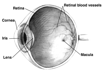
Each of these medicines may
have local and systemic side effects. Adherence to medication protocol can be
confusing and expensive; if side effects occur, the patient must be willing
either to tolerate these, or to communicate with the treating physician to
improve the drug regimen.
Poor compliance with
medications and follow-up visits is a major reason for vision loss in glaucoma
patients. Patient education and communication must be ongoing to sustain
successful treatment plans for this lifelong disease with no early symptoms.
The possible neuroprotective
effects of various topical and systemic medications are also being
investigated.
[edit] Commonly used medications
- Prostaglandin analogs like latanoprost
(Xalatan), bimatoprost (Lumigan) and travoprost
(Travatan) increase uveoscleral outflow of aqueous humor.
- Topical beta-adrenergic receptor
antagonists such as timolol, levobunolol (Betagan), and betaxolol
decrease aqueous humor production by the ciliary body.
- Alpha2-adrenergic agonists such as brimonidine
(Alphagan) work by a dual mechanism, decreasing aqueous production and
increasing uveo-scleral outflow.
- Less-selective sympathomimetics
like epinephrine
and dipivefrin
(Propine) increase outflow of aqueous humor through trabecular meshwork
and possibly through uveoscleral outflow pathway, probably by a
beta2-agonist action.
- Miotic agents (parasympathomimetics) like pilocarpine
work by contraction of the ciliary muscle, tightening the trabecular meshwork and allowing increased
outflow of the aqueous humour.
- Carbonic anhydrase inhibitors
like dorzolamide
(Trusopt), brinzolamide (Azopt), acetazolamide
(Diamox) lower secretion of aqueous humor by inhibiting carbonic anhydrase
in the ciliary body.
ANTIGLAUCOMA DRUGS
The aqueous humor is a nourishing liquid that is
produced by the ciliary body and flows from the posterior chamber (behind the iris) to
the anterior chamber (in front of the iris). It is removed via the canal of
Schiemm, which is located adjacent to the union of the sclera
and cornea in the anterior chamber. When the normal flow and drainage of
aqueous humor is inhibited, a serious ocular condition called glaucoma occurs.
Figure 56-6 illustrates
the main structures of the eye and an enlargement of the canal of Schiemm showing aqueous flow.
Glaucoma is an eye disorder characterized
by excessive intraocular pressure (TOP)
created by abnormally elevated levels of aqueous humor. This occurs
when the aqueous humor is not drained through
the canal of Schiemm as quickly as it is formed by the ciliary body. The
accumulated aqueous humor creates a backward pressure
that pushes the vitreous humor against the retina. Continued pressure on the retina destroys its neurons,
leading to impaired vision and eventual blindness. Unfortunately glaucoma is often without early symptoms and therefore many people are not
diagnosed until some permanent sight loss has occurred. Glaucoma occurs in African-Americans at 3 to 4 times the
incidence in Caucasians, and blindness results in 6 times as many African-Americans as Caucasians. In addition, it
has been shown that African-Americans and Caucasians may respond
differently to different surgical treatments for glaucoma. There are several categories of glaucoma that are classified according to underlying cause. The
three main categories are primary, secondary, and congenital glaucoma Effective
treatment of glaucoma involves reducing IOP by either increasing the drainage of or decreasing the production of aqueous humor. Some drugs may do
both. Effective drug therapy can delay and possibly even prevent the
development of glaucoma. Drugs used to reduce IOP include the following:
• Beta-b lockers
• Carbonic anhydrase inhibitors
• Osmotic diuretics
• Parasympathomimetics, direct-acting
• Parasympathomimetics,
ndirect-acting (cholinesterase inhibitors)
• Prostaglandins
• Sympathomimetics
PARASYMPATHOMIMETICS
Both the direct- and indirect-acting
parasympatho-mimetk drugs work in different ways to mimic the PSNS neurotransmitter
acetylcholine (ACh). In the eye they cause miosis (pupillary
constriction). For this reason they are also called miotics. They lower
IOP by an average of 20% to 30%. Examples of ophthalmic drugs that fall into these two drug
categories are found in Box 56-1.
Mechanism of Action and Drug Effects
The direct-acting miotics are able to directly stimulate PSNS receptors because their structures closely
resemble that of the PSNS neurotransmitter ACh. Direct-acting miotics are administered topically to the eye. This
route of administration drastically limits systemic absorption of the drug by keeping the cholinergic effects
localized to the site of
administration in the eye. The cholinergic response produced by these
drugs causes pupillary contraction (miosis),
which leads to a reduction of IOP secondary to an increased outflow of aqueous humor (Fig. 56-8).
The indirect-acting
miotics work by inhibiting acetyl-cholinesterase (AChE), the enzyme
that breaks down ACh. This results in higher levels of ACh and allows
ACh to remain at the receptor longer. This results in
miosis, aqueous humor drainage, and reduced IOP.
ACh is the endogenous
mediator of nerve impulses in the PSNS. It stimulates cholinergic
receptors. This results in several effects in the eye: miosis,
vasodilation, contraction of ciliary muscles, and reduced IOP. The
action of ACh is short lived. It is rapidly hydrolyzed by
cholinesterases (AChE and pseuclocholinesterase) to choline and acetic acid. Direct-acting
miotics have effects similar to those of ACh, but their actions are more
prolonged.
The indirect-acting
miotics bind with and inactivate cholinesterases (AChE and
pseudocholinesterase), thus inhibiting hydrolysis of ACh. As a result ACh
accumulates at cholinergic nerve endings, enhancing cholinergic neurotransmission. Echothiophate is an
indirect- acting, agent that has an organophosphate structure and acts by
|
|

phosphorylating cholinesterase. This effect is normally
irreversible until new cholinesterase enzymes are synthesized by the body.
This production of new enzymes may take days or even weeks.
The drug effects of
both direct- and indirect-acting miotics alter various eye muscles,
IOP, aqueous humor flow, and vasodilation of blood vessels in and around
the eye. The drug effects that alter the muscles of the eye
result in contraction of the iris sphincter, which produces constriction of the pupil
(miosis) and contraction of the ciliary muscle, resulting in spasm (paralysis)
of visual accommodation
by the lens.
Drug-induced pressure
alterations within the eye act to reduce IOP in both normal and glaucomatous eyes. They do this by
facilitating aqueous humor outflow by causing
contraction of the ciliary muscle, thus widening the area from which
this fluid escapes. IOP is also reduced by constriction of the pupil, which
causes the iris to stretch and thus relieves
blockage of the area where the fluid
leaves the inner eye. This effect is less pronounced in individuals with
dark eyes (brown or hazel) than in those
with light eyes (blue) because the pigment absorbs the drugs and dark eyes have more pigment.
Miotic agents also cause vasodilation of blood
vessels of the conjunctiva, iris, and ciliary body, resulting in increased permeability
of the blood-aqueous barrier, which may lead to vascular congestion and ocular inflammation. Blood-aqueous barrier is the
name of an anatomic mechanism that
normally prevents exchange of fluids
between eye chambers and the blood. This con-gestion and inflammation is more common with long-acting
anticholinesterases (indirect-acting miotics).
Indications
The direct- and indirect-acting miotics are used
for open-angle glaucoma, angle-closure glaucoma, ocular surgery, convergent strabismus
(condition where one eye points toward the other ["cross-eye"]), and
ophthalmologic examinations. The miotics are used topically on the eye to
reduce elevated IOP in the treatment of primary open-angle
glaucoma. This reduction in IOP is the result of
contraction of the ciliary muscle and widening of the exit route of the aqueous humor. Acute
(congestive) angle-closure glaucoma may be relieved temporarily by
pilocarpine or occasionally by carbachol to acutely decrease extremely high
IOP.
Some of the miotic
drugs may be used for ocular surgery. Pilocarpine, carbachol, and acetylcholine
are used
to reduce IOP and to protect the lens by causing miosis before certain types of laser surgery on the iris. Miotics may be used to counteract the mydriatic effects
(dilation effects) of sympathomimetic agents such as
hy-droxyamphetamine and phenylephrine that are used for ophthalmoscopic examinations. Table 56-3 lists the miotic drugs and their
indications.
Contraindications
Contraindications of miotics include known drug
allergy and any serious active eye disorder with which induction of miosis might be
harmful. An ophthalmologist will usually make this judgment
Side Effects and Adverse Effects
Most of the side effects and adverse effects associated with the use of
cholinergic and anticholinesterase drugs (miotics)
are local and limited to the eye. There are, however, some systemic effects that can occur, especially if sufficient amounts of drug pass into the
bloodstream.
The most common
ocular side effects that may occur with the use of miotic drugs are blurred vision and accommodative spasms. Other undesirable effects
include conjunctivitis, lacrimation, twitching eyelids, poor low-light vision, and pain. Prolonged use can result
in iris cysts; lens opacities; and,
rarely, retinal detachment.
Systemic effects that
may occur with the use of miotic drugs are caused by PSNS stimulation.
Cholinesterase inhibitors
usually produce more pronounced effects than direct-acting
agents. The most common side effects include
bronchodilation, lacrimation, temporary stinging on instillation, decreased
night vision, nausea, vomiting, salivation, sweating, gastrointestinal
(GI) stimulation, and urinary incontinence.
Toxicity and Management of
Overdose
Occasionally toxic effects may develop after
the use of topically applied miotic drugs. Toxicity produced by miotics is an extension
of their systemic effects and is more common with prolonged use of
high doses. Most severe and prolonged effects are seen with long-acting anti-cholinesterases. Excessive PSNS effects are
treated with 0.4 to 2 mg of atropine administered intravenously or intramuscularly
for adults and 0.04 to 0.08 mg/kg administered
intravenously or intramuscularly for children. If required, repeat doses can be given every 5 minutes intravenously and every 15 minutes intramuscularly.
In addition, pralidoxime (Protopam),
a cholinesterase reactivator, may be required to reverse paralysis
induced by either of the organophosphate
anu'cholinesterases (demecarium and echothiophate).
Interactions
Direct- and indirect-acting miotics are
capable of interactions with several
categories of drugs. The miotic drugs, when given with topical epinephrine,
timolol, and carbonic anhydrase inhibitors, have additive lowering
effects on IOP. Systemic cholinesterase inhibitors have additive effects when given with mitotic drugs. Indirect-acting miotics may potentate the effects
of anesthetic agents.
DRUG PROFILE
Direct-acting and indirect-acting miotics have a
variety of uses. The principal use is that of relief of symptoms caused by
glaucoma and increased IOP. The three most commonly used direct-acting miotics are
acetylcholine, carbachol, and pilocarpine. These drugs have
actions similar to those of ACh. The two currently available indirect-acting
miotics are demecarium and echothiophate. These agents work by preventing
cholinesterase from breaking down ACh, allowing it to produce a
prolonged PSNS response.
Because the safe use
of miotics in pregnancy has not been established, many of the miotics are
classified as pregnancy category C agents. Because of the potential risks of cholinesterase inhibition in
general, the manufacturers of some anticholinesterase agents (e.g., demecarium,
isoflurophate) state that the drugs are con-traindicated
in women who are or may become pregnant.
Direct-acting miotic
drugs are contraindicated in patients who have shown a hypersensitivity
reaction to them, those with acute inflammatory conditions in the anterior
chamber, and those with pupillary block glaucoma.
Indirect-acting miotics are contraindicated in patients who have
shown a hypersensitivity reaction to them and in patients with acute
inflammatory conditions and glaucoma associated with iridocyclitrs (inflammation of iris
and ciliary body). In addition, echothiophate is not suitable for treatment of
most cases of angle-closure glaucoma.
SYMPATHOMIMETICS
Sympathomimetic
drugs such as brimonidine, apraclonidine, dipivefrin, and epinephrine are used for the
treatment of glaucoma and ocular
hypertension. Dipivefrin is a prodrug
of epinephrine. When instilled into the eye it is hydrolyzed to
epinephrine. Its advantage over epinephrine
is that it has enhanced lipophilkity (fat solubility) and can better
penetrate into the tissues of the anterior chamber of the eye. On a weight
basis dipivefrin is 4 to 11 times as potent
as epinephrine in reducing IOP and 5
to 12 times as potent as epinephrine in pupil dilation (mydriasis).
Apraclonidine and brimonidine are structurally
and pharmacologically related to clonidine. Apraclonidine is relatively
selective for alpha2 receptors.they mimic the sympathetic
neurotransmitters norepi-nephrine and epinephrine and stimulate the dilator muscle to contract. This stimulation results in
increased pupil size (mydriasis; illustrated in Fig. 56-11). Dilation is seen within minutes of instillation of the ophthalmic
drops and lasts for several hours,
during which time the IOP is reduced. This occurs secondary to either
enhanced outflow or decreased production of
aqueous humor, which is also a
drug-related effect.
Mechanism of Action and Drug Effects
These agents reduce IOP in patients with normal
IOF and in
patients with elevated IOP, such as those with glaucoma. Dipivefrin 0.1% reduces mean IOP approximately 15% to 25%. The exact
mechanism by which the sympathomimetic drugs lower IOP is unknown. They
are believed to work by stimulating both
alpha2 and beta2 receptors, causing the dilator
muscle to contract, resulting in mydriasis. During mydriasis there is an
increase in aqueous humor outflow, resulting in a decrease in IOP (Fig. 56-12). These effects appear to be dose
dependent.
Apraclonidine reduces
IOP 23% to 39%. By stimulating alpha, and beta, receptors, apraclonidine prevents constriction of the blood vessels of the eye and
reduces pressure, resulting in reduced aqueous humor formation. Brimonidine works in a similar fashion as
apraclonidine.
Dipivefrin is a more
lipophilic agent and therefore has more localized effects in the
eye. However, both dipivefrin and epinephrine can cause drug effects outside the eye. Because these two drugs mimic the effects of
the SNS neurotransmitters, they can
cause increased cardiovascular effects such as increased heart rate or blood
pressure.
Indications
Both epinephrine and dipivefrin may be
used to reduce elevated IOP in the
treatment of chronic, open-angle glaucoma, either as initial therapy or as
chronic therapy. Apraclonidine is primarily used to inhibit
periopera-tive IOP increases. Increases in IOP during ophthalmic surgery are usually mediated via increased
catechol
TREATMENT OF EAR DISORDERS
Some of the minor ailments that affect the outer ear can be treated with over-the-counter (OTC) medications, but persistent, painful conditions always require a
physician's care. Middle ear disorders are rarely treated with OTC medications unless a physician prescribes
them after referral. The drugs commonly used to treat the relatively minor
disorders of the external and middle ear are called otic agents and are
topically applied.
|
|
They are listed as follows:
• Antibiotics
• Antifungals
• Antiinflammatory agents
• Local analgesics
• Local anesthetics
• Steroids
• Wax emulsifiers
More serious problems than those
previously mentioned may require treatment
with potent, systemically administered medications such as antimicrobial
agents, analgesics, antiinflammatory
drugs, and antihistamines. 
ANTIBACTERIAL AND
ANTIFUNGAL OTIC AGENTS
Many of the antibacterial and antifungal
agents that are given systemically also
come in topical formulations that are
applied to the external ear. Chloramphenicol and gen-tamicin are two such agents that are commonly used

DRUG PROFILES
chloramphenicol
Chloramphenicol (Chloromycetin
Otic) has the advantage of possessing a very broad spectrum of activity. It is effective against Staphylococcus aureus,
Escherichia coli, Pseudomonas
aeruginosa, Enterobacter aerogenes, Haemophilus
influenzae, and many other
bacteria. It can cause adverse
effects very similar to those caused by many of the topically applied
antibiotics. These include burning,
redness, rash, swelling, and other signs of topical irritation. Chloramphenicol
is available as a 0.5% solution that
is usually instilled in the ear. Two to three drops 3 times daily is the recommended dosage for both adults and children.
ANTIBIOTIC AND
STEROID COMBINATION
PRODUCTS
The steroid most commonly used in combination with antibiotics is hydrocortisone, which is added to
reduce the inflammation and itching associated with ear infections. The antibiotics contained in the most popular
combination products and the various trade names of these products are listed in Table 57-1. These products
are most commonly used for the treatment of bacterial infections of the external auditory canal caused by
susceptible bacteria such as S. aureus,
E. coli, Klebsiella spp., and others.
MISCELLANEOUS OTIC AGENTS
A wide variety of
single-agent and combination-a gent products are used to treat fungal and
bacterial infections, inflammation, ear pain, and other minor or superficial problems of the external ear. An additional
problem is the accumulation and
eventual impaction of earwax, or cerumen,
which can be the cause of many of these symptoms. Products that soften and help to eliminate earwax
are re-ferred to as wax errtulsifiers
and are therefore also discussed with the miscellaneous agents.
DRUG PROFILES
Although fungal infections of the
ear are uncommon, otic agents are available for their treatment. These agents may also have antibacterial and
even antiviral properties. Examples include benzalkonium chloride, benzethonium
chloride, and chloroxylenol. The agents contained
in two commonly used antifungal combination
products and the trade names of each are listed in Table 57-2. These
combination products include drugs from several classes of agents—corticosteroids,
local anesthetics, and topical antiseptics—and hence the therapeutic effect is
a combination of the respective effects of the individual agents. They
are used for their collective properties as antibacterial, antifungal, local anesthetic, and antiinflammatory otic drugs.
LOCAL ANESTHETIC
AGENTS
Numerous otic
combination products contain local anesthetic agents. Many common
ear disorders are associated with some degree of pain and inflammation, and the numbing or
anesthetic effect provided by local anesthetic agents makes them beneficial for
pain relief. The
same characteristics of the local anesthetics that were discussed apply to the topically applied local anesthetics
used in these preparations.
Proper ear function is essential for hearing and balance. Hearing is one
of the most critical senses. Loss of hearing greatly compromises a patient's
ability to interact with people and the environment.[1] Loss of
balance makes such common tasks as walking and driving difficult to impossible.
Unfortunately, several ototoxic medications can affect hearing and/or balance
through one or more mechanisms.
Incidence
How common is ototoxicity? Its incidence is largely unknown. Although a
reported 130 medications can produce ototoxicity, some are seldom seen by the
average retail pharmacist (e.g., ethacrynic acid, dihydrostreptomycin,
kanamycin, sisomycin).[1]

How
Medications Cause Ototoxicity
There are three major locations where medications exert ototoxicity.[2]
The first location is the cochlea; medications that affect it are thought to
exhibit cochleotoxicity. Ototoxicity involving the cochlea produces hearing
loss, usually commencing with high frequencies but often eventually progressing
to the lower frequencies that encompass speech.[3] The hearing loss
may be one-sided or bilateral and may fluctuate in severity. Cochlear damage
may also manifest as tinnitus. The tinnitus may be constant or fluctuate.
Patients with preexisting tinnitus may notice the problem worsening or the
appearance of a new sound that was not present before the medication was
administered.
The vestibulum is a second site of action for ototoxic medications;
these are known as vestibulotoxic medications. Vestibulotoxicity usually
manifests as balance-related problems (e.g., disequilibrium). The patient
reports a spinning sensation that is often aggravated by motion and is
associated with nausea.
The stria vascularis is the third site of action for ototoxic
medications. The stria vascularis is a type of epithelium that is uniquely able
to produce endolymph in the cochlea. Excessive endolymph is responsible
Aminoglycosides. Aminoglycoside antibiotics (e.g., kanamycin, neomycin, amikacin,
streptomycin, gentamicin) exhibit cochleotoxicity but also affect the stria
vascularis, causing vestibular problems.[3,4] They produce damage
through the ability to generate free radicals in the inner ear.[5]
Babies have suffered congenital deafness when their mothers took kanamycin or
streptomycin during pregnancy.[6] Neomycin is the worst offender
relating to cochleotoxicity.[7]
Topical Otic Preparations. Treatment of otic disease can be accomplished with
the use of systemic or topical preparations. Systemic therapy cannot achieve
the concentrations allowed with the use of ototopical drops.[8]
Topical drops also have the advantages of rapid delivery, good compliance, and
lower cost. Furthermore, some drops are combinations of two or more
ingredients, increasing the efficacy of the product. If the product is used for
otitis externa, the danger in the application of potentially ototoxic
medications is that the patient might have a perforation in the eardrum. This
perforation might have occurred as a result of trauma, otitis media, or
following placement of ventilation tubes. If a perforation is present,
instillation of preparations with ototoxic potential could lead to inner ear
damage. Topical medications, such as those containing neomycin/polymyxin B, may
produce vestibular and/or cochlear toxicity when the patient has a tympanic
membrane perforation.[9,10]
Aminoglycosides are especially toxic when instilled into the ear.
Ironically, this aspect of their toxicity has therapeutic use in patients with
intractable Meniere's disease. In a treatment known as vestibular ablation,
the physician prescribes gentamicin/ betamethasone otic solution in a dose of
three drops instilled four times daily until the onset of dizziness. Patients
usually experience remission of Meniere's disease by day 12.[8]
Gentamicin is not exclusively vestibulotoxic, as it possesses some cochleotoxic
activity.[5] Researchers report that 90% to 100% of patients
experience vertigo control with gentamicin instillation, while only 30% also
suffer hearing loss.[5]
Otitis externa may be treated with quinolones, such as ofloxacin otic
drops (Floxin Otic), without fear of ototoxicity.[8,10,11]
Loop Diuretics. Loop diuretics (e.g., furosemide, ethacrynic acid, bumetanide) affect
the potassium gradient of the stria vascularis, as well as the electrical
potential of the endocochlear structure.[2,3] These medications
produce tinnitus and hearing loss. The hearing loss may be perceptible to
patients or may be apparent only with audiometric testing. Their toxicity is
dose-related.[12] Thus, ototoxicity is more likely when the patient
receives a rapid infusion of injectable loop diuretics in renal failure, which
allows the medications to accumulate. Furosemide-related ototoxicity is usually
reversible but may be permanent in rare instances (e.g., in patients with renal
failure).[7] Ethacrynic acid is virtually obsolete, partly due to
the potential for ototoxicity, especially when it was given intravenously to
patients whose regimen also included aminoglycosides.[7]
Antineoplastics. Cisplatin affects the cochlea and stria vascularis through its ability
to generate free radicals within the inner ear.[13] Researchers have
examined various compounds with possible otoprotective activity that might be
administered concomitantly with cisplatin to prevent ototoxicty.[13]
However, none of those investigated (e.g., alpha-tocopherol, d-methionine,
salicylate, iron chelators) is clearly effective.
Salicylates. Salicylates impact the cochlea. In high doses, they cause tinnitus and
loss of hearing; both are usually seen only with higher doses and regress upon
discontinuation in most instances.[7]
The relationship between salicylate serum concentrations and the level
of hearing loss is linear. Serum concentrations below 20 to 50 mg/dL produce
little risk of hearing loss.[2] Concentrations exceeding this level
expose the patient to a possible hearing loss of 30 decibels or above.
Hearing loss could occur with topical administration of counterirritants
containing methyl salicylate.[14] For this reason, it is preferable
to consider the use of therapeutic heat wraps as a safer alternative for knee
or back pain or for pains in the shoulder-to-arm area, particularly in patients
with risk factors that would predispose them to ototoxicity.
Quinine.
Quinine was once widely sold as a nonprescription product, but the FDA found
its traditional use for nocturnal leg cramps to be ineffective and also issued
an opinion that it is outdated as an antimalarial. Thus, there is no longer any
justification for stocking or selling it to any patient at any time, which is
critical advice considering its potential for causing tinnitus, loss of
hearing, or vertigo.[3] The hearing loss may be irreversible.
Patients who take 200 to 300 mg over a sustained period experience a 20% risk
of hearing loss.[2]
Tea Tree Oil. Tea tree oil is an alternative medical treatment claimed to be
effective for bacteria and fungi. Although there is little evidence to support
any use of tea tree oil, some have recommended its placement into the ears to
treat otitis media or otitis externa. In one article, researchers discovered
that it may be toxic to the cochlea, producing deficiency in the high-frequency
region of hearing.[15] Therefore, while alternative medicines in
general must be used with caution, otic instillation of tea tree oil appears
unwarranted due to the lack of information on efficacy and should also be
avoided to prevent possible cochleotoxicity.
Additive Ototoxicity. If more than one medication with ototoxic potential is administered to
the same patient, the effect can be additive.[3] For this reason, if
a patient is already taking a potentially ototoxic medication, any addition to
the regimen should be examined carefully to detect additional ototoxins.
Importance of Dose and Dosing Interval. If a medication has ototoxic potential,
its blood levels should remain as low as possible. This may require assessing
blood levels frequently and adjusting the dosage downward if blood levels
exceed those required to gain the desired therapeutic effect.
Otitis media is an inflammation of the middle ear:
the space behind the ear drum. It is one of the two conditions that are
commonly thought of as ear infections, the other being otitis
externa. Otitis media is very common in childhood, and includes acute and
chronic conditions; all of which involve inflammation of the ear drum (tympanic
membrane), and are usually associated with a buildup of fluid in the space
behind the ear drum (middle ear space).
There are several kinds of
otitis media:
1.
Acute
otitis media is an infection
that produces pus,
fluid, and inflammation within the middle ear. It is frequently associated with
signs of upper respiratory infection, such as a runny nose
or stuffy nose. It
is painful, but usually self-limiting. The most serious, but rare, complication
Mastoiditis
- an infection of the bone around the ear.
2.
Otitis
media with effusion, (or Glue
Ear) formerly termed serous Otitis Media or secretory Otitis Media, is Middle
Ear Effusion of any duration that lacks the associated signs and symptoms of
infection (eg, fever, otalgia, irritability), but causes hearing problems.
Otitis Media with Effusion usually follows an episode of Acute Otitis Media.
3.
Chronic
suppurative otitis media
is when discharge from the infection persists for more than two weeks.
Treatment
Most episodes simply require
analgesics to manage the pain and fever. Acute otitis media will usually settle
without treatment. Whilst antibiotics were previously routinely immediately started,
this practice is diminishing. Antibiotics do shorten the illness by around 1/3
compared to the illness's natural history, but this is a small gain for most
children. However, very young children, those with bilateral otitis, and those
with a high fever are likely to have a more severe course and hence benefit
more from antibiotics[2][3][4]
Many guidelines now suggest
deferring the start of antibiotics for 24 to 72 hours.[5] This
results in 2 out of 3 children avoiding the need to start antibiotics,[6] and no
adverse effect on longterm outcomes for those whose treatment is deferred.[7] First line
antibiotic treatment, if warranted, is amoxicillin. If the bacteria is
resistant, then Augmentin or another penicillin derivative plus beta
lactimase inhibitor is second line.
In chronic cases or with
effusions present for months, surgery is sometimes performed to insert a grommet (called a
"tympanostomy tube") into the eardrum to allow
air to pass through into the middle ear, and thus release any pressure buildup and
help clear excess fluid within.
Prior to the invention of antibiotics,
severe acute otits media was mainly remedied surgically by Myringotomy.
An outpatient procedure, it consists of making a small incision in the tympanic
membrane to relieve pressure build-up.
For chronic cases (glue ear),
it is possible to use the Valsalva
maneuver to reestablish middle ear ventilation.
1. http://www.youtube.com/watch?v=8W0UzjmSPWc&feature=related
2. http://www.youtube.com/watch?v=gGrDAGN5pC0&feature=related
3. http://www.youtube.com/watch?v=--bZUeb83uU&feature=related
4. http://www.youtube.com/watch?v=WXOBJEXxNEo&feature=related
5. http://www.youtube.com/watch?v=Lt0BjNwF6IU&feature=related
6. http://www.youtube.com/watch?v=fjjEiVeYRNg&feature=related
7. http://www.youtube.com/watch?v=mut1VYu9tzk&feature=related
8. http://www.youtube.com/watch?v=6zEiH7X1Jz4&NR=1
9. http://www.youtube.com/watch?v=kUiEEski4Aw&feature=related