Purulent
Diseases of Soft Tissues and Bones: diagnosis and treatment in the Outpatient
Department of Family Doctor,
bleeding, hemorrhage, transient and terminal blood arrest. Wounds
and Wound Process. Prevention of infection development in the
wound. Treatment of clear wounds. Patient’s referral arrangement. Principles
of antibiotic therapy in prevention and management of surgical infection.
Medical and Labour Expert Examination (MLEE)..
Soft tissue infections
Introduction
Soft
tissue infections are classified by anatomic extent and pathophysiologic
process. Thus, they may be focal or diffuse; the most severe have associated
tissue necrosis; and when this is combined with systemic toxic effects the
results are devastating. But most cases are easily diagnosed and treated.

Figure 1 Schema of approach to soft tissue infection.
Awareness of the harbingers of
necrotizing infection and knowledge of the complicating factors in toxic
infections are essential to avoid loss of limb and life. Although most treatment
has been empirical, strategies for diagnosis and treatment are emerging, that yield improved
results (fig. 1).
Presentation
and pathogenesis
Simple
focal infections
These
infections arise in the skin or in the adenexal skin structures, particularly the
follicles. The commonest examples are impetigo contagiosa and folliculitis. Impetigo
contagiosa is an infection of minor skin abrasions on the face or limbs of
children caused by S. aureus or Strep. pyogenes. In folliculitis
staphylococcal pyodermas of the hair follicles may extend to form furuncles or
coalesce to form carbuncles.
Necrotizing focal
infections
Focal necrotizing
infections occur far less commonly. The best examples are Meleney's
Progressive bacterial Synergistic Gangrene and Fournier's Idiopathic
Scrotal Gangrene. Progressive synergistic gangrene is characterized by
three concentric zones of erythema, cyanosis, and necrosis at sites of
synergistic infection of operative wounds or pressure sores by a
microaerophilic nonhemolytic streptococcus and a hemolytic S. aureus or
gram-negative bacillus. Idiopathic scrotal gangrene as described by Fournier
is caused by anaerobic streptococci and differs from the perineal form of
Gram-negative bacterial synergistic gangrene commonly given this name today.
The onset is dramatic and progression to gangrene of the scrotum occurs within
24 hours.
Toxic non-necrotizing
focal infections
These infections occur
still less often and include the Staphylococcal scalded skin syndrome (SSSS)
and toxic shock syndrome (TSS). SSSS occurs most frequently in young
children and sometimes in neonates (pemphigus neonatorum). It presents as a
diffuse rash that follows a staphylococcal infection, and progresses to
generalized bullae that break and slough. An exfoliative toxin has been
identified that splits the epidermis so that the bullae develop. In TSS
fever and a generalized macular rash develops in a patient with vaginitis
associated with menstruation and the use of superabsorbent tampons. Enterotoxin
F produced by group I phage strains of S. aureus is responsible for this
constellation of symptoms and for the multiple organ failure that commonly
ensues.
Diffuse non-necrotizing infection: cellulitis
Cellulitis is a diffuse infection of the skin and subcutaneous tissues
characterized by local spreading erythema, warmth, tenderness and swelling.
Most infections are mild and are caused by S. aureus or group A
streptococci. Systemic signs such as fever, chills, and leucocytosis indicate severe
cellulitis that usually requires intravenous antibiotic therapy. Patients
should be investigated for underlying diseases including diabetes mellitus and
peripheral arterial occlusive disease that worsen the prognosis.
Infections at some sites are regarded as high risk
cellulitis because they are prone to serious complications. These include
facial and orbital cellulitis, post adenectomy and postvenectomy cellulitis of
the extremities, and infected animal and human bites of the hands. Facial
cellulitis that originates in the teeth or gums is odontogenic. It occurs
typically in the lower face of older children. Most facial cellulitis is
non-odontogenic and occurs in the upper face of infants and younger children.
Typical examples are erysipelas and Haemophilus influenzae type B (HIB)
cellulitis. The peau d'orange cellulitis of erysipelas follows
streptococcal upper respiratory infection that invades the dermis and
lymphatics of the cheek. Similar infection by Haemophilus influenzae
type B results in the characteristic “bruised cheek” appearance of HIB
cellulitis now uncommon with the use of H. influenzee vaccine.
Postadenectomy and postvenectomy cellulitis are caused by lymphedema that is
secondarily infected by non-group A streptococci. High risk hand infections
include erysipeloid and infected human and animal bites. Erysipeloid is
diagnosed when a spreading violaceous wound appears at the site of a minor
wound on the hand of a worker handling fish, meat, or poultry. The infection is
caused by the gram positive bacillus Erysipelothrix rhusiopathae. Animal
and human bites are dangerous when secondary infection occurs and
tenosynovitis develops in deep tendons. Pasteurella multocida in cat
bites, and Eikenella corrodens in human bites are common pathogens.
Delay in seeking treatment and anaerobic conditions in deep hand infections
favor tissue necrosis and rapid progression.
The spread of infection in cellulitis is associated with
toxins and enzymes produced by the offending organisms. Further, a disparity
has been noted between the low frequency of positive culture of deep aspirates
of these wounds and the marked inflammation observed. This has recently been
attributed to cytokines such as interleukin-1 and tumor necrosis factor
produced by specific dendritic cells of Langerhans in the stratum spinosum of
the skin when they are exposed to bacterial components. The inflammatory
response may therefore persist even when the bacteria have been killed by
antibiotics.
Diffuse necrotizing cellulitis
These conditions are described fully in the chapter that
follows. In general, they include necrotizing infections in which inflammatory
cells are absent or sparse on biopsy, particularly clostridial myonecrosis, and
those conditions in which inflammatory cells are abundant on histologic
examination or Gram's stain of deep aspirates. Meleney's streptococcal
gangrene, clostridial cellulitis, necrotizing fasciitis as described by
Diagnosis
A systematic approach
to diagnosis that includes, needle aspiration, blood sampling for risk
assessment, aerobic and anaerobic culture and diagnostic imaging is employed
except in the simplest and most obvious cases.
Needle aspiration
Needle aspiration for
sampling of tissue fluid or collections is preferable to superficial culture.
It supplies the best information for improving on empirical therapy with
semisynthetic penicillins.
Blood sampling
Blood sampling aids in
assessment of the risk of the infection to the patient, and is an important
reminder of the importance on outcome of conditions affecting the immune state
of the patient . Diabetes, severe malnutrition and organ failure are all
important.
Culture
Culture of the tissue
aspirate is positive for organisms in only 20 to 30% of patients, but allows
revision of antibiotic therapy in difficult cases. Blood culture was formerly
done routinely, but is seldom positive, and should only be obtained by specific
indication.
Diagnostic imaging
Plain roentgenographs
of the affected site should be obtained except in mild disease. They may
provide the only evidence of gas in the tissues, complicating deep infection
such as osteomyelitis, or of a retained foreign body. Isotope imaging performed
with technetium (99mTc) pyrophosphate detects areas of increased
blood flow or newbone formation. Gallium citrate and indium chloride indicate
sequestered polymorphonuclears cells and macrophages in areas of inflammation.
By combining the two or using phased 99mTc pyrophosphate scans one
may distinguish between soft tissue and bome inflammation. But the best method
of identifying osteomyelitis complicating soft tissue infection of the
extremities is by use of magnetic resonance imaging (MRI) with gadolinium
contrast. MRI with also differentiates necrotizing from non-necrotizing soft
tissue infection by failure of the necrotic fascia to enhance with gadolinium.
Treatment
Strategies for optimal
treatment follow naturally from the schema suggested in figure 1. Simple focal
lesions are readily treated with oral or topical antibiotics, such as mupirocin
2% ointment applied three times per day, and cloxacillin 250 or 500 mg every 6
hours for 7 days, local cleansing of the wounds, and attention to hygiene. On
the other hand, focal necrotizing disease and focal lesions with toxic symptoms
require intravenous fluid resuscitation and combined antibiotics such as
amoxycillin 1 to
Necrotizing soft-tissue infections
Necrotizing soft-tissue infections (NSTI) encompass a diverse
disease process characterized by extensive, rapidly progressive soft tissue
inflammation and necrosis. NSTIs are increasingly more common, and continue to
be associated with a fulminant course and high mortality rates. These
infections comprise a spectrum of diseases ranging from necrosis of the skin to
life-threatening infections involving the fascia and muscle with systemic
toxicity. They vary in predisposing and causative factors, anatomic location,
offending bacteria, and tissue level of involvement.
The nomenclature of NSTI can be confusing with many terms
used to describe the variable presentations. The original description of “hospital
gangrene” was made by Joseph Jones in 1871
following his experience during the U.S. Civil War. Additional terms
used in the past included Fournier gangrene, acute hemolytic streptococcus
gangrene, and gas gangrene (clostridial myonecrosis). The term “necrotizing
fasciitis” was popularized by Wilson in 1952, and remains in use today.
The terminology of NSTI is of secondary concern when surgeons
are challenged by a patient with this aggressive illness. Primary emphasis must
be focused on rapid recognition and appropriate aggressive treatment. A high
index of suspicion leading to early diagnosis, aggressive surgical intervention
and appropriate antimicrobial therapy is essential to reducing the mortality of
NSTIs.
Presentation and diagnosis
The presentation of NSTI is highly variable and can range
from early sepsis with obvious skin involvement to minimal cutaneous
manifestations with a disproportionate (even alarming) underlying necrotizing
fasciitis. The usual clinical presentation begins with localized pain and a
deceptively benign appearance. Clinical “clues” which may assist in
establishing an early diagnosis are: edema beyond the area of erythema, small
skin vesicles, crepitus and the absence of lymphangitis. Additional local signs
suggesting deep infection may include cyanosis or bronzing of the skin,
induration, dermal thrombosis, epidermolysis or dermal gangrene. Classic signs
of fever, diffuse crepitus, and shock are late signs. Once large blisters and
gangrene develop, the infectious process is already at an advanced stage.
Early recognition is the sine qua non for the assessment of
risk factors. Multiple risk factors increase the probability of a
life-threatening infection. These co-morbid conditions include diabetes
mellitus, peripheral vascular disease, malnutrition, malignancy,
immunocompromised states (AIDS, steroid therapy), obesity, chronic alcohol or
intravenous drug abuse. In the urban setting, intravenous and subcutaneous
injection of illicit substances has become a more prevalent risk factor and
should raise one's suspicion. Our recent experience with NSTIs (1990–1995)
revealed that 67% of patients were actively practicing parental drug use.
The etiologies for NSTI are not always obvious, and often the
initiating event is surprisingly minor. The most common etiologies include
postoperative wound complications, penetrating and blunt trauma, cutaneous
infections, intravenous or subcutaneous illicit substance injection,
peri-rectal abscesses, strangulated hernias, and idiopathic causes.
Establishing an early diagnosis depends on a high index of
suspicion and a willingness to proceed with appropriate treatment. Plain x-rays
may demonstrate subcutaneous air or a foreign body but are usually not
necessary. Interest in the use of MRI scanning
and frozen section biopsy to aid in diagnosis has been reported. Early
and aggressive surgical intervention should not be delayed by these ancillary
studies. At exploration, the diagnosis can be confirmed by the presence of
necrotic fascia, the absence of gross pus and easy finger dissection along the
fascial planes.
Bacteriology
The bacteriology of
NSTI is well recognized, and the infectious process is independent of specific
bacteria . Most NSTI are polymicrobial, involving organisms behaving
synergistically. In our series of patients treated 78% of cases were
polymicrobial, and 2.8 organisms were recovered per patient. Anaerobes, skin
flora, and gram negative rods are commonly encountered.
Table I Bacteriologic findings in patients with necrotizing soft-tissue
infections
Organism
|
No of patients
|
Anaerobes
|
|
Mixed anaerobes
|
16
|
Clostridium
speciesa
|
8
|
Diphtheroids
|
3
|
Bacteroides fragilis
|
2
|
Bacteroides buccae
|
2
|
Peptostreptococcus
sp
|
1
|
Total
|
32
|
Gram-positive cocci
|
|
Streptococcus viridans
|
8
|
Hemolytic group A streptococci
|
7
|
Enterococcus faecalis
|
6
|
Total
|
21
|
Gram-negative rods
|
|
Mixed gram-negative rods
|
6
|
Proteus mirabilis
|
4
|
Escherichia coli
|
3
|
Eikenella corrodens
|
3
|
Enterobacter
sp
|
1
|
Serratia marcescens
|
1
|
Total
|
18
|
Skin flora
|
|
Staphylococcus coagulase negative
|
14
|
Staphylococcus aureus
|
14
|
Mixed skin flora
|
4
|
Total
|
12
|
A Includes
C perfringens, C septicum, C sordellii, C tetani, and C botulinum.
Monomicrobial infections are usually caused by hemolytic
group A streptococcus, Staphylococcus aureus, or clostridial species.
Group A streptococcal NSTI not uncommonly involve younger patients, the
extremities, and are associated with a streptococcal toxic shock-like syndrome.
There has been a recent rise in cases of necrotizing fasciitis caused by group
B streptococcus. Marine vibrio species have also been associated with NSTI.
The sharp increase in NSTI caused by parental drug abuse is
at least in part attributable to Black Tar Heroin. This substance is
manufactured in
Treatment
The treatment of NSTI
must be aggressive and rapid. All infections should be treated as potentially
life-threatening emergencies. Previous studies have documented the efficacy of
a unified approach to successful treatment. The essential elements of treatment
are resuscitation, antimicrobial therapy, surgical debridement, and supportive
care. Aggressive resuscitative measures are required early with invasive
monitoring to achieve fluid, electrolyte, and hemodynamic stability. Massive
volumes of fluid are often required secondary to third spacing and sepsis.
Antimicrobial therapy
consists of broad-spectrum antibiotics. Empiric coverage with high dose
penicillin G, an aminoglycoside, and clindamycin or metronidazole is
recommended. Single agent coverage with imipenem-cilastin is an alternative
choice. Antibiotics can be adjusted once cultures are reported. The tetanus
status of the patient must be addressed.
Surgical debridement
is paramount, and must be performed early and aggressively. It should not be
delayed if the patient is in septic shock. Debridement is best performed under
general anesthesia and all necrotic tissue must be excised. Debridement is
considered adequate when finger dissection no longer easily separates the
subcutaneous tissue from the fascia. The deep muscle should be inspected.
Parallel counter incisions may be made if indicated to exclude additional
spread of infection. Amputation may be necessary for massive involvement of an
extremity. The wound is packed open and kept moist with saline or 0.25% Dakin's
solution. Patients should undergo re-exploration and debridement every 24 hours
(or earlier if indicated) until progression of the necrosis has been halted.
Post-operatively,
patients are monitored in the intensive care unit until deemed stable. Early
nutritional support is initiated, and physical therapy can be started once
sepsis has resolved. Coverage of the wound should be delayed until the
infection has clinically resolved and healthy granulation tissue is present.
Split-thickness skin grafts usually provide coverage, although more extensive
wounds may require a flap procedure.
Controversy surrounds
the routine use of hyperbaric oxygen (HBO) in treating NSTI. Supporters claim a
decrease in mortality when infections involve anaerobic bacteria, specifically
the clostridial species. However, no survival benefit has been demonstrated by
a prospective, randomized trial for HBO in treating nonclostridial NSTI. Its
use should not delay appropriate and adequate surgical debridement. For these
reasons, HBO should be considered as an adjunctive therapy and in no way should
diminish the importance of proper surgical management.
Mortality
Despite advances in intensive medical care, mortality rates
for NSTI remain substantial. Recent large series continue to demonstrate rates
in excess of 20%. The number of risk factors present, extent of infection,
delay in first debridement, and degree of organ system dysfunction at admission
have been shown to be predictors of outcome. Retrospective reviews have clearly demonstrated that a delay in
presentation, debridement, or both contribute to the high mortality rates. Time
is a critical element in successful management of NSTI, and delays must be
avoided.
Wound healing
is an integral step to the restoration of tissue after injury. Surface wounds
are often the only external evidence of surgical intervention and as such are
the patients' visual link to their procedures. The major`ity of wounds heal
without difficulty. However, wound complications such as infection, disruption,
or delayed closure can be devastating.
Phases of normal healing
Although wound healing is a dynamic cascade of events
initiated by injury and extending well beyond the restoration of tissue
continuity, it may be divided into distinct phases as characterized by both the
predominant cellular population and cellular function.
Tissue disruption causes bleeding and initiates the
coagulation cascade. Platelet activation, in the form of degranulation and
adhesion, leads to hemostasis and chemotaxis of inflammatory cells. These are
the hallmarks of the initial phase of healing. The inflammatory phase of
healing (injury to 5 days) is defined by neutrophil infiltration with
subsequent replacement by macrophages and lymphocytes. Each population of cells
acts in response to specific cytokines that are temporally released as the
normal healing process progresses. Neutrophils function primarily to clean the
wound environment by production of superoxides that kill bacteria and by
phagocytosis of necrotic material. Although optimal healing requires that all
different populations of cells be present, only macrophages are an absolute
necessity. The fibroplastic phase is the second phase of wound healing
(days 4 through 12). Macrophages produce growth factors and other cytokines
which then promote fibroblast migration, proliferation and collagen synthesis.
It is during this phase that tissue continuity is restored; angiogenesis and
epithelialization are also achieved. The maturation and remodeling of
the scar begins during the fibroplastic phase and is characterized by
reorganization of the previously synthesized collagen. Collagen is broken down
by matrix metalloproteinases (MMPs) and net collagen in the wound is the result
of a balance between collagenolysis and collagen synthesis.
Role of growth factors
in normal healing
Growth factors are
polypeptide substances which function to stimulate cellular migration,
proliferation and function. They are often named for the cells from which they
were first derived (i.e. platelet-derived growth factor, PDGF) or for
their initially identified function (i.e. fibroblast growth factor, FGF).
These names are at times misleading because growth factors have multiple
functions (and their functions continue to be defined). Most growth factors are
extremely potent and produce their effects naturally in nanomolar
concentrations. They may act in an autocrine manner (where the growth
factor acts on the cell producing it), a paracrine manner (by release
into the extracellular environment where it acts on the immediately neighboring
cells) or in an endocrine manner (where the effect of the substance is
distant to the site of release and the substance is carried to the effector
site through the blood). Temporal release of growth factors may be as
important as concentration in determining effect. As these polypeptides exert
their effects by cell-surface receptor binding, the appropriate receptor on the
responding cells must be present at the time of release in order that the
effect occur.
Table Origin and action of growth
factors
Growth factor
|
Wound cell origin
|
Noted pre-clinical cellular effects
|
Platelet-derived growth factor (PDGF)
|
platelets, macrophages, monocytes, smooth muscle cells, endothelial
cells
|
·
chemotaxis: fibroblasts, smooth muscle, monocytes, neutrophils
·
mitogenesis: fibroblasts, smooth muscle cells
·
stimulation of angiogenesis
·
stimulation of collagen synthesis
|
Fibroblast growth factor (FGF)
|
fibroblasts, endothelial cells, smooth muscle cells, endothelial cells
|
·
stimulation of angiogenesis (by stimulation of endothelial cell
proliferation and migration)
·
mitogenesis: mesoderm and neuroectoderm
·
stimulate fibroblasts, keratinocytes, chondrocytes, myoblasts
|
Keratinocyte growth factor (KGF)
|
fibroblasts
|
·
significant homology with FGF; stimulates keratinocytes
|
Epidermal growth factor (EGF)
|
platelet, macrophages, monocytes (also identified in salivary glands,
duodenal glands, kidney and lacrimal glands)
|
·
stimulates proliferation and migration of all epithelial cell types
|
Transforming growth factor alpha (TGF-α)
|
keratinocytes, platelets, macrophages
|
·
homology with EGF; binds to EGF receptor
·
mitogenic and chemotactic for epidermal and endothelial cells
|
Transforming growth factor beta (TGF-β)
|
platelets, T-lymphocytes, macrophages, monocytes, neutrophils
|
·
three isoforms: β1, β2, β3
·
stimulates angiogenesis
·
TGF-β1 stimulates wound matrix production (fibronectin, collagen glycosaminoglycans);
regulation of inflammation
·
TGF-β3 inhibits scar formation
|
Insulin-like growth factors (IGF-1, IGF-2)
|
platelets (IGF-
|
·
likely the effector of growth hormone action promotes
protein/extra-cellular matrix synthesis
·
increase membrane glucose transport
|
Platelet-derived wound healing formula (PD-WHF)
|
platelets
|
·
growth factor “soup”; assessed in clinical scenarios
|
Vascular endothelial growth factor (VEGF)
|
macrophages, fibroblasts, keratinocytes
|
·
similar to PDGF
·
mitogen for endothelial cells (not fibroblasts)
·
stimulates angiogenesis
|
GM-CSF
|
macrophage/monocyte, endothelial cell, fibroblast
|
·
stimulates macrophage differentiation/proliferation
|
Impaired healing in sepsis
The global physiologic changes characteristic of sepsis or
septic inflammatory response are governed by a heightened production and
release of inflammatory cytokines with their concomitant dysregulation. This
extended production of acute phase proteins may be likened to a light switch
stuck in the on position. Frequently the most readily identifiable cause is a
focal infection (or septic source), but the hyper-dynamic state of inflammatory
response may be the result of overwhelming injury as well. Excessive
inflammatory mediators are responsible, either directly or indirectly, for
fever, hypotension, elevated cardiac output, depressed tissue perfusion and
continued protein catabolism.
There are several ways in which wound healing is impaired
during sepsis. The key to survival following severe traumatic or infectious
insult is the maintenance of blood flow to essential organs. This occurs at the
expense of non-essential structures such as viscera, skeletal muscle and
cutaneous tissue. Prolonged insult results in impaired cellular oxygen
perfusion (tissue hypoxia) in the setting of an increased metabolic
state; ultimately there is progression to multiple organ failure. Locally,
hypoxia is exacerbated by the edema which results from leaky capillaries.
Hypoxia itself is a powerful stimulant of the healing response. The
accumulation of toxic metabolites and excessive inflammatory cytokines are a
direct result. Macrophages activated by the hypoxic stimulus produce
pro-inflammatory cytokines (such as TNF-α; and IL-1β). These then
lead to altered of MMP synthesis with increased collagenolysis and decreased
collagen synthesis; the resultant decreased wound collagen content physically
weakens the wound leading to acute wound failure. Hypoxia also impairs leukocyte
killing of bacteria, angiogenesis, collagen synthesis and thus contributing to
depression of healing. It is important to recognize that growth factors
influence normal cellular function, but do not correct deficits caused by
inadequate oxygen delivery.
As wound healing is a restorative process, tissue repair is
anabolic and requires an adequate nutrient supply. Sepsis induces an
overwhelmingly catabolic state where nutrient allocation is detrimental to
healing tissues. Additionally, poor nutritional status significantly impairs
immune response, which in itself is closely linked to the wound healing
cascade.
It is still unclear the degree to which local or systemically
produced inflammatory cytokines result in wound impairment. Excessive or
prolonged inflammation enhances scarring; conversely, anti-inflammatory therapy
(such as steroids and NSAIDs) adversely effect wound healing. There are no
studies examining the effects of inflammatory cytokine modulation and clinical
wound outcomes in humans. Nor are there consistent data to support modulation
of the inflammatory responses seen in septic syndromes by monokine (or
anti-monokine) therapy that correlate with positive outcome.
Dietary enhancement with immunomodulators such as arginine,
nucleic acids and omega-3 fatty acids have demonstrated some benefit in
decreasing ICU stay and total ventilator days, but does not appear to benefit
mortality. Its role in wound healing or failure has not been examined.
Supplemental dietary arginine, which is known to both qualitatively enhance
wound collagen deposition and T-cell reactivity in normal and physiologically
stressed humans, has not been assessed for affect on clinical wound outcome.
Optimization of oxygen delivery, providing adequate nutrition
and treating proven infected sources are the therapeutic mainstays for the
septic patients. They are also appropriate for reducing the effect of sepsis on
the healing wound. At present, the greatest benefit to wound healing during
sepsis is to eliminate the sepsis. Concerns about wound healing in septic or
inflammatory states are directed most frequently towards the acute
post-surgical wound. Wound failures in these patients (i.e. abdominal
dehiscence or anastomotic disruption) are devastating and frequently fatal
occurrences. Unfortunately it is difficult to predict which wounds will become
clinically significant prior to the event.
Surgical site infection
Surgical site infection inhibits
healing by maintaining an inflammatory state in the healing tissues.
Additionally, the inflammatory response may be especially damaging secondary to
excessive neutrophil accumulation, superoxide and protease release, tissue
necrosis and pus formation. This also contributes to acute wound failure.
Appropriate wound care consisting of debridement of necrotic tissues and
drainage of purulent collection combined with systemic antibiotic therapy
remain the most important therapies for surgical site infection.
Antibiotic prophylaxis
Epidemiology
Approximately 1
million patients suffer from wound infections each year in the
Pathogenesis
The
development of wound infection requires a local inoculum which is sufficient to
overcome the local host defense. The development of wound infection depends on
microbial virulence factors, the local environment, systemic factors, e.g.,
comorbidity, and surgical technique. Antibiotic prophylaxis plays an important
part in prevention of wound infections. The efficacy of antibiotic prophylaxis
has been demonstrated to be significant; however, antibiotic prophylaxis cannot
be a substitute for any other preventive measure. The scientific basis for the
perioperative use of antibiotics was established by Burke.Polk and Stone have
confirmed the hypothesis in clinical studies and laid the ground for antibiotic
prophylaxis in surgery.
Classification and Risk factors
It has been demonstrated in large clinical studies that the
classification of operative wounds, e.g., clean, clean-contaminated,
contaminated, and dirty, is associated with a different risk for the occurence
of wound infections. The classification of postoperative wounds is based on a
definition of the National Academy of Sciences, National Research Council
(NRC), Division of Medicine, Ad Hoc Committee on Trauma. The risk of wound
infections in trauma patients is considered to be similar to equivalent classes
of elective operations. There is a widely accepted agreement that antibiotic
prophylaxis in clean-contaminated, contaminated and dirty wounds is warranted.
Table
I Classification of operative wounds
and risk of infection
|
Classification |
Criteria |
Risk
(%) |
|
Clean |
Elective,
not emergency, nontraumatic, primarily closed; no acute inflammation; no
breka in technique; respiratory, gastrointestinal, biliary and genitourinary
tracts not entered |
<
2 |
|
Clean-contaminated |
Urgent
or emergency case that is otherwise clean; elective opening of respiratory,
gastrointestinal, biliary or genitourinary tract with minimal spillage (e.g.,
appendectomy) not encountering infected urine or bile; minor technique break |
<10 |
|
Contaminated |
Nonpurulent
inflammation; gross spillage from gastrointestinal tract; entry into biliary
or genitourinary tract in the presence of infected bile or urine; major break
in technique; penetrating trauma < 4 hours old; chronic open wounds to be
grafted or covered |
Approx.
20 |
|
Dirty |
Purulent
inflammation(e.g., abscess); preoperative perforation of respiratory,
gastrointestinal, biliary or genitourinary tract; penetratinbg trauma > 4
hours old |
Approx.
40 |
Controversy exists about the
necessity of antibiotic prophylaxis in clean operations. The argument against
the prophylaxis is the low wound infection rate of 2% and less. However, it is
well recognized that 40% of wound infections occur after clean operations. and
in some clean operations the effect of wound infection may be devastating for
the patient. We have observed in clean operation, e.g. hyperthermic perfusion
of melanoma patients, an infection rate of 13% which was mainly due to a
prolonged operation time and the use of immunosupressive agents. There is
evidence that the infection rate in clean operations may be reduced by
antibiotic prophylaxis by 17%.
Risk factors associated with increased risk of infection may
be systemic or local factors. Systemic factors include diabetes, corticosteroid
use, obesity, age, malnutrition, recent surgery, massive transfusion,
comorbidity (more than three diagnoses), and American Society of
Anaesthesiologists (ASA) class 3, 4 or 5. Local factors are considered to be
foreign body, electrocautery, injection with epinephrine, wound drains, hair
removal with razor, previous irradion. Remote infection, the length of operation
(> 2 hours), and three comorbid diagnoses were identified to be independent
risk factors. However, meticulous surgical technique remains the mainstay of
infection control.
Prevention and management of wound infection
Guidance from WHO’s Department of Violence and Injury
Prevention and Disability and the
Department of Essential Health Technologies
Introduction
Open injuries have a potential for serious bacterial wound
infections, including gas gangrene and tetanus, and these in turn may lead to long
term disabilities, chronic wound or bone infection, and death. Wound infection
is particularly of concern when injured patients present late for definitive
care, or in disasters where large numbers of injured survivors exceed available
trauma care capacity. Appropriate management of injuries is important to reduce
the likelihood of wound infections. The following core principles and protocols
provide guidance for appropriate prevention and management of infected wounds.
Core
Principles
• Never
close infected wounds Systematically
perform wound toilet and surgical debridement (described in Protocol 1 given
below). Continue the cycle of surgical debridement and saline irrigation until
the wound is completely clean.
• Do not close contaminated wounds and clean wounds that are more than six hours
old. Manage these with surgical toilet, leave open and then close 48 hours
later. This is known as delayed primary closure.
• To prevent wound infection: Restore breathing and blood circulation as soon
as possible after injury. Warm the victim and at the earliest
opportunity provide high-energy nutrition and pain relief.
Do
not use tourniquets.
Perform
wound toilet and debridement as soon as possible (within 8 hours if possible). Respect universal precautions to avoid
transmission of infection.
Give
antibiotic prophylaxis to victims with deep wounds and other indications
(described in Protocol 3).
• Antibiotics do not reach the source of the
wound infection. Antibiotics only reach the area around the wound; they are
necessary but not sufficient and need to be combined with appropriate
debridement and wound toilet as described above.
• Use of topical antibiotics and washing wounds
with antibiotic solutions are not recommended.
1. An infected wound is a wound with pus present.
Department of Violence and Injury Prevention and Disability
World Health Organization
Protocols
Protocol 1: Wound toilet and surgical debridement
Apply one of these two
antiseptics to the wound:
o Polyvidone-iodine
10% solution apply undiluted twice daily.
The application to large open wounds may produce systemic
adverse effects.
o Cetrimide 15% +
chlorhexidine gluconate 1.5%
Note: The freshly prepared aqueous solution (0.05%) of
Chlorhexidine gluconate 5% is not recommended in emergency situations (risk of
flakes according to water quality)
1. Wash the wound with
large quantities of soap and boiled water for 10 minutes, and then irrigate the wound with saline.
2. Debridement:
mechanically remove dirt particles and other foreign matter from the wound and use surgical techniques to cut away damaged
and dead tissue. Dead tissue does not bleed when cut. Irrigate the wound again.
If a local anaesthetic is needed, use 1% lidocaine without epinephrine.
3. Leave the wound
open. Pack it lightly with damp saline disinfected or clean gauze and cover the
packed wound with dry dressing. Change the packing and dressing at least
daily.
Protocol 2: Management of tetanus-prone wounds
1. Wounds are
considered to be tetanus-prone if they are sustained either more than 6 hours
before surgical treatment of the wound or at any interval after injury and show
one or more of the following: a puncture-type wound, a significant degree of
devitalized tissue, clinical evidence of sepsis, contamination with soil/manure likely
to contain tetanus organisms, burns, frostbite, and high velocity missile
injuries.
2. For patients with
tetanus-prone injuries, WHO recommends TT or Td and TIG.
3. When tetanus
vaccine and tetanus immunoglobulin are administered at the same time, they should be administered using separate syringes
and separates sites.
Tetanus vaccine
ADULT and CHILDREN over 10 years:
• Active immunization
with tetanus toxoid (TT) or with tetanus and diphtheria vaccine (Td) 1 dose (0.5 ml) by intramuscular or deep
subcutaneous injection. Follow up: 6weeks, 6 months.
CHILDREN under 10
years:
Diphtheria and
tetanus vaccine (DT) 0.5 ml by intramuscular or deep subcutaneous injection.
Follow up at least 4 weeks and 8 weeks.
Tetanus immune globulin (TIG)
In addition to wound toilet and absorbed tetanus vaccine.
Also consider if antibacterial prophylaxis
(Protocol 3 below) is indicated.
ADULT and CHILD
• Tetanus
immunoglobulin (human) 500 units/vial
250 units by intramuscular injection, increased to 500 units
if any of the following conditions apply: wound older than 12 hours; presence,
or risk of, heavy contamination; or if patient weights more than
Note: national recommendations may vary
Protocol 3: Antibiotic prophylaxis and treatment
Antibiotic prophylaxis
Antibiotic prophylaxis is indicated in situations or wounds
at high risk to become infected such as:
contaminated wounds, penetrating wounds, abdominal trauma,
compound fractures, lacerations greater than
• Penicillin G ADULT: IV 8-12 million IU once. CHILD: IV
200,000 IU/kg once.
• Metronidazole ADULT:
IV 1,500 mg once (infused over 30 min). CHILD: IV 20 mg/kg once.
Antibiotic treatment
If infection is present or likely, administer antibiotics via
intravenous and not intramuscular route.
Penicillin G and metronidazole for 5-7 days provide good
coverage.
• Penicillin G ADULT: IV 1 - 5 MIU every 6 hours. After 2 days it is possible to use oral
Penicillin: Penicillin V 2 tablets every 6 hours.
CHILD: IV 100mg/kg daily divided doses (with higher doses in severe infections),
In case of known allergy to penicillin use erythromycin.
In case of sudden allergy reaction (seldom):
IM adrenaline 0.5 - 1.0 mg to
adults. 0.1 mg/
• Metronidazole ADULT: IV 500 mg every 8 hours (infused over
20 minutes).
CHILD: IV 7.5 mg/kg every 8 hours.
Selection of antibiotics
Antibiotics
used for prophylaxis should be active against the most likely infecting
organism with relevant tissue penetration. Antibiotics may be applied topical,
systemical or enteral. They should show a low toxicity, a low incidence of
allergy, and should be involved in the selection of virulent organisms.
The antibiotic should
be administered ideally 30 minutes before incision in order to achieve relevant
tissue concentration. In operations lasting longer than three hours a second
dosage is recommended. There is no evidence to support a prolongation of
antibiotic administration to 24 or 48 hours in most instances. Single dose is
cheaper and does not increase the risk of the developement of bacterial
resistance. The most commonly administered drug in the United States is
cefazolin (Ancef, Kefzol).Gram-negative and anaerobic pathogens are likely to
influence wound infections in operations of the alimentary tract or the
hepatobiliary system and should be covered by antibiotic prophylaxis.
Cefotetan, cefoxitin, ceftizoxime with or without metronidazole are possible
options in these operations. Quinolones have been mentioned by some authors;
however, there is no evidence to support the use of these compounds in
antibiotic prophylaxis.
Recommendation for specific procedures
Table
Antibiotic prophylaxis in specific
procedures
|
Specific
operations |
Expected
organism |
Antibiotic
of choice |
Dosage
in adults |
|
Esophagus |
S.
aureus, streptococci |
Cefazolin |
1- |
|
|
|
Amoxycillin
+ clavulanate |
|
|
Thoracic |
S.
aureus. S. epidermidis |
Cefazolin |
1- |
|
Gastroduodenal |
Gram-positive
cocci, enteric gram-negative bacilli |
Cefazolin |
1- |
|
Colorectal |
Enteric
gram-negative bacilli, anaerobes |
Oral:
neomycin and erythromycin base |
1g
orally (3 times daily) |
|
|
|
Parenteral:
cefotetan or cefoxitin |
1- |
|
|
|
Amoxycillin+clavulanate |
|
|
Appendectomy |
Enteric
gram-negative bacilli, anaerobes |
Cefotetan
or cefoxitin |
1- |
|
Biliary |
Enteric
gram-negative bacilli |
Cefazolin |
1- |
|
|
|
Amoxycillin
+ clavulanate |
|
|
Vascular |
S.
aureus, S. epidermidis, enteric gram-negative bacilli |
Cefazolin |
1- |
|
Breast
and hernia |
S.
aureus, S. epidermidis |
Cefazolin |
1- |
According to : Woods RK and Dellinger
EP 1998 based on recommendations of the Surgical Infection Society of North
America (1991)
- Cutaneous
and soft tissue procedures
There is no general recommendation for antibiotic
prophylaxis in soft tissue or cutaneous procedures available.
- Head and
neck procedures
In case of operations of the esophagus antibiotic
prophylaxis is recommended. Cefazolin is commonly used.
- General
thoracic procedures
Antibiotic prophylaxis is routinely used in most
institutions, preferably with cefazolin. The evidence is based mostly on
studies of pulmonary resection for lung cancer. The rationale for antibiotic
prophylaxis in thoracic procedures is not yet clear.
- Gastrointestinal
tract procedures
Prophylaxis is
recommended for most operations in the gastrointestinal tract. The increasing
number of pathogens in the lower gastrointestinal tract is a strong argument
for antibiotic coverage. However, despite low microbial count in the stomach,
duodenum or small bowel, antibiotic prophylaxis may be indicated when there
is a situation with decreased gastric acidity, previous use of antacids,
histamine blockers or proton pump inhibitors, stasis, upper gastrointestinal
bleeding, morbid obesity or advanced malignancy. The levels of intragastric
flora were increased in patients in whom gastric pH was increased or gastric
motility impaired. These patients had postoperative infection rates of greater
than 20%. Several studies have confirmed that antibiotic prophylaxis in high
risk patients may reduce the infection rate from 35% to 0–5%. Cefazolin may be
recommended for operations of the upper gastrointestinal tract which is
associated with one of the afore mentioned factors.
In colorectal operation there is an increased
risk of wound infection due to the large number of pathogens. It has been
demonstrated that antibiotic prophylaxis covering gram-negative aerobes and
anaerobic bacteria may reduce the incidence of wound infections from 50% to
less than 9%. Antibiotic prophylaxis may be given either orally or parentally. However,
preoperative mechanical bowel preparation with purgatives, e.g., polyethylene
glycol, mannitol or magnesium citrate, and enemas are a cornerstone of the
infection prophylaxis. In general, the addition of oral antibiotics may reduce
the risk of infection to approximately 9% which is similar to the risk of
infection when parenteral antibiotics are given alone.
In the United States it is common practice to use both
oral and parenteral antibiotic prophylaxis. For intraluminal prophylaxis
erythromycin base or metronidazole and neomycin or kanamycin (3 times 1g per
dose per day) are given the day before the operation. Second generation
cephalosporins, e.g., cefotetan and cefoxitin, are administered parenterally 30
minutes before incision.
In appendectomy cefotetan or cefoxitin may be
the antibiotic of choice for prophylaxis. Single dose is equally effective as
multiple doses. In combined topical and systemic antibiotic prophylaxis the
wound infection rate was reduced to 5% which is equal to wound infection rate
after systemic antibiotic prophylaxis. The use of topical povidone-iodine alone
is not recommended. Preincisional or intraincisional administration of
metronidazole was able to reduce the wound infection rate. Single dose
cefamandole is as effective as cefamandole plus carbenicillin to reduce the
rate of wound infections. Bauer et al. were able to show a significant
reduction in wound infections after normal appendectomy, acutely inflammed
appendix and gangrenous appendix by cefoxtin antibiotic prophylaxis. Lau et al.
have studied the effect of bacteriology on septic complications in
appendicitis. The most effective agent against anaerobes was metronidazole, the
most effective agent against aerobes aminoglycosides and cephalosporins.
Moxalactam was considered to be the best single agent against aerobes and
anaerobes.
Some authors accept metronidazole combined with an
aminoglycoside or a quinolone for prophylaxis. In a recent systematic review of
randomised controlled trials for antimicrobial prophylaxis in colorectal
surgery it was again confirmed that prophylactic antibiotics reduce the wound
infection rate, however it was impossible to say which antibiotic is the best.
Certain regimens appear to be inadequate, e.g., metronidazole alone,
doxycycline alone, piperacillin alone, oral neomycin plus erythromycin alone).
There is no convincing evidence that new-generation cephalosporins are more
effective than first-generation cephalosporins. The authors found infection
rates for the same antibiotic to be as low as 2% and as high as 30%. There is
evidence that bowel prep, decontamination by oral nonabsorbable antibiotics and
systemic antibiotic prophylaxis covering aerobic and anaerobic pathogens is the
best regimen for prevention of wound infections. Most studies favour
prophylaxis in appendectomy and gastroduodenal surgery when bacterial growth
may be likely (Grade A/B).
- Biliary
tract procedures
Antibiotic
prophylaxis for biliary tract operations is considered optional by some authors
and depends largely on the identification of risk factors, e.g., advanced age,
common duct disease, cholecystitis, jaundice, previous biliary tract surgery.
Cefazolin is accepted as effective antibiotic prophylaxis. The addition of
mezlocillin may be useful in selected cases. The clinical success rate in the
1g cefotetan, 2g cefotetan and 2g cefoxitin group was 97%, 98% and 97%,
respectively. Topical cefamandole achieved similar results as systemic
antibiotic prophylaxis. The authors concluded that the application of topical
cefamandole is sufficient prophylaxis in biliary surgery. In a comparison of 1g
cefotaxime to 4 doses of 2g cefoxitin cefotaxime was superior to cefoxitin.
Hjortrup et al. found no difference in wound infection rate after single dose
ceftriaxone and 2 doses of cefuroxime. The wound infection rate after cefazolin
and ceftizoxime in elective biliary surgery was comparable (7% versus 8%) Kellum et al. found no difference in clinical
success between first generation cephalosporins and third generation
cephalosporins (51).One
dose of ceftriaxone achieved similar results than one perioperative and three
postoperative doses of cefazolin. Cefamandole and ampicillin prophylaxis
achieved similar results in biliary surgery (wound infection rate 1.8% and
3.2%, respectively). Sarr et al. compared systemic and topical antibiotic
regimens. There was no difference in the wound infection rate when topical
antibiotics were given alone, combined with cefoxtin or combined with
penicillin, tobramycin and clindamycin. In a meta-analysis Meijer et al.
compared all available clinical trials from 1965 to 1988. Wound infection rates
in controls ranged from 3% to 47%. The overall difference in infection rate was
9% in favour of antibiotic prophylaxis (95% CI 7–11%). There was no difference
between first and third generation cephalosporins nor single versus multiple
doses. There is evidence that antibiotic prophylaxis in biliary surgery reduces
the wound infection rate. In general, the administration of systemic
antibiotics is acepted. (Grade A/B)
- Vascular
procedures
Antibiotic prophylaxis is recommended in incisions of
the groin, in procedures using synthetic material, and in procedures affecting
the aorta. Cefazolin is the appropriate antibiotic as in most instances S.
aureus and S. epidermidis are isolated. Single-shot may be accepted although
there is evidence that two doses may be superior. There was no difference
detected in patients who received topical (incisional) cephradine, systemic
cephradine or both. Hasselgren et al. have compared the effect of cefuroxime
1.5g every 8 hours at the day of operation, three days versus placebo and found
a reduction in wound infection rate from 16.7% in the placebo group to 3.8% in
the day 1 only group and 4.3% in the three day group. Controversy exists whether
multiple-dose antibiotic prophylaxis produces better results. Hall et al. have
shown that multiple-dose regimen with ticarcillin 3.0g/clavulanate 0.1g
(Timentin) was superior to single dose. In a retrospective review of a
randomized trial of two antibiotics (cephalothin sodium versus oxacillin
sodium) there was no difference in wound infection rate. In summary, there is
evidence that antibiotic prophylaxis in vascular surgery is relevant for the
reduction in wound infection. It is not yet decided that multiple dose of
antibiotics are superior to single dose. (Grade A/B)
- Breast and
hernia procedures
It has been demonstrated in several studies that
antibiotic prophylaxis in groin or breast surgery may lead to a reduction in
wound infection despite the intrinsically low risk. The application of
synthetic mesh in hernia surgery may be alleviated by the administration of
prophylactic antibiotics. The efect of antibiotic prophylaxis may not be
visible in other clean operations. A reduction of complications was seen only
in axillary lymph node dissection whereas after inguinal lymph node dissection
the complication rate was 69% in the antibiotic group and 62% in the placebo
group.
- Laparoscopic
procedures
The role of antibiotic prophylaxis in laparoscopic
procedures needs to be determined. In a retrospective study incisional
infections were discovered in 11 of 556 cases, of whom 10 had received
prophylactic antibiotics. In a prospective randomized study in 53 patients no
incisional infection was discovered. In 150 patients undergoing elective
laparoscopic cholecystectomy there was no difference in the infection rate in
the cefotetan group, the cefazolin group and the intravenous placebo group. The
overall infection rate was 2.4%. Cefotaxime was given randomly as antibiotic
prophylaxis.The wound infection rate in the treatment group was 7%, in the
placebo group 10% (not significant). In a prospective open study in 253
patients cefuroxime was administered as antibiotic prophylaxis; 2 of 253
patients suffered from wound infection. In summary, there is no evidence to
support the antibiotic prophylaxis in laparoscopic cholecystectomy (Grade A/B).
- Trauma
surgery
In trauma
patients a major problem is the morbidity due to infections. Single-antibiotic
use may suffice for prophylaxis in penetrating abdominal trauma. 12 hours of
antibiotic prophylaxis were comparable to fiveday antibiotic treatment in the
prevention of postoperative infections. The decision for short term prophylaxis
may depend on the operative findings, e.g., colonic injury. There is an
increased risk when the following factors are present: injury to the liver,
pancreas, or colon; abdominal trauma index > 25, and/or prolonged surgery).
A revised Trauma Index value above 20 and a colon injury represent an increased
risk for intra-abdominal infection. Griswold et al. compared the efficacy of
three different antibiotics (cefoxitin, ceftizoxime, mezlocillin) in patients
with a trauma index of 8.8 and an infection rate of 10.3% to patients with a
trauma index of 28.2 and an infection rate of 42.3%. Only in patients with
ceftizoxime there was no increase in the infection rate observed. Cefoxitin
prophylaxis is as safe and effective in preventing infections as a triple drug
treatment. The overall infection rate was 14.5% in the cefoxitin treated
patients versus 18.0% in the triple drug treated patients. Cefoxitin is as
effective as clindamycin/tobramycin and superior to cefamandole. However, an
improved coverage of enterococcal and Bacteroides by ampicillin/sulbactam may
lower the abdominal surgical wound infection rate when compared to cefoxitin.
Harlan Stone has demonstrated the efficacy of antibiotic prophylaxis to reduce
infections in closed tube thoracostomy. The incidence of posttraumatic empyema
ranges between 0 and 18%. Administration of antibiotics for longer than 24
hours did not reduce the risk compared with a shorter duration. A clear
management guideline for prophylactic use in penetrating abdominal trauma was
rececently published by the AAST.Single preoperative dose of antibiotics with
coverage of aerobic and anaerobic pathogens is the standard for treating trauma
patients with penetrating abdominal trauma. In the presence of injury to hollow
viscus a continuation of antibiotics is recommended for 24 hours only.
Aminoglycosides have suboptimal activity in patients with serious injury and
should not be used. The use of cephalosporins may avoid serious side effects).
(Grade A and B).
OSTEOMYELITIS

Osteomyelitis is a bone
infection caused by bacteria or other germs.
- Osteomyelitis
is an infection of the bone
- Many species
of organisms have been implicated in the etiology of osteomyelitis, with Staphylococcus
aureus being the most commonly isolated organism
- Patients with
osteomyelitis typically present with localized pain and swelling
accompanied by nonspecific symptoms, including chills, fever, and malaise
- Several
diagnostic modalities are used to determine the presence of osteomyelitis,
including laboratory tests, radiographic imaging, radionuclide studies,
and cross-sectional imaging. The gold standard for diagnosing
osteomyelitis is bone biopsy and culture
- Treatment of
osteomyelitis involves both antimicrobial therapy, with administration of
antibiotics for at least 4 to 6 weeks, and surgical intervention, which
involves debridement, dead space management, and bone stabilization
- Complications
of osteomyelitis include abscess formation, sepsis, bone deformity,
limited range of movement, and motor and sensory deficits
- Approximately
20% to 30% of patients with osteomyelitis experience recurrence within 2
years, even with appropriate medical and surgical treatment
Description
- Osteomyelitis
is an inflammation or infection of the bone and may include the marrow,
cortex, and periosteum; surrounding soft tissues are often involved as
well
- The condition
may arise from trauma, bacteremia, surgery, or orthopedic implants that
disrupt the integrity of the bone, as well as from overlying infections,
such as those associated with diabetic foot ulcerations
- There are two
major systems for classifying osteomyelitis. The Waldvogel classification
system is based on the pathogenesis of the infection, whereas the Cierny-Mader
staging and classification system categorizes the disease according to the
extent of involvement and the patient's physiologic status, which is
valuable in determining treatment and prognosis.
Pathogenesis
The primary septic focus is often a septic skin
lesion such as scabies, a septic tooth or other lesion. The child is often
undernourished and the disease is often associated with poverty. There may be a
history of minor trauma, the organisms seed to the metaphysis and form a small
abscess, perhaps in the pre-existing haematoma. The pus builds up in the
metaphysis and later escapes under the periosteum.

|
Pathogenesis of acute
osteitis, showing how the pus escapes from the bone |
By this
time there is a general septicemia and the child is toxically ill.In some areas
such as the hip and knee the metaphysis is partially intra capsular and escape
of this pus can cause a septic arthritis to complicate the original
osteomyelitis. The growth plate acts as a barrier and the pus cannot cross it
directly into the joint.
Acute
Osteomyelitis is a disease of children. In adults only the vertebrae can be
infected. In children all long bones can be affected especially the proximal
femur and about the knee. The pelvis and vertebrae are also often affected in children.
The
septicemia can seed organisms to other bones and other systemic complications
such as meningitis, bronchopneumonia and pericarditis are common. The child may
present primarily to the paediatric casualty with these complaints and the bone
infection can be overlooked.
The
causative organism in 95% of cases is Staphylococcus aureus.
Haemophyllis influenza is common under 2 years of age. In the immune
suppressed, virtually any organism can be the cause.

|
|
|
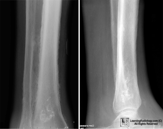
The first X Ray signs
of osteomyelitis |
|
|
|
Early Osteomyelitis |

Osteomyelitis is classified according
to the mechanism of infection (hematogenous or contiguous) and the presence of
vascular insufficiency:
- Hematogenous osteomyelitis
- Occurs when bone tissue is seeded by pathogenic organisms during
the course of bacteremia
- Accounts for 20% of cases of osteomyelitis in adults
- The vertebrae are the most common site of hematogenous infection
in adults, but the long bones, pelvis, and clavicle may also be affected
- Vertebral osteomyelitis is divided into the following two
categories:
- Pyogenic infections, which are most commonly caused by S.
aureus (40%-45% of all cases)
- Nonpyogenic (granulomatous) infections, which are most commonly
caused by Mycobacterium tuberculosis
- Vertebral osteomyelitis occurs most commonly in men between 60
and 70 years of age and involves the lumbar spine
- Osteomyelitis secondary to a contiguous focus of infection
- Occurs after a traumatic bone injury or as a result of the spread
of infection from a nearby source (eg, soft tissue infection)
- Common associated factors include a history of surgical reduction
and internal fixation of fractures, prosthetic devices, open fractures,
and chronic soft tissue infections; decubitus ulcer; burn; or regional
soft tissue infection
- More common in older patients, who generally develop infections
following cellulitis or arthroplasties; infection in younger patients
usually occurs as a result of trauma or surgery
- Most often affects the tibia and femur
- Osteomyelitis associated with vascular insufficiency
- Caused by impaired blood supply to susceptible tissues
- Usually occurs in older patients and in patients with diabetes
mellitus or severe atherosclerosis
- In patients with diabetes, the small bones of the feet are most
often involved; neuropathy may also be present
- The risk of developing osteomyelitis is greater in patients with
large (>
Osteomyelitis may be classified as
acute, subacute, or chronic, depending on the time to clinical presentation
relative to the introduction of infection.
- Acute osteomyelitis is characterized as a suppurative infection
presenting with edema, small vessel thrombosis, and vascular congestion
within 2 weeks of onset
- Subacute osteomyelitis may be more indolent, presenting 1 to
several months after infection
- Chronic osteomyelitis is the result of longstanding infection,
which may take months or years to develop or which has been suppressed by
the host ('remission') or partially treated so that it remains relatively
dormant for long periods before becoming clinically apparent. Chronic
osteomyelitis is characterized by the presence of necrotic bone
(sequestrum); new bone formation; drainage or sinus tracts; and the
presence of leukocytes, lymphocytes, and histiocytes. It can be recognized
in patients with a history of osteomyelitis who experience a recurrence of
pain, erythema, and swelling, along with a draining sinus
Cierny-Mader staging and classification system
Osteomyelitis
is categorized according to the portion of bone affected; the physiologic
status of the patient; and risk factors that affect immunity, metabolism, and
vascularity. The first part of the system categorizes osteomyelitis according
to anatomic type, as follows:
- Stage 1: medullary osteomyelitis
- Limited to the medullary cavity
- Often caused by a solitary
organism
- Causes include hematogenous
spread and infections from orthopedic devices (intramedullary rods)
- Stage 2: superficial
osteomyelitis
- Involves the cortex
- Often caused by an adjacent
soft tissue infection
- Exposed, infected outer
necrotic surface of bone is observed at the base of a soft tissue wound
- Local ischemia is seen
- Stage 3: localized osteomyelitis
- May involve both the medulla
and cortex, but the bone generally remains stable, as the infection does
not involve its entire diameter
- Stage 4: diffuse osteomyelitis
- Extensive disease
- May occur on both sides of a
nonunion or a joint
- Involves the entire thickness
of the bone, with loss of stability
The
second part of the system describes the patient's physiologic status, as
deficiencies of leukocyte recruitment, phagocytosis, or vascular supplies may
promote osteomyelitis and contribute to its chronicity. The physiologic class
of the infected patient is often more important than the anatomic type because
the state of the host is the strongest predictor of treatment failure.
- Class A: normal host
- Normal physiologic, metabolic,
and immune functions
- Associated with a much better
prognosis
- Class B: host factors limit
normal immune response and healing
- Immunocompromised, either
locally (Bl), systemically (Bs), or both (Bls)
- Local factors include problems
of perfusion (peripheral vascular disease, vasculitis, venous stasis,
lymphedema)
- Systemic factors include
hypoxemia, illnesses associated with impaired immune function (chronic
renal or hepatic insufficiency, malignancy, diabetes), or use of
immunosuppressive medication (steroids)
- The goal of treatment is to
remove the factors that lead to the development of osteomyelitis
- Class C: health of host does not
allow full treatment
- Treatment poses a greater risk
than the infection itself
- Surgery may not be possible
because of the patient's debilitated or immunocompromised status
Epidemiology
- There are several recent trends in the epidemiology of
osteomyelitis. Acute hematogenous osteomyelitis is decreasing in
incidence, whereas the incidence of osteomyelitis due to direct
inoculation or contiguous focus of infection is increasing. This is
attributed to the increase in both trauma (due to motor vehicle accidents)
and orthopedic surgical procedures
- Osteomyelitis secondary to open fractures occurs in 3% to 25% of
cases, usually in young men in their twenties and thirties
- Foot ulcers occur in 2% of patients with diabetes every year, 15%
of whom will develop osteomyelitis. Recurrent infection occurs in up to
36% of patients with diabetes
- Vertebral osteomyelitis is responsible for 2% to 4% of all cases
of osteomyelitis, with an annual incidence of 5.3 cases per million
persons. Men are more commonly affected than women, with a mean age at
presentation of 61 years
Causes and risk factors
Causes:
- The focus for hematogenous
osteomyelitis may vary from a mild skin infection to bacterial
endocarditis; it is also a complication among intravenous drug users
- Osteomyelitis secondary to a
contiguous focus of infection may be caused by the direct inoculation of
bacteria through trauma, from spread of adjacent soft tissue infection, or
introduction of infection during preoperative or intraoperative
procedures. Predisposing factors include surgical reduction and internal
fixation of fractures, prosthetic devices, open fractures, and chronic
soft tissue infections
- Osteomyelitis secondary to
vascular insufficiency is often associated with diabetes mellitus.
Infection often results from minor trauma to the feet, such as infected
nail beds or skin ulceration. Inadequate tissue perfusion limits local
tissue response to injury
- Multiple organisms are
responsible for osteomyelitis in different populations. The causative
organism is related to the age, clinical history, and immune status of the
patient (see Table 1). S. aureus is the most common cause in all
cases
Table Organisms Commonly Implicated
in Osteomyelitis in Different Patient Populations
|
Category
of Osteomyelitis |
Population |
Causative
Organism(s) |
|
Hematogenous osteomyelitis |
Patients of all ages |
S. aureus |
|
Neonates |
Enterobacteriaceae,
group B streptococci |
|
|
Infants and children |
Haemophilus influenzae
type B |
|
|
Intravenous drug users |
S. aureus, Pseudomonas aeruginosa, Candida species |
|
|
Patients with sickle cell disease |
Streptococcus pneumoniae, Salmonella species |
|
|
HIV-infected patients |
Bartonella henselae |
|
|
Patients with nosocomial infections |
S. aureus, Enterobacteriaceae, Candida species, Aspergillus (in
immunocompromised patients) |
|
|
Vertebral osteomyelitis (hematogenous and
contiguous focus) |
Adults (most commonly) |
S. aureus |
|
Patients with urinary tract infections |
Aerobic gram-negative bacilli, Enterococcus species |
|
|
Intravenous drug users |
S. aureus, P. aeruginosa |
|
|
Patients undergoing spinal surgery |
Coagulase-negative staphylococci, S. aureus, aerobic gram-negative
bacilli |
|
|
Patients with infections of intravascular
devices |
Candida
species, staphylococci |
|
|
Patients living in endemic regions |
M. tuberculosis, Brucella species,
regional fungi (coccidioidomycosis, blastomycosis, histoplasmosis), Coxiella burnetii (Q
fever) |
|
|
Contiguous-focus osteomyelitis |
Patients exposed to contaminated soil |
Clostridium
species, Bacillus
species, Stenotrophomonas
maltophilia, Nocardia
species, atypical mycobacteria, Aspergillus
species, Rhizopus
species, Mucor
species |
|
Patients with orthopedic devices |
S. aureus,
coagulase-negative staphylococci, Propionibacterium
species |
|
|
Patients with decubitus ulcers |
Enterobacteriaceae, P. aeruginosa,
enterococci, anaerobes, Candida
species |
|
|
Patients with a history of cat bites |
Pasteurella multocida |
|
|
Patients with a history of human bites
(including clenched-fist injury) |
Eikenella corrodens, Moraxella species |
|
|
Patients with puncture injuries on the foot |
P. aeruginosa |
|
|
Patients with periodontal infection |
Actinomyces
species |
|
|
Osteomyelitis associated with vascular
insufficiency |
Patients with diabetes |
Polymicrobial: S. aureus, β-hemolytic streptococci, Enterococcus faecalis,
aerobic gram-negative bacilli |
HIV
= human immunodeficiency virus
Risk
factors:
- Diabetes mellitus
- Immunocompromise
- Neuropathy
- Vascular insufficiency
- Intravenous drug use
- Open fractures
- Local trauma
- Orthopedic hardware (including
prosthetic joints)
- Hemodialysis
- Sickle cell disease
- Dental infections
- Urinary tract infections
- Catheter-related bloodstream infection
Associated disorders
- Occurs more frequently in
patients with diabetes mellitus, vascular insufficiency, or
immunosuppression
- Recent history of surgical
procedure or joint or bone trauma
Screening
Not
applicable.
Primary prevention
Summary
approach
- Patients with diabetes should have a complete
examination of the lower extremities annually and inspection of the feet
for wounds at interim routine follow-up visits. Measures to prevent
diabetic foot ulcers should be emphasized. A high index of suspicion
should be maintained for the contiguous spread of local diabetic foot
infections to the bone, with continuous evaluation for signs and symptoms
of the development of osteomyelitis
- Patients with open fractures who are able to
receive antibiotics within 6 hours of injury and prompt surgical treatment
have a reduced risk of developing osteomyelitis
- The use of prophylactic antibiotics prior to
bone surgery has been shown to prevent wound infections
- Scrupulous care should be taken to avoid
health care–associated osteomyelitis, with careful attention to
intravascular and urinary catheters, surgical incisions, and other wounds
Population
at risk
- Patients with
diabetes mellitus
- Patients
undergoing orthopedic surgery, including placement of prostheses and other
clean surgery and management of open fractures
Preventive
measures
In patients with diabetes mellitus:
- Measures to
prevent diabetic foot include excellent foot hygiene, glycemic control,
and use of protective footwear
- Patients
should be instructed to examine their feet daily and to seek prompt
medical care for new wounds or other injuries to the feet
- A complete
evaluation of the lower extremities should be done annually, and the feet
should be inspected for wounds at periodic follow-up visits in the interim
- Patients with
Charcot joints or other abnormalities that result in friction with shoes
may require specially adapted shoes
- In patients
undergoing foot surgery or amputation, the use of protective footwear
postoperatively is helpful in preventing subsequent ulceration and
infection
In patients with open fractures:
- Administration
of antibiotics within 6 hours of injury and prompt surgical treatment are
associated with a reduced risk of developing osteomyelitis
- A continued
24-hour regimen of penicillin or first-generation or second-generation
cephalosporins is also beneficial
In patients undergoing bone surgery:
- Administration
of prophylactic antibiotics has proven to be successful in the prevention
of infection following surgery, particularly in patients with noncompound
hip fractures and those receiving total hip and knee prostheses
- In
patients undergoing clean bone surgery, intravenous antibiotics are
administered 30 minutes before skin incision and up to 24 hours following
the procedure. A first-generation or second-generation cephalosporin is
appropriate in many cases; vancomycin may be used in patients who are
allergic to cephalosporin and in settings with a high prevalence of
methicillin-resistant staphylococci
- In
patients undergoing surgery for closed fractures, the use of penicillin,
first-generation cephalosporins (eg, cefazolin), or
second-generation cephalosporins (eg, cefamandole, cefuroxime)
has led to a reduction in postsurgical infection
- Standard
preoperative procedures, such as the use of antimicrobial shower, shaving,
and topical disinfectants, should be followed. Observation of such
procedures, together with the use of surgical rooms with laminar airflow
and prophylactic antibiotic therapy, has led to a reduction in the
postsurgical rate of infection to 0.5% to 2%, depending on the type of
joint replacement
BLEEDING
Bleeding
is the name commonly used to describe blood loss. It can refer to blood loss
inside the body (internal bleeding) or blood loss outside of the body (external
bleeding).
Blood loss can occur in almost any
area of the body. Typically, internal bleeding occurs when blood leaks out
through damage
to a blood vessel or organ. External bleeding occurs either when blood exits
through a break in the skin, or when blood exits through a natural opening in
the body, such as the mouth, vagina, or rectum.
Bleeding is the loss of blood. Bleeding may be:
- Inside the
body (internally)
- Outside the
body (externally)
Bleeding may occur:
- Inside the
body when blood leaks from blood vessels or organs
- Outside the
body when blood flows through a natural opening (such as the vagina,
mouth, or rectum)
- Outside the
body when blood moves through a break in the skin
Considerations
Get emergency medical help for severe bleeding. This is very
important if you think there is internal bleeding. Internal bleeding can
very quickly become life threatening. Immediate medical care is needed.
Serious injuries don't cause heavy bleeding. Sometimes, relatively
minor injuries can bleed a lot. An example is a scalp wound.
You may bleed a lot if you take blood-thinning
medication or have a bleeding disorder such as hemophilia. Bleeding in such
people requires immediate medical attention.
The most important step for external bleeding is to apply
direct pressure. This will stop most external bleeding.
Always wash your hands before (if possible) and after giving
first aid to someone who is bleeding. This helps prevent infection.
Try to use latex gloves when treating someone who is
bleeding. Latex gloves should be in every first aid kit. People allergic to
latex can use a nonlatex glove. You can catch viral hepatitis if you touch
infected blood. HIV can be spread if infected blood gets into an open wound, even
a small one.
Although puncture wounds usually don't bleed very much, they
carry a high risk of infection. Seek medical care to prevent tetanus or other
infection.
Abdominal and chest wounds can be very serious because of the
possibility of severe internal bleeding. They may not look very serious, but
can result in shock.
- Seek
immediate medical care for any abdominal or chest wound.
- If
organs are showing through the wound, do not try to push them back into
place.
- Cover the
injury with a moistened cloth or bandage.
- Apply only
very gentle pressure to stop the bleeding.
Blood loss can cause blood to collect under the skin, turning
it black and blue (bruised). Apply a cool compress to the area as soon as
possible to reduce swelling. Wrap the ice in a towel and place the towel over
the injury. Do not place ice directly on the skin.
Causes
Bleeding can be caused by injuries or may be spontaneous.
Spontaneous bleeding is most commonly caused by problems with the joints,
or gastrointestinal or urogenital tracts.
Symptoms
- Blood coming from an open wound
- Bruising
- Shock, which may cause any of the following
symptoms:
- Confusion or decreasing alertness
- Clammy skin
- Dizziness or light-headedness
after an injury
- Low blood pressure
- Paleness (pallor)
- Rapid pulse, increased heart rate
- Shortness of breath
- Weakness
Symptoms of internal
bleeding may also include:
- Abdominal pain and swelling
- Chest pain
- External bleeding through a natural opening
- Blood in the stool (appears
black, maroon, or bright red)
- Blood in the urine (appears red,
pink, or tea-colored)
- Blood in the vomit (looks bright
red, or brown like coffee-grounds)
- Vaginal bleeding (heavier than
usual or after menopause)
- Skin color
changes that occur several days after an injury (skin may black, blue,
purple, yellowish green)

First Aid
First aid is
appropriate for external bleeding. If bleeding is severe, or if you think there
is internal bleeding or the person is in shock, get emergency help.
1.
Calm and reassure the person. The sight
of blood can be very frightening.
2.
If the wound affects just the top
layers of skin (superficial), wash it with soap and warm water and pat dry.
Bleeding from superficial wounds or scrapes is often described as
"oozing," because it is slow.
3.
Lay the person down. This reduces the
chances of fainting by increasing blood flow to the brain. When possible, raise
up the part of the body that is bleeding.
4.
Remove any loose debris or dirt that
you can see from a wound.
5.
Do NOT remove an object such as a
knife, stick, or arrow that is stuck in the body. Doing so may cause more
damage and bleeding. Place pads and bandages around the object and tape the
object in place.
6.
Put pressure directly on an outer wound
with a sterile bandage, clean cloth, or even a piece of clothing. If nothing
else is available, use your hand. Direct pressure is best for external
bleeding, except for an eye injury.
7.
Maintain pressure until the bleeding
stops. When it has stopped, tightly wrap the wound dressing with adhesive tape
or a piece of clean clothing. Place a cold pack over the dressing. Do not peek
to see if the bleeding has stopped.
8.
If bleeding continues and seeps through
the material being held on the wound, do not remove it. Simply place another
cloth over the first one. Be sure to seek medical attention.
9. If
the bleeding is severe, get medical help and take steps to prevent shock. Keep
the injured body part completely still. Lay the person flat, raise the feet
about

Bleeding from most injuries can be
stopped by applying direct pressure to the injury. This keeps from cutting off
the blood supply to the affected limb.

Bleeding from most injuries can be
stopped by applying direct pressure to the injury. This keeps from cutting off
the blood supply to the affected limb. When there is severe bleeding, where a
major artery has been severed, pressure may be insufficient and a tourniquet
may be necessary.

When there is severe bleeding where a
major artery has been severed, pressure may be insufficient and a tourniquet
may be necessary. Tourniquets are an effective way of stopping bleeding from an
extremity. They do, however, stop circulation to the affected extremity and
should ONLY be used when other methods, such as pressure dressings, have failed
(or are likely to fail). Pressure from tourniquets must be relieved
periodically to prevent damage to the tissue from lack of oxygen.
DO NOT
- DO NOT apply
a tourniquet to control bleeding, except as a last resort. Doing so may
cause more harm than good. A tourniquet should be used only in a
life-threatening situation and should be applied by an experienced person
- If continuous
pressure hasn't stopped the bleeding and bleeding is extremely severe, a
tourniquet may be used until medical help arrives or bleeding is
controllable.
- It
should be applied to the limb between the bleeding site and the heart and
tightened so bleeding can be controlled by applying direct pressure over
the wound.
- To
make a tourniquet, use bandages 2 to
- Check
the tourniquet every 10 to 15 minutes. If the bleeding becomes
controllable, (manageable by applying direct pressure), release the
tourniquet.
- DO NOT peek
at a wound to see if the bleeding is stopping. The less a wound is
disturbed, the more likely it is that you'll be able to control the
bleeding
- DO NOT probe
a wound or pull out any embedded object from a wound. This will usually
cause more bleeding and harm
- DO NOT remove
a dressing if it becomes soaked with blood. Instead, add a new one on top
- DO NOT try to
clean a large wound. This can cause heavier bleeding
- DO NOT try to
clean a wound after you get the bleeding under control. Get medical help
When to Contact a Medical Professional
Seek medical help if:
- Bleeding
can't be controlled, required the use of a tourniquet, or was caused by a
serious injury
- The wound
might need stitches
- Gravel or
dirt cannot be removed easily with gentle cleaning
- You think
there may be internal bleeding or shock
- Signs of
infection develop, including increased pain, redness, swelling, yellow or
brown fluid, swollen lymph nodes, fever, or red streaks spreading from the
site toward the heart
- The injury
was due to an animal or human bite
- The patient
has not had a tetanus shot in the last 5-10 years
Prevention
Use
good judgment and keep knives and sharp objects away from small children.
Stay
up-to-date on vaccinations, especially the tetanus immunization.
The body’s response to blood loss
Introduction
Maintenance of circulating blood
volume is one of the most important homeostatic mechanisms that the body must
support. Poor blood supply to the tissues ultimately affects every cell in the
body and organ dysfunction will quickly develop if tissue perfusion fails.
Haemorrhagic shock is defined as a failure of adequate tissue perfusion
resulting from a loss of circulating blood volume. Homeostatic and subcellular
mechanisms Significant blood loss from any cause initiates a sequence of stress
responses that are intended to preserve flow to vital organs and to
signal cells to expend internal energy stores. Haemorrhagic shock reduces wall
tension in the large intrathoracic arteries, which activates baroreceptors.
Adrenergic reflexes that have neural and circulating hormonal components
are also activated. Neural effects are immediate; hormonal changes may be
rapid, but some time is needed for them to take full effect. The two major neural
components are sympathetic fibres from the stellate ganglion, which stimulate
the heart, and sympathetic fibres from regional ganglia, which cause
peripheral arterial vasoconstriction. The circulating ‘stress hormones’ derive
mainly from the hypothalamic −pituitary adrenomedullary axis. They lead
to secretion of epinephrine and norepinephrine from the adrenal medulla and corticosteroids from the adrenal cortex, renin
from the kidney, and glucagon from the pancreas. These hormones signal the
liver to break down glycogen in order to release glucose into the plasma,
promote release of fatty acids from adipose tissue via lipolysis, and stimulate
the breakdown of tissue glycogen stores. At a cellular level, blood loss
first affects the mitochondria. Mitochondria function at the lowest
oxygen tension in the body, but consume almost all the body’s supply of oxygen.
More than 95% of aerobic chemical energy comes from mitochondrial combustion of
fuel substrates (fats, carbohydrates, ketones) using oxygen to produce carbon
dioxide (CO2 ) and water. Most cellular energy transfer derives from
acetyl coenzyme A (aCoA) formed by one of two pathways: α - oxidation or decarboxylation of pyruvate. Either fatty acids or ketones are
produced by α -oxidation. Pyruvate derives from either glycolysis or
lactate dehydrogenation. ACoA is consumed by the tricarboxylic acid (TCA) cycle and forms reduced
pyridine and flavin nucleotides. These pass electrons along a series of
proteins in the inner mitochondrial membrane, culminating in the reduction of
molecular oxygen to form water. The mitochondrion thus harnesses energy from this
process to form adenosine triphosphate from adenosine diphosphate plus inorganic phosphate. This forms the basis of all
cellular energy production. In the early stage of shock, the skeletal muscle
and splanchnic organs are affected more by oxygen deprivation than by a lack of
delivery of fuel substrate. As a result, shock rapidly stalls transfer of
electrons in the mitochondria and ‘jams’ the pathways of acetyl coenzyme A
input into the TCA cycle. Lactic acid results and is transported actively
across the cell membrane by way of a specific protein transporter. In the
resuscitated haemorrhagic shock patient, lactate production may also occur from
skeletal muscle, not entirely because of low mitochondrial oxygenation, but
because the delivery of pyruvate from glycolysis overwhelms the ability of
dehydrogenase enzymes in the TCA cycle to dispose of pyruvate. Pyruvate is then
converted into lactate, a condition known as aerobic glycolysis. Thus, it can
be seen that elevated concentrations of lactate in the blood are a sentinel
marker
of widespread inadequate tissue perfusion. When adequate resuscitation
has been achieved, lactate levels return to normal. Global physiological
effects The physiological responses that
the body mounts to increasing blood loss are well defined by the American College
Class I haemorrhage (0–15% total
blood volume) This represents up to 750 ml blood loss. Minimal physiological
changes occur at this level. A patient may exhibit mild anxiety, but heart
rate, blood pressure and peripheral circulation largely remain unchanged. Urine
output is only slightly decreased. The body can compensate well for this degree
of haemorrhage. This situation is mimicked by a blood donation.
Class II haemorrhage (15–30% total blood volume) Beyond 750 ml blood
lost, sympathetic nerve stimulation occurs as described above. A modest
elevation in heart rate and a decrease in the pulse pressure, as the diastolic
pressure rises, are to be expected. A degree of peripheral ‘shut down’, i.e. cool
extremities and digital cyanosis, accompanied by pallor, occur. Inexperienced
staff may fail to recognize the diastolic pressure rise associated with
increasing blood loss. A blood pressure of 120/70 changing to a pressure of
120/95 may be a sign of the impending cardiovascular
collapse associated with a blood loss in excess of 1500 ml. This physiological change is a direct result of the
vasoconstriction induced by sympathetic nerve stimulation of the vascular tree.
Hyperventilation to a respiratory rate of over 20 breaths/min can also be
expected at this stage as the tissues are starting to suffer a lack of
oxygenation and increasing production of lactic acid stimulates the respiratory
centre.
Class III haemorrhage (30–40% total blood volume) Now haemorrhagic shock
is well established and a critical phase is reached, with a drop in systolic
and diastolic blood pressure. Mental confusion and anxiety are normal,
respiratory rate is high, peripheral perfusion is very poor, and the patient appears
pale and diaphoretic. The pulse rate is invariably well over 100 beats/min and
the production of urine is negligible. Under these circumstances, widespread
failure of tissue oxygenation is occurring, with diversion of what blood
flow there is to essential organs such as brain, kidney and heart.
Class IV haemorrhage (over 40% of blood volume) This situation is
immediately life-threatening and within 15 min a mortality of 50% is to be
expected. Blood pressure has dropped to be, in some cases, unrecordable, and
the pulse is barely palpable. Vital organ perfusion is failing. Other guides to
recognition of haemorrhagic shock Not
all haemorrhagic shock is as obvious as the ATLS classification would
suggest. A change in arterial acid base status will often precede any significant
decrease in cardiac output with haemorrhage. The arterial and venous blood
bicarbonate level and base deficit will decrease early in haemorrhage even
if pH and blood pressure remain in the normal range. Distinguishing simple
haemorrhage from haemorrhagic shock is possible by noting that when the base
deficit becomes pathologically low, widespread tissue hypoperfusion may
be suspected. Patients with tachycardia, a low base deficit, and low
urine output should be suspected of having haemorrhagic shock. The base
deficit is defined as the amount of strong base that would have to
be added to a litre of blood to normalize the pH. In the clinical setting the
base deficit is indirectly calculated from the pH and arterial CO2 partial pressure (PaCO2 ) and is
normally higher than − 2
mM . The brainstem chemoreceptor responds to acidaemia
by increasing respiratory rate, leading to reduced PaCO2. After
approximately one third of the total blood volume is acutely lost,
cardiovascular reflexes can no longer cause adequate filling of the
arterial tree, and hypotension supervenes. Arterial hypotension is generally defined as an
arterial blood pressure below 90 mmHg, but this threshold should be increased
to 100 mmHg in patients with known systemic hypertension and in patients over
age 60 years. With the development of hypotension, the patient can no longer
hyperventilate sufficiently to maintain a normal arterial pH, and
acidaemia occurs. Blood gases can therefore provide very useful information
regarding the early detection of acute blood loss. Treatment of haemorrhagic
shock Conventional wisdom regarding shock has been directed at rapid correction
of the source of haemorrhage and rapid restoration of normal circulating blood
volume and perfusion. The ATLS curriculum advocates the immediate establishment
of largebore intravenous access and infusion of resuscitative fluids,
characteristically up to 2 l
of Hartmann’s solution, followed by blood. The goal is to return the blood
pressure to normal. If blood pressure subsequently falls again, this is taken
as a sign of serious injury, which must be addressed by immediate surgery.
Maintenance of blood pressure during the period of active haemorrhage is
presumed to reduce the risk of organ-system failure and irreversible shock. This
approach may not be as sensible as it seems. The analogy of pouring water into
a bucket with a hole at the bottom can be made. The greater the volume of water
added to the bucket, the more rapidly it runs from the hole. In the traumatized
patient, this means greater blood loss, a longer duration of bleeding, and
disruption of the normal plasma composition including dilution of clotting
factors. Aggressive fluid administration and higher blood pressure
potentially increase the extent of haemorrhage. Surgical control must be
rapidly achieved. What is not understood at the present is the priority that
must be given to these two conflicting goals: maintenance of perfusion
and limitation of haemorrhage. Deliberate hypotension is employed in elective
head and neck surgery, prostate resections, and major orthopaedic procedures as
a means of reducing the blood loss and need for transfusion. Trauma patients,
however, may have already lost a substantial amount of blood before hospital
arrival and be in some degree of shock. Identifying the optimal balance between
fluid resuscitation and haemorrhage control is one of the most
difficult and controversial areas in trauma care today. Delayed or
limited resuscitation until surgical control is available may improve outcome. The
effects of persisting inadequate circulating
volume Untreated or inadequately treated haemorrhagic shock will ultimately
have serious consequences for the patient. Life hreatening problems can occur a
considerable time after the traumatic event. With limited or inadequate
restoration of circulating volume, initiation of inflammatory changes can
be expected. Shock causes neutrophil activation and liberation of adhesion
molecules, which promote binding of neutrophils to lung and vascular
endothelium. Within the lung, this initiates capillary leak that can produce
the adult respiratory distress syndrome. Within the circulation,
inflammatory cytokines are liberated during resuscitation, and membrane injury
occurs in many cells. In the liver, damage from inflammation and reactive
oxygen species from neutrophils is compounded by persistent microischaemia.
During resuscitation from haemorrhagic shock, the normal balance of
vasodilatation by nitric oxide versus vasoconstriction by endothelins becomes
distorted, producing patchy centrilobular ischaemic damage in the liver, which
may produce an immediate rise in blood transaminase levels. A growing body of
evidence suggests that re-transfusion from haemorrhage exerts greater injury to
the heart than the actual hypotensive insult. Depending on the degree of
hypotensive insult, the kidney may manifest acute spasm of the preglomerular
arterioles, causing acute tubular necrosis. Systemic metabolic changes can
impair fuel delivery to the heart and brain, secondary to depressed hepatic
glucose output, impaired hepatic ketone production, and inhibited peripheral
lipolysis. Activated, sticky neutrophils can also directly damage organs by
liberating toxic reactive oxygen species, N-chloramines, and proteolytic enzymes. Neutrophils
can also plug capillaries and cause microischaemia. Although much of the knowledge
about the inflammatory responses in shock has evolved from the study of
septic shock, the consensus is that any low-perfusion state that produces
widespread cellular hypoxia can trigger systemic inflammation.
Haemorrhagic shock is one such low-flow state. Multiple tissues (e.g.
macrophages, endothelial and epithelial cells, muscle cells) are signalled to
enhance transcription of messenger ribonucleic acid (mRNA) coding for
cytokines, including tumour necrosis factors (TNF-α , TNF-α) and
interleukins (IL-1, IL-6). The effects of intravascular cytokine release on the
body are manifest as systemic inflammatory response syndrome (SIRS). SIRS
can progress to sepsis syndrome and ultimately septic shock. One additional
mechanism by which this is suggested to occur is a breakdown in the integrity
of the gut mucosa caused by inadequate perfusion of the gut wall. This leads to
bacterial translocation of Gram-negative gut bacteria into the bloodstream,
thus inducing cytokine release and septic shock. The prevention of inadequate
circulation by the timely surgical intervention and restoration of circulating
volume will ameliorate the above pathological processes.
Conclusions
The consequences of inadequate circulating blood volume are
life-threatening. Recognition of haemorrhage by the classical physiological
signs is important for clinicians to diagnose shock and prevent both short- and
long-term complications of this condition. These interventions centre on
surgical control of bleeding and rapid restoration of blood volume.
Lower gastrointestinal tract bleeding: a problem based approach
Clinical presentation
McGuire
and Haynes reviewed the literature from 1956 to 1971 and found 473 cases
of massive lower GI bleeding. More than three-fourths of patients were
successfully treated nonoperatively with a 22% rebleeding rate but only a 3%
mortality. One hundred and three of four hundred and seventy-three (22%)
underwent operations, 28 of the 62 (45%) hemicolectomies rebled and nine of the
62 (15%) died. Only three of 34 (9%) patients undergoing subtotal colectomy
died and none rebled. McGuire recently reviewed the natural history of 79
patients with 108 episodes of acute lower GI bleeding. Bleeding stopped
spontaneously in 76% but 24% required emergency surgery. In the 66 patients
with lower gastrointestinal bleeding who required no more than three units
blood transfusion over a 24 hour period, 98.5% stopped spontaneously. When
greater than three units were transfused in a 24 hour time period, 25 of 42
(60%) patients needed emergency surgery. Patients with acute LGIB can
demonstrate a decrease in hemoglobin as well as changes in hemodynamics (50%),
orthostatic changes (30%), syncope (10%), or cardiovascular collapse (9%).
History and physical examination
Information such as a prior history of peptic ulcer disease,
diverticulosis, aortic vascular surgery, radiation therapy, use of medications
such as non-steroidal anti-inflammatory and anti-coagulants, or recent
endoscopy with biopsy may indicate the origin of bleeding. Diagnostic and
resuscitative manuevers must occur simultaneously if hemodynamic instability is
present. Initial laboratories should include complete blood count, type and
crossmatch, platelet count and coagulation parameters. Digital rectal
examination with stool guaiac is a routine part of the physical examination in
the initial evaluation of all patients with gastrointestinal bleeding as 80%
will have blood per rectum.
Diagnosis and therapeutics
Exclusion of an upper gastrointestinal source of bleeding per
rectum including nasogastric tube aspiration and/or upper endoscopy is
mandatory prior to evaluation of potential lower gastrointestinal tract
sources. Aspiration of the nasogastric tube excludes all but a small percentage
of upper GI sources. Sigmoidoscopy/anoscopy has been routinely performed early
in the course of evaluation of LGIB to rule out anorectal sources of hemorrhage.
Colonoscopy permits examination of the entire colon and potential therapeutic
interventions in LGIB. The diagnostic yield for colonoscopy is related to the
rate of bleeding and the ability to obtain an adequate bowel preparation.
Overall diagnostic yield for a total of 1561 patients undergoing urgent
colonoscopy in acute LGIB was 68%, with an incomplete procedure rate of 2%, and
a reported complication rate of 1.3%. Endoscopic techniques to control
hemorrhage have typically been attempted far less frequently in LGIB than in
UGIB with reported rates of utilization of 27% and 51% respectively. When
colonoscopy is non diagnostic, unavailable or incomplete the workup of acute
lower GI bleeding after exclusion of upper GI bleeding, may include some form of
nuclear medicine scan. If results of these scans demonstrate acute hemorrhage
then selective angiography is indicated. The diagnostic inaccuracy of 20% in
pooled data from tagged red blood cell scanning in localization of acute LGIB
has led to the recommendation that surgical resection cannot be solely based on
the results of nuclear scans. Even in cases where a positive TRBC scan was
noted, a wide range (7–61%) of angiograms were noted to be abnormal. Segmental
colectomy may subsequently be planned in those patients in whom site of
bleeding is demonstrated by angiogram and when there is clinical evidence of
ongoing hemorrhage.
The need for this myriad of diagnostic studies evolved from
the premise that subtotal colectomy was to be avoided and that the benefits of
segmental colectomy outweighed the risks of angiography and associated delay in
surgical intervention. Subtotal colectomy was thought to be associated with a
higher mortality rate and an increased incidence of disabling diarrhea than
hemicolectomy. The view that subtotal colectomy with ileorectal anastamosis
leads to a higher morbidity and mortality rates is certainly debatable.
Early studies, predating current sophisticated critical care management,
demonstrated a low mortality after subtotal colectomy for lower GI bleeding. Fazio
et al. reviewed 163 patients undergoing elective ileorectal anastamosis and
found only a 1.2% leak rate and 1.9% operative mortality. A higher leak rate
(28–33%) for ileorectal anastamosis, however, has been noted in the emergency
setting. The surgeon must strongly consider fecal diversion, avoiding
construction of a tenous anastamosis in hypoperfused patients undergoing any
emergency colectomy, to minimize the risk for anastamotic dehiscence. It now
appears that severe disabling diarrhea rarely occurs after subtotal colectomy
with ileorectal anastamosis. Most patients’ bowel habits are relatively normal
following subtotal colectomy, averaging 2 to 4 bowel movements per day after
emergency or elective surgery for cancer, ulcerative colitis or acute lower GI
bleeding. Preserving a length of remaining ileum, (greater than
Angiography has been used to identify sites of intestinal
bleeding for more than three decades supposedly to minimize the extent of
colonic resection. In addition, the use of therapeutic angiography (vasopressin
and embolization) has been thought to possibly minimize the need for operative
intervention. While angiography does occasionally localize the origin of
hemorrhage, studies are negative in more than one-half of patients requiring
emergency operation. Furthermore, the angiogram can be misleading, showing only
some of multiple bleeding site, leading to an inadequate segmental resection.
Surprisingly, selective angiography has been shown to be
clinically beneficial in only 8%–28% of patients. Nath and colleagues
reported that in only 10 of 49 patients evaluated, angiography led to a
segmental resection. Similarly, Britt et al. found that just 11 of 40
patients undergoing angiography for lower GI bleeding received a segmental
resection. Browder and co-workers reported that 8 of 50 patients had
emergent operations but four of the eight underwent subtotal colectomy for
multiple bleeding sites. Unfortunately, complication rates of angiography are
not insignificant with reported overall rate of 9.3%. Major complications
include femoral artery thrombosis, hematoma, contrast reactions, renal failure,
and transient ischemic attacks. Furthermore, it is unclear how the delay in
performing a definitive operative intervention may impact on the development of
organ failure and death in these populations alternatively therapeutic
angiography has been utilized with variable success to stop bleeding with vasoconstrictive
agents and embolization. Angiography has also been used to induce an identify
occult bleeding sites by employing anticoagulants, fibrinolytic agents, and
vasodilators. In a review of 83 patients with intra-arterial infusion of
vasopressin for colonic bleeding, it was found that 41% rebled. The long term
consequences of angiographic control of hemorrhage are unknown, but the colon
appears to be at increased risk of ischemia and colonic stricture after
transcatheter embolization.
In summary, mortality rates for lower gastrointestinal
bleedings are less than five percent and hemorrhage related deaths are actually
infrequent. After resuscitation, placement of a nasogastric tube to exclude an
upper gastrointestinal source, and screening anoscopy/sigmoidoscopy to rule out
the presence of anorectal pathology is required. (Fig.) It is our preference to
proceed to subtotal colectomy when transfusion requirements exceed three units.
However, an acceptable alternative includes TRBC followed by angiography and
possible segmental colectomy. A positive TRBC requires verification by
angiography typically and subsequent attempt to halt hemorrhage with
vasopressin infusion. If angiography is negative in the setting of a positive
TRBC, provocative tests utilizing anti-coagulants or vasodilators may be
attempted. If after these attempts no site is localized on angiography,
subtotal colectomy is recommended. If no active bleeding is evident clinically
(< 4 unit transfusion over 24 hours), whole gut lavage should be performed,
and colonoscopy should be carried out. If no lesion is identified on
colonoscopy and the patient continues to actively bleed total abdominal
colectomy is recommended. Patients who have > 3 unit blood transfusion
requirement during a 24 hour period should be considered to be immediate
surgical candidates.

Diagnostic algorithm.
Management of massive blood loss: a
template guideline
The management of acute massive blood
loss is considered and a template guideline is formulated, supported by a review
of the key literature and current evidence. It is emphasized that, if avoidable
deaths are to be prevented, surgeons, anaesthetists, haematologists and blood-bank
staff need to communicate closely in order to achieve the goals of secure
haemostasis, restoration of circulating volume, and effective management of
blood component replacement.
Complications of major blood loss and
massive transfusion may jeopardize the survival of patients from many
specialties, and challenge haematological and blood transfusion resources.
Avoidable deaths of patients with
major haemorrhage are well recognized,1 2 and locally agreed and/or speciality-specific
guidelines3 are needed to ensure effective management. Current
The guideline is presented in this
article (Table) as a simple template which may be modified to take into account
local circumstances and displayed in clinical areas. The left-hand column of
the template outlines the key steps or goals, the centre column adds procedural
detail and the right-hand column provides additional advice and information.
Acute massive blood loss: template
guideline
Table
Acute massive blood
loss: template guideline
|
Goal |
Procedure |
Comments |
|
Restore circulating volume |
Insert wide bore peripheral cannulae |
|
|
|
Give adequate volumes of warmed crystalloid, ?colloid, |
Monitor central venous pressure |
|
|
blood |
Blood loss is often underestimated |
|
|
Aim to maintain normal blood pressure and urine output |
Refer to Advanced Trauma Life Support guidelines |
|
|
>30 ml h –1 |
Keep patient warm |
|
Contact key personnel |
Clinician in charge |
Nominated coordinator should take responsibility for |
|
|
Duty anaesthetist |
communication and documentation |
|
|
Blood bank |
|
|
|
Duty haematologist |
|
|
Arrest bleeding |
Early surgical or obstetric intervention |
|
|
|
Interventional radiology |
|
|
Request laboratory investigations |
FBC, PT, APTT, fibrinogen; blood bank sample, biochemical
profile, blood gases or pulse oximetry |
Take samples at earliest opportunity as results may be affected
by colloid infusion |
|
|
Ensure correct sample identity |
Misidentification is commonest transfusion risk |
|
|
Repeat FBC, PT, APTT, fibrinogen every 4 h or after 1/3 blood
volume replacement |
May need to give components before results available |
|
|
Repeat after blood component infusion |
|
|
Request suitable red cells |
Un crossmatched group O Rh negative |
Rh positive is acceptable if patient is male or |
|
|
In extreme emergency |
postmenopausal female |
|
|
No more than 2 units |
|
|
|
Un crossmatched ABO group specific |
Laboratory will complete cross match after issue |
|
|
When blood group known |
|
|
|
Fully cross matched |
Further cross match not required after replacement of 1
blood volume (8–10 units) |
|
|
If irregular antibodies present |
|
|
|
When time permits |
|
|
|
Use blood warmer and/or rapid infusion device. |
Blood warmer indicated if flow rate
>50 ml kg–1 h–1 in adult |
|
|
Employ blood salvage if available and appropriate |
Salvage contraindicated if wound heavily contaminated |
|
Request platelets |
Allow for delivery time from blood centre |
Target platelet count: |
|
|
Anticipate platelet count <50×109 litre–1 after
2 × blood volume replacement |
>100×109 litre–1 for multiple/CNS trauma
or if platelet function abnormal |
|
|
|
>50×109 litre–1 for other situations |
|
Request FFP |
Aim for PT and APTT <1.5× control mean |
PT and APTT >1.5× control mean correlates with |
|
(12–15 ml kg–1 body weight=1 litre or
4 units for an adult) |
Allow for 30 min thawing time |
increased surgical bleeding |
|
Request cryoprecipitate |
Replace fibrinogen and factor VIII |
Fibrinogen <0.5 strongly associated with microvascular |
|
(1–1.5 packs/10 kg body weight) |
|
bleeding |
|
|
Aim for fibrinogen >1.0 g litre–1 Allow for delivery time plus 30 min thawing time |
Fibrinogen deficiency develops early when plasma poor red blood cells used for replacement |
|
Suspect DIC |
Treat underlying cause if possible |
Shock, hypothermia, acidosis leading to risk of DIC |
|
|
|
Mortality from DIC is high |
The accompanying commentary is not
intended to be an exhaustive review, but provides key references on which the
recommendations are based.
The Hospital Transfusion Committee
has a central role in ensuring the optimum and safe use of blood components.
The development of protocols for the management of massive transfusion is an
important part of the remit of such committees and it is hoped that this
article will facilitate this process.
Previous SectionNext
SectionCommentary
Background
Massive blood loss is usually defined
as the loss of one blood volume within a 24 h period,7 normal blood volume
being approximately 7% of ideal body weight in adults and 8–9% in children.
Alternative definitions include 50% blood volume loss within 3 h or a rate of
loss of 150 ml min–1.8 Such definitions emphasize the importance of the early
recognition of major blood loss and the need for effective action to prevent
shock and its consequences.
Priorities for treatment are:
• restoration of blood volume to
maintain tissue perfusion and oxygenation;
• achieving haemostasis by:
• treating any surgical source of
bleeding;
• correcting coagulopathy by the judicious
use of blood component therapy.
A successful outcome requires prompt
action and good communication between clinical specialties, diagnostic
laboratories, blood-bank staff and the local blood centre. Blood component
support takes time to organize and the blood centre may be up to 2 h away from
the hospital.
Early consultation with surgical,
anaesthetic and haematology colleagues is advisable, and the importance of good
communication and cooperation in this situation cannot be overemphasized. A member
of the clinical team should be nominated to act as the coordinator responsible
for overall organization, liaison, communication and documentation. This is a
critical role for a designated member of the permanent clinical staff. The
Hospital Transfusion Committee should provide a forum in which a rapid
communication cascade can be agreed and massive transfusion episodes reviewed.
Resuscitation
Prolonged oligaemic shock carries a
high mortality rate because of organ failure and disseminated intravascular
coagulation. Restoration of circulating volume is initially achieved by rapid
infusion of crystalloid or colloid through large-bore (14 gauge or larger)
peripheral cannulae.9 The use of albumin and non-albumin colloids versus
crystalloids for volume replacement has recently been the subject of debate
after two controversial meta-analyses,10 11 and the use of colloid is not
recommended in the latest American College of Surgeons Advanced Trauma Life
Support Guidelines.12 Further trials are required before firm recommendations
can be made.
Red cell transfusion is likely to be
required when 30–40% of blood volume is lost; the loss of over 40% of blood
volume is immediately life-threatening.12 Hypothermia increases the risk of
disseminated intravascular coagulation and other complications12 13 and may be
prevented by prewarming the resuscitation fluids, patient-warming devices such
as warm air blankets, and the use of temperature-controlled blood warmers.
Blood loss is usually underestimated,
and it must be remembered that haemoglobin and haematocrit values do not fall
for several hours after acute haemorrhage.9
For acutely anaemic patients, the
American Society of Anesthesiologists Task Force on Blood Component Therapy has
concluded, on the basis of the available evidence, that transfusion is rarely
indicated when the haemoglobin concentration is >
Intraoperative blood salvage may be
of great value in reducing the requirement for allogeneic blood, but bacterial
contamination of the wound is a relative contraindication.15
Investigations
Blood samples should be sent to the
laboratory at the earliest possible opportunity for blood grouping, antibody
screening and compatibility testing, as well as for baseline haematology,
coagulation screening, including fibrinogen estimation and biochemistry
investigations.
When dealing with an evolving
process, it is important to check the parameters frequently (at least four-hourly
and after each therapeutic intervention) to monitor the need for and the
efficacy of component therapy.
Expert advice should be sought from a
haematologist regarding appropriate investigations, their interpretation and
the optimum corrective therapy.
Previous SectionNext SectionBlood
component therapy
Red cells
In an extreme situation it may be
necessary to use group O un-crossmatched red cells if the blood group is
unknown. In an emergency, premenopausal females whose blood group is unknown should
be given ORh(D) negative red cells in order to avoid sensitization and the risk
of haemolytic disease of the newborn in subsequent pregnancy. It is acceptable
to give ORh(D) positive cells to males and postmenopausal females of unknown
blood group.16 Group-specific red cells should be given at the earliest
possible opportunity as group O blood is a scarce resource.
It is important to bear in mind that
most transfusion-related morbidity is due to incorrect blood being
transfused.17 It is therefore essential that protocols are in place for the
administration of blood and blood components18 and that these are adhered to
even in an emergency situation.
All blood components supplied by the
Platelets
Expert consensus argues that
platelets should not be allowed to fall below the critical level of
50×109 litre–1 in acutely bleeding patients.20 A higher target level of
100×109 litre–1 has been recommended for those with multiple high-energy
trauma or central nervous system injury.21 22 Empirical platelet transfusion
may be required when platelet function is abnormal, as is found after
cardiopulmonary bypass.
A platelet count of 50×109
litre–1 is to be anticipated when approximately two blood volumes have been
replaced by plasma-poor red cells,23 but there is marked individual variation.
In assessing the requirement for platelets, frequent measurements are needed,
and it may be necessary to request platelets from the blood centre at levels
above the desired target in order to ensure their availability when needed.
Fresh frozen plasma (FFP) and
cryoprecipitate
Most clinical studies and guidelines
have been based on the use of whole blood or plasma-reduced red cells, which
contain some residual coagulation factor activity. Nowadays, red cell
replacement is likely to be in the form of plasma-poor red cells suspended in
optimal additive solution, in which coagulation factor activity is negligible.
Under these circumstances, coagulation factor deficiency is the primary cause
of coagulopathy. The level of fibrinogen falls first; the critical level of
Laboratory tests of coagulation
should be monitored frequently and interpreted with advice from a clinical
haematologist; laboratories should have in place standard operating procedures
to ensure that clinical staff are contacted appropriately. Experienced
laboratory staff should be empowered to issue blood components in the first
instance using a locally agreed algorithm. It may be necessary to request
components before results are available, depending on the rate of bleeding and
the laboratory turnaround time. Although ‘formula replacement’ with fresh
plasma is not recommended, it has been suggested that infusion of FFP should be
considered after one blood volume has been lost.25 The dose should be large
enough to maintain coagulation factors well above the critical level, bearing
in mind that the efficacy may be reduced because of rapid consumption.21 25
FFP alone, if given in sufficient
quantity, will correct fibrinogen and most coagulation factor deficiencies, but
large volumes may be required. If fibrinogen levels remain critically low (<
The Guidelines on Oral
Anticoagulation of the British Committee for Standards in Haematology recommend
prothrombin complex concentrate as an alternative to FFP when major bleeding
complicates anticoagulant overdose.26 It should be remembered, however, that
these preparations are potentially thrombogenic and the role of specific
coagulation factor concentrate outwith hereditary bleeding disorders is
unproven.
Disseminated intravascular
coagulation (DIC)
DIC is a feared complication in the
acutely bleeding patient. It carries a considerable mortality rate, and once established
it is difficult to reverse. At particular risk are: patients with prolonged
hypoxia or hypovolaemia; patients with cerebral or extensive muscle damage; and
patients who become hypothermic after infusion of cold resuscitation fluids.
Laboratory evidence of DIC should be sought before microvascular bleeding
becomes evident so that appropriate and aggressive action can be taken to
address the underlying cause. Frequent estimation of platelet count,
fibrinogen, PT and APTT is strongly recommended; measurement of fibrinogen
degradation products or d-dimers may be useful. Prolongation of PT and APTT
beyond that expected by dilution, together with significant thrombocytopenia
and fibrinogen of <
Treatment consists of platelets, FFP
and cryoprecipitate, given sooner rather than later, in sufficient dosage but
avoiding circulatory overload.
Previous SectionNext
SectionDiscussion
A guideline has been defined as ‘a
systematically developed statement that assists in decision-making about
appropriate health care for specific clinical situations’.27 Successful
implementation will depend on local ownership by adaptation to local
circumstances and accessibility at the point of clinical activity.
In developing this template
guideline, we have examined such sound scientific evidence as is available,
reviewed the relevant literature and professional consensus statements, and
taken into account discussion and comment from contributors and delegates at
the National Blood Service Northern Zone Symposium on Massive Transfusion.
The
recommendations contained in these guidelines must be regarded as Grade C,
based as they are on uncontrolled observational studies and a consensus of
expert opinion (Level 3 evidence). Well-designed case–control studies and
randomized clinical trials are lacking in this important area of transfusion
medicine.
A
recent cohort study shows a
significantly improved survival rate in massively transfused patients over a
10-yr period and associates this with more effective and efficient rewarming
techniques, aggressive resuscitation and component therapy, and improved
blood-banking.
There
is a need for further studies to clarify these issues and provide firm evidence
on which future recommendations can be based.