CANCER AND SARCOMA OF THE MANDIBLE: ORIGIN AND HISTOLOGICAL STRUCTURE, CLASSIFICATION, CLINICAL FEATURES, DIFFERENTIAL DIAGNOSIS, TREATMENT, COMPLICATIONS AND PREVENTION.
CANCER AND SARCOMA OF THE UPPER JAW: THE ORIGIN AND HISTOLOGICAL STRUCTURE, CLASSIFICATION, CLINICAL FEATURES, DIFFERENTIAL DIAGNOSIS, TREATMENT, COMPLICATIONS AND PREVENTION.
The mouth and throat
This booklet is about cancers that occur in the mouth (oral cavity) and the part of the throat at the back of the mouth (oropharynx). The oral cavity and oropharynx have many parts:
- Lips
- Lining of your cheeks
- Salivary glands (glands that make saliva)
- Roof of your mouth (hard palate)
- Back of your mouth (soft palate and uvula)
- Floor of your mouth (area under the tongue)
- Gums and teeth
- Tongue
- Tonsils
Understanding cancer
Cancer begins in cells, the building blocks that make up tissues. Tissues make up the organs of the body.
Normally, cells grow and divide to form new cells as the body needs them. When cells grow old, they die, and new cells take their place.
Sometimes this orderly process goes wrong. New cells form when the body does not need them, and old cells do not die when they should. These extra cells can form a mass of tissue called a growth or tumor.
Tumors can be benign or malignant:
- Benign tumors are not cancer:
- Benign tumors are rarely life-threatening.
- Generally, benign tumors can be removed, and they usually do not grow back.
- Cells from benign tumors do not invade the tissues around them.
- Cells from benign tumors do not spread to other parts of the body.
- Malignant tumors are cancer:
- Malignant tumors are generally more serious than benign tumors. They may be life-threatening.
- Malignant tumors often can be removed, but sometimes they grow back.
- Cells from malignant tumors can invade and damage nearby tissues and organs.
- Cells from malignant tumors can spread to other parts of the body. The cells spread by breaking away from the original cancer (primary tumor) and entering the bloodstream or lymphatic system. They invade other organs, forming new tumors and damaging these organs. The spread of cancer is called metastasis.
Oral
cancer
Oral cancer is part of a group of cancers called head and neck cancers. Oral
cancer can develop in any part of the oral cavity or oropharynx. Most oral
cancers begin in the tongue and in the floor of the mouth. Almost all oral
cancers begin in the flat cells (squamous cells) that cover the surfaces of the
mouth, tongue, and lips. These cancers are called squamous cell carcinomas.
When oral cancer spreads (metastasizes), it usually travels through the lymphatic system. Cancer cells that enter the lymphatic system are carried along by lymph, a clear, watery fluid. The cancer cells often appear first in nearby lymph nodes in the neck.
Cancer cells can also spread to other parts of the neck, the lungs, and other parts of the body. When this happens, the new tumor has the same kind of abnormal cells as the primary tumor. For example, if oral cancer spreads to the lungs, the cancer cells in the lungs are actually oral cancer cells. The disease is metastatic oral cancer, not lung cancer. It is treated as oral cancer, not lung cancer. Doctors sometimes call the new tumor "distant" or metastatic disease.
Oral cancer: Who's at risk?
Doctors cannot always explain why one person develops oral cancer and another does not. However, we do know that this disease is not contagious. You cannot "catch" oral cancer from another person.
Research has shown that people with certain risk factors are more likely than others to develop oral cancer. A risk factor is anything that increases your chance of developing a disease.
The following are risk factors for oral cancer:
- Tobacco: Tobacco use accounts for most oral cancers. Smoking cigarettes, cigars, or pipes; using chewing tobacco; and dipping snuff are all linked to oral cancer. The use of other tobacco products (such as bidis and kreteks) may also increase the risk of oral cancer. Heavy smokers who use tobacco for a long time are most at risk. The risk is even higher for tobacco users who drink alcohol heavily. In fact, three out of four oral cancers occur in people who use alcohol, tobacco, or both alcohol and tobacco.
- Alcohol: People who drink alcohol are more likely to develop oral cancer than people who don't drink. The risk increases with the amount of alcohol that a person consumes. The risk increases even more if the person both drinks alcohol and uses tobacco.
- Sun: Cancer of the lip can be caused by exposure to the sun. Using a lotion or lip balm that has a sunscreen can reduce the risk. Wearing a hat with a brim can also block the sun's harmful rays. The risk of cancer of the lip increases if the person also smokes.
- A personal history of head and neck cancer: People who have had head and neck cancer are at increased risk of developing another primary head and neck cancer. Smoking increases this risk.
|
Quitting tobacco reduces the risk of oral cancer. Also, quitting reduces the chance that a person with oral cancer will get a second cancer in the head and neck region. People who stop smoking can also reduce their risk of cancer of the lung, larynx, mouth, pancreas, bladder, and esophagus. There are many resources to help smokers quit:
|
Some studies suggest that not eating enough fruits and vegetables may increase the chance of getting oral cancer. Scientists also are studying whether infections with certain viruses (such as the human papillomavirus) are linked to oral cancer.
If you think you may be at risk, you should discuss this concern with your doctor or dentist. You may want to ask about an appropriate schedule for checkups. Your health care team will probably tell you that not using tobacco and limiting your use of alcohol are the most important things you can do to prevent oral cancers. Also, if you spend a lot of time in the sun, using a lip balm that contains sunscreen and wearing a hat with a brim will help protect your lips.
What are the symptoms of oral cancer?
Early detection
Your regular checkup is a good time for your dentist or doctor to check your
entire mouth for signs of cancer. Regular checkups can detect the early stages
of oral cancer or conditions that may lead to oral cancer. Ask your doctor or
dentist about checking the tissues in your mouth as part of your routine exam.
Symptoms
Common symptoms of oral cancer include:
- Patches inside your mouth or on your lips that are white, a mixture of red and white, or red
- White patches (leukoplakia) are the most common. White patches sometimes become malignant.
- Mixed red and white patches (erythroleukoplakia) are more likely than white patches to become malignant.
- Red patches (erythroplakia) are brightly colored, smooth areas that often become malignant.
- A sore on your lip or in your mouth that won't heal
- Bleeding in your mouth
- Loose teeth
- Difficulty or pain when swallowing
- Difficulty wearing dentures
- A lump in your neck
- An earache
Anyone with these symptoms should see a doctor or dentist so that any problem can be diagnosed and treated as early as possible. Most often, these symptoms do not mean cancer. An infection or another problem can cause the same symptoms.
Diagnosis of oral cancer
If you have symptoms that suggest oral cancer, the doctor or dentist checks your mouth and throat for red or white patches, lumps, swelling, or other problems. This exam includes looking carefully at the roof of the mouth, back of the throat, and insides of the cheeks and lips. The doctor or dentist also gently pulls out your tongue so it can be checked on the sides and underneath. The floor of your mouth and lymph nodes in your neck also are checked.
If an exam shows an abnormal area, a small sample of tissue may be removed. Removing tissue to look for cancer cells is called a biopsy. Usually, a biopsy is done with local anesthesia. Sometimes, it is done under general anesthesia. A pathologist then looks at the tissue under a microscope to check for cancer cells. A biopsy is the only sure way to know if the abnormal area is cancerous.
|
If you need a biopsy, you may want to ask the doctor or dentist some of the following questions:
|
Treatment for oral cancer
Staging
If the biopsy shows that cancer is present, your doctor needs to know the stage (extent) of your disease to plan the
best treatment. The stage is based on the size of the tumor, whether the cancer
has spread and, if so, to what parts of the body.
Staging may require lab tests. It also may involve endoscopy. The doctor uses a thin, lighted tube (endoscope) to check your throat, windpipe, and lungs. The doctor inserts the endoscope through your nose or mouth. Local anesthesia is used to ease your discomfort and prevent you from gagging. Some people also may have a mild sedative. Sometimes the doctor uses general anesthesia to put a person to sleep. This exam may be done in a doctor's office, an outpatient clinic, or a hospital.
The doctor may order one or more imaging tests to learn whether the cancer has spread:
- Dental x-rays: An x-ray of your entire mouth can show whether cancer has spread to the jaw.
- Chest x-rays: Images of your chest and lungs can show whether cancer has spread to these areas.
- CT scan: An x-ray machine linked to a computer takes a series of detailed pictures of your body. You may receive an injection of dye. Tumors in the mouth, throat, neck, or elsewhere in the body show up on the CT scan.
- MRI: A powerful magnet linked to a computer is used to make detailed pictures of your body. The doctor can view these pictures on a monitor and can print them on film. An MRI can show whether oral cancer has spread.
Treatment
Many people with oral cancer want to take an active part in making decisions
about their medical care. It is natural to want to learn all you can about your
disease and your treatment choices. However, shock and stress after the diagnosis can make it hard to think of everything
you want to ask the doctor. It often helps to make a list of questions before
an appointment. To help remember what the doctor says, you may take notes or
ask whether you may use a tape recorder. You may also want to have a family
member or friend with you when you talk to the doctor -- to take part in the
discussion, to take notes, or just to listen.
Your doctor may refer you to a specialist, or you may ask for a referral. Specialists who treat oral cancer include oral and maxillofacial surgeons, otolaryngologists (ear, nose, and throat doctors), medical oncologists, radiation oncologists, and plastic surgeons. You may be referred to a team that includes specialists in surgery, radiation therapy, or chemotherapy. Other health care professionals who may work with the specialists as a team include a dentist, speech pathologist, nutritionist, and mental health counselor.
Getting
a second opinion
Before starting treatment, you might want a second opinion about the diagnosis
and the treatment plan. Some insurance companies require a second opinion;
others may cover a second opinion if you or your doctor requests it.
There are a number of ways to find a doctor for a second opinion:
- Your doctor may refer you to one or more specialists. At cancer centers, several specialists often work together as a team.
- The Cancer Information Service, at 1-800-4-CANCER, can tell you about nearby treatment centers.
- A local or state medical or dental society, a nearby hospital, or a medical or dental school can usually provide the names of specialists in your area.
- The American Board of Medical Specialties (ABMS) has a list of doctors who have had training and exams in their specialty. You can find this list in the Official ABMS Directory of Board Certified Medical Specialists. The directory is available in most public libraries. Or you can look up doctors at http://www.abms.org. (Click on Who's Certified.)
- The American Dental Association (ADA) Web site provides a list of dentists by specialty and location. The ADA Member Directory is available on the Internet at http://www.ada.org.
- The NCI provides a helpful fact sheet on how to find a doctor called "How To Find a Doctor or Treatment Facility If You Have Cancer." It is available on the Internet at http://cancer.gov/publications.
|
You may want to ask the doctor these questions before treatment begins:
|
Preparing for treatment
The choice of treatment depends mainly on your general health, where in your
mouth or oropharynx the cancer began, the size of the tumor, and whether the
cancer has spread. Your doctor can describe your treatment choices and the
expected results. You will want to consider how treatment may affect normal
activities such as swallowing and talking, and whether it will change the way
you look. You and your doctor can work together to develop a treatment plan
that meets your needs and personal values.
You do not need to ask all your questions or understand all the answers at once. You will have other chances to ask your doctor to explain things that are not clear and to ask for more information.
Methods of treatment
Oral cancer treatment may include surgery, radiation therapy, or chemotherapy. Some patients have a combination of treatments.
At any stage of disease, people with oral cancer may have treatment to control pain and other symptoms, to relieve the side effects of therapy, and to ease emotional and practical problems. This kind of treatment is called supportive care, symptom management, or palliative care. Information about supportive care is available on NCI's Web site at http://cancer.gov and from NCI's Cancer Information Service at 1-800-4-CANCER.
You may want to talk to the doctor about taking part in a clinical trial, a research study of new treatment methods. The section on "The Promise of Cancer Research" has more information about clinical trials.
Surgery
Surgery to remove the tumor in the mouth or throat is a common treatment for
oral cancer. Sometimes the surgeon also removes lymph nodes in the neck. Other
tissues in the mouth and neck may be removed as well. Patients may have surgery
alone or in combination with radiation therapy.
|
You may want to ask the doctor these questions before having surgery:
|
Radiation
therapy
Radiation therapy (also called radiotherapy) is a type of local therapy. It
affects cells only in the treated area. Radiation therapy is used alone for
small tumors or for patients who cannot have surgery. It may be used before
surgery to kill cancer cells and shrink the tumor. It also may be used after
surgery to destroy cancer cells that may remain in the area.
Radiation therapy uses high-energy rays to kill cancer cells. Doctors use two types of radiation therapy to treat oral cancer:
- External radiation: The radiation comes from a machine. Patients go to the hospital or clinic once or twice a day, generally 5 days a week for several weeks.
- Internal radiation (implant radiation): The radiation comes from radioactive material placed in seeds, needles, or thin plastic tubes put directly in the tissue. The patient stays in the hospital. The implants remain in place for several days. Usually they are removed before the patient goes home.
Some people with oral cancer have both kinds of radiation therapy.
|
You may want to ask the doctor these questions before having radiation therapy:
|
Chemotherapy
Chemotherapy uses anticancer drugs to kill cancer cells. It is called systemic
therapy because it enters the bloodstream and can affect cancer cells throughout
the body.
Chemotherapy is usually given by injection. It may be given in an outpatient part of the hospital, at the doctor's office, or at home. Rarely, a hospital stay may be needed.
|
You may want to ask the doctor these questions before having chemotherapy:
|
Side effects of treatment for oral cancer
Because treatment often damages healthy cells and tissues, unwanted side effects are common. These side effects depend mainly on the location of the tumor and the type and extent of the treatment. Side effects may not be the same for each person, and they may even change from one treatment session to the next. Before treatment starts, your health care team will explain possible side effects and suggest ways to help you manage them.
The NCI provides helpful booklets about cancer treatments and coping with side effects. Booklets such as Radiation Therapy and You, Chemotherapy and You, and Eating Hints for Cancer Patients may be viewed, downloaded, and ordered from http://cancer.gov/publications. These materials also may be ordered by calling the Cancer Information Service at 1-800-4-CANCER.
The National Institute of Dental and Craniofacial Research (NIDCR) also provides helpful materials. Head and Neck Radiation Treatment and Your Mouth, Chemotherapy and Your Mouth, and other booklets are available from NIDCR. See "National Institute of Dental and Craniofacial Research Information Resources" for a list of publications.
Surgery
It takes time to heal after surgery, and the time needed to recover is
different for each person. You may be uncomfortable for the first few days
after surgery. However, medicine can usually control the pain. Before surgery,
you should discuss the plan for pain relief with your doctor or nurse. After
surgery, your doctor can adjust the plan if you need more pain relief.
It is common to feel tired or weak for a while. Also, surgery may cause tissues in your face to swell. This swelling usually goes away within a few weeks. However, removing lymph nodes can result in swelling that lasts a long time.
Surgery to remove a small tumor in the mouth may not cause any lasting problems. For a larger tumor, however, the surgeon may remove part of the palate, tongue, or jaw. This surgery may change your ability to chew, swallow, or talk. Also, your face may look different after surgery. Reconstructive or plastic surgery may be done to rebuild the bones or tissues of the mouth. (See "Reconstruction.")
Radiation
therapy
Almost all patients who have radiation therapy to the head and neck area
develop oral side effects. That is why it is important to get the mouth in good
condition before cancer treatment begins. Seeing a dentist two weeks before
cancer treatment begins gives the mouth time to heal after dental work.
The side effects of radiation therapy depend mainly on the amount of radiation given. Some side effects in the mouth go away after radiation treatment ends, while others last a long time. A few side effects (such as dry mouth) may never go away.
Radiation therapy may cause some or all of these side effects:
- Dry mouth: Dry mouth can make it hard for you to eat, talk, and swallow. It can also lead to tooth decay. You may find it helpful to drink lots of water, suck ice chips or sugar-free hard candy, and use a saliva substitute to moisten your mouth.
- Tooth decay: Radiation can cause major tooth decay problems. Good mouth care can help you keep your teeth and gums healthy and can help you feel better.
- Doctors usually suggest that people gently brush their teeth, gums, and tongue with an extra-soft toothbrush and fluoride toothpaste after every meal and before bed. If brushing hurts, you can soften the bristles in warm water.
- Your dentist may suggest that you use fluoride gel before, during, and after radiation treatment.
- It also helps to rinse your mouth several times a day with a solution made from 1/4 teaspoon baking soda and 1/8 teaspoon salt in one cup of warm water. After you rinse with this solution, follow with a plain water rinse.
- Sore throat or mouth: Radiation therapy can cause painful ulcers and inflammation. Your doctor can suggest medicines to help control the pain. Your doctor also may suggest special rinses to numb the throat and mouth to help relieve the soreness. If your pain continues, you can ask your doctor about stronger medicines.
- Sore or bleeding gums: It is important to brush and floss teeth gently. You may want to avoid areas that are sore or bleeding. To protect your gums from damage, it is a good idea to avoid the use of toothpicks.
- Infection: Dry mouth and damage to the lining of the mouth from radiation therapy can cause infection to develop. It helps to check your mouth every day for sores or other changes and to tell your doctor or nurse about any mouth problems.
- Delayed healing after dental care: Radiation treatment may make it hard for tissues in the mouth to heal. It helps to have a thorough dental exam and complete all needed dental treatment well before radiation therapy begins.
- Jaw stiffness: Radiation can affect the chewing muscles and make it difficult for you to open your mouth. You can prevent or reduce jaw stiffness by exercising your jaw muscles. Health care providers often suggest opening and closing the mouth as far as possible (without causing pain) 20 times in a row, 3 times a day.
- Denture problems: Radiation therapy can change the tissues in your mouth so that dentures do not fit anymore. Because of soreness and dry mouth, some people may not be able to wear dentures for as long as one year after radiation therapy. After the tissues heal completely and your mouth is no longer sore, your dentist may need to refit or replace your dentures.
- Changes in the sense of taste and smell: During radiation therapy, food may taste or smell different.
- Changes in voice quality: Your voice may be weak at the end of the day. It may also be affected by changes in the weather. Radiation directed at the neck may cause your larynx to swell, causing voice changes and the feeling of a lump in your throat. Your doctor may suggest medicine to reduce this swelling.
- Changes in the thyroid: Radiation treatment can affect your thyroid (an organ in your neck beneath the voice box). If your thyroid does not make enough thyroid hormone, you may feel tired, gain weight, feel cold, and have dry skin and hair. Your doctor can check the level of thyroid hormone with a blood test. If the level is low, you may need to take thyroid hormone pills.
- Skin changes in the treated area: The skin in the treated area may become red or dry. Good skin care is important at this time. It is helpful to expose this area to the air while protecting it from the sun. Also, avoid wearing clothes that rub the treated area, and do not shave the treated area. You should not use lotions or creams in the treated area without your doctor's advice.
- Fatigue: You may become very tired, especially in the later weeks of radiation therapy. Resting is important, but doctors usually advise their patients to stay as active as they can.
Although the side effects of radiation therapy can be distressing, your doctor can usually treat or control them. It helps to report any problems that you are having so that your doctor can work with you to relieve them.
Chemotherapy
Chemotherapy and radiation therapy can cause some of the same side effects,
including painful mouth and gums, dry mouth, infection, and changes in taste.
Some anticancer drugs can also cause bleeding in the mouth and a deep pain that
feels like a toothache. The problems you have depend on the
type and amount of anticancer drugs you receive, and how your body reacts to
them. You may have these problems only during treatment or for a short time
after treatment ends.
Generally, anticancer drugs affect cells that divide rapidly. In addition to cancer cells, these rapidly dividing cells include the following:
- Blood cells: These cells fight infection, help your blood to clot, and carry oxygen to all parts of the body. When drugs affect your blood cells, you are more likely to get infections, bruise or bleed easily, and feel very weak and tired.
- Cells in hair roots: Chemotherapy can lead to hair loss. The hair grows back, but sometimes the new hair is somewhat different in color and texture.
- Cells that line the digestive tract: Chemotherapy can cause poor appetite, nausea and vomiting, diarrhea, or mouth and lip sores. Many of these side effects can be controlled with drugs.
Nutrition
Eating well during cancer treatment means getting enough calories and protein to prevent weight loss, regain strength, and rebuild healthy tissues. But eating well may be difficult after treatment for oral cancer. Some people with cancer find it hard to eat because they lose their appetite. They may not feel like eating because they are uncomfortable or tired. A dry or sore mouth or changes in smell and taste also may make eating difficult.
If your mouth is dry, you may find that soft foods moistened with sauces or gravies are easier to eat. Thick soups, puddings, and milkshakes often are easier to swallow. Nurses and dietitians can help you choose the right foods. Also, the National Cancer Institute booklet Eating Hints for Cancer Patients contains many useful ideas and recipes. The "National Cancer Institute Information Resources" section tells how to get this publication.
After surgery or radiation therapy for oral cancer, some people need a feeding tube. A feeding tube is a flexible plastic tube that is passed into the stomach through an incision in the abdomen. In almost all cases, the tube is temporary. Most people gradually return to a regular diet.
To protect your mouth during cancer treatment, it helps to avoid:
- Sharp, crunchy foods like taco chips
- Foods that are hot, spicy, or high in acid like citrus fruits and juices
- Sugary foods that can cause cavities
- Alcoholic drinks
Reconstruction
Some people with oral cancer may need to have plastic or reconstructive surgery
to rebuild the bones or tissues of the mouth. Research has led to many advances
in the way bones and tissues can be replaced.
Some people may need dental implants. Or they may need to have grafts (tissue moved from another part of the body). Skin, muscle, and bone can be moved to the oral cavity from the chest, arm, or leg. The plastic surgeon uses this tissue for repair.
If you are thinking about reconstruction, you may wish to consult with a plastic or reconstructive surgeon before your treatment begins. You can have reconstructive surgery at the same time as you have the cancer removed, or you can have it later on. Talk with your doctor about which approach is right for you.
Rehabilitation
The health care team will help you return to normal activities as soon as
possible. The goals of rehabilitation depend on the extent of the disease and
type of treatment. Rehabilitation may include being fitted with a dental
prosthesis (an artificial dental device) and having dental implants. It also
may involve speech therapy, dietary counseling, or other services.
Sometimes surgery to rebuild the bones or tissues of the mouth is not possible. A dentist with special training (a prosthodontist) may be able to make you a prosthesis to help you eat and talk normally. You may need special training to learn to use it.
If oral cancer or its treatment leads to problems with talking, speech therapy will generally begin as soon as possible. A speech therapist may see you in the hospital to plan therapy and teach speech exercises. Often speech therapy continues after you return home.
Follow-up care for oral cancer
Follow-up care after treatment for oral cancer is important. Even when the cancer seems to have been completely removed or destroyed, the disease sometimes returns because undetected cancer cells remained in the body after treatment. The doctor monitors your recovery and checks for recurrence of cancer. Checkups help ensure that any changes in your health are noted. Your doctor will probably encourage you to inspect your mouth regularly and continue to have exams when you visit your dentist. It is important to report any changes in your mouth right away.
Checkups include exams of the mouth, throat, and neck. From time to time, your doctor may do a complete physical exam, order blood tests, and take x-rays.
People who have had oral cancer have a chance of developing a new cancer in the mouth, throat, or other areas of the head and neck. This is especially true for those who use tobacco or who drink alcohol heavily. Doctors strongly urge their patients to stop using tobacco and drinking to cut down the risk of a new cancer and other health problems.
The NCI has prepared a booklet for people who have completed their treatment to help answer questions about follow-up care and other concerns. Facing Forward Series: Life After Cancer Treatment provides tips for making the best use of medical visits. It describes how to talk to your health care team about creating a plan of action for recovery and future health.
Support for people with oral cancer
Living with a serious disease such as oral cancer is not easy. You may worry about caring for your family, keeping your job, or continuing daily activities. You may have concerns about treatments and managing side effects, hospital stays, and medical bills. Doctors, nurses, and other members of the health care team can answer your questions about treatment, working, or other activities. Meeting with a social worker, counselor, or member of the clergy can be helpful if you want to talk about your feelings or discuss your concerns. Often, a social worker can suggest resources for financial aid, transportation, home care, or emotional support.
Support groups also can help. In these groups, patients or their family members meet with other patients or their families to share what they have learned about coping with the disease and the effects of treatment. Groups may offer support in person, over the telephone, or on the Internet. You may want to talk with a member of your health care team about finding a support group. The NCI's fact sheets "Cancer Support Groups: Questions and Answers" and "National Organizations That Offer Services to People With Cancer and Their Families" tell how to find a support group. See "National Cancer Institute Information Resources" for ordering information.
The Cancer Information Service can provide information to help patients and their families locate programs, services, and publications.
The promise of cancer research
Doctors all over the country are conducting many types of clinical trials.
These are research studies in which people volunteer to take part. In clinical
trials, doctors are testing new ways to treat oral cancer. Research has already
led to advances, and researchers continue to search for more effective
approaches.
People who join clinical trials may be among the first to benefit if a new approach is shown to be effective. And if participants do not benefit directly, they still make an important contribution to medical science by helping doctors learn more about the disease and how to control it. Although clinical trials may pose some risks, researchers do all they can to protect their patients.
Researchers are testing anticancer drugs and combinations of drugs. They are studying radiation therapy combined with drugs and other treatments. They also are testing drugs that prevent or reduce the side effects of radiation therapy.
What is Jaw Cancer?
Jaw cancer is the growth of malignant cells on the jaw bones mainly osteosarcomas. The jaw is the most common site for cyst growth, which are usually benign. The jaw is composed of the maxilla and the mandible. The maxilla is the upper portion of the jaw while the mandible supports the floor of the mouth.
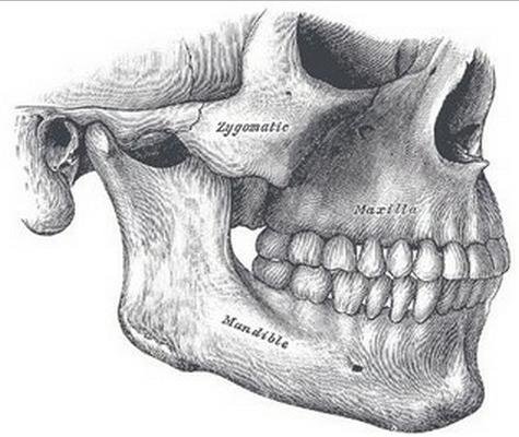
The Jaw Bones
Other types of malignant cells that arise in the jaw bone are Ewing’s sarcomas or giant cell tumors. Cancer that arises from the jaw bone is termed primary jaw cancer. However, malignant cells can also spread to the jaw from other cancers in the neck and head, termed as secondary jaw cancer. Jaw cancer results in pain in the area with difficulty in opening the mouth. Malignant cells in the jaw tend to spread quickly to adjacent organs.

A tumor that Has Grown on the Mandible
Signs & Symptoms
Signs and symptoms of jaw cancer are related to the growth of tumor on the area which restricts movement and circulation to the area. These include:
Acute pain while eating or drinking
This is the most common symptom of jaw cancer due to the compression of the tumor on the surrounding nerves. The tumor also creates disruption on the temporo-mandibular joint. The jaw pain may be characterized as slowly progressing, radiating to the face and neck and increasing in intensity while the jaw is moved. There is also tenderness around the area.
Paresthesia in the jaw
Compression of the nerves in the jaw results in tingling sensation and numbness in the area.
Swelling in the jaw
The tumor does not only compress nerves, but also obstruct the blood flow in the area causing swelling.
Mass in the jaw area
Oral examination can reveal an abnormal growth around the jaw. The patient may also feel an uneven alignment over the maxilla and mandible as a result of the mass or tumor. Tumors may grow inside leading to oral masses or outside the jaw leading to facial masses.
Teeth Looseness
Jaw tumors affect the sockets of the teeth that lead to loosening. Sometimes, dentists are the ones who initially diagnose jaw tumors because of affectations of the teeth during dental check-ups. Jaw cancer pain may also be mistaken as a toothache by a patient that’s why the first consult is made from dentists. Other symptoms include sore throat, sores around the jaw and difficulty of swallowing or chewing
Causes & Risk Factors
The development of jaw cancer is associated with mutations in the oncogenes. The risk factors forjaw cancer are similar to those of oral cancer. The following outlines the risk factors of malignancy in the jaw:
Smoking and tobacco use
Smoking and tobacco use have been one of the most common risk factor for oral and jaw cancers. The carcinogens contained in cigarettes are potent reasons for the development of malignant cells.
Alcohol drinking
Constant irritation of the oral mucosa from alcoholic drinks may lead to secondary jaw cancers.
Betel nut chewing
People fond of chewing betel nut are also at risk for secondary jaw cancers as a result of oral cancers.
Chronic irritation
Conditions that may cause constant irritation of the mandibular joint may also cause malignancies in the area.
HPV infection
Human papilloma virus specifically type 16 of the virus is a definite causative factor for jaw and oral cancers. The virus travels from the mouth into the tonsils, pharynx and the bones on the jaws which cause cellular changes on the area.
Staging
The staging of jaw cancer follows the general staging of malignancies.
Stage 0 or Carcinoma in Situ
The initial stage involves the growth of the cancer cells within cell membranes of the jaw tissues.
Stage I or Localized Growth
This stage involves the growth of malignant cells in the tissues in the joint. The cells may not be present on the subcutaneous and muscle tissues.
Stage II or Locally Advanced Growth
Stage II involves growth of the tumor in the jaw bone including the subcutaneous, muscle tissues, ligaments and tendons.
Stage III or Regional Spread
Regional spread involves the spread of malignant cells on adjacent tissues such as the oral cavity and surrounding lymph areas.
Stage IIIA
This sub stage involves the occurrence of gross tumor with microscopic seeding on adjacent tissues with no lymph spread.
Stage IIIB
This involves gross tumor spread on surrounding tissues with seeding of less than 2 cm in size.
Stage IIIC
This involves gross tumor spread on surrounding areas with seeding of more than 2 cm in size. The lymph nodes on the neck and upper chest may be affected.
Stage IV or Metastatic Cancer
The last stage of jaw cancer involves the spread of the malignant cells throughout the rest of the body due to lymphatic spread. Patients who are diagnosed in this stage usually have poor prognosis and lesser survival rate.
Diagnosis
Diagnosis of jaw cancer involves physical examination, radiographic studies and biopsy.
Physical Examination
Examination of the mouth and head may show masses on the oral cavity or on the face. The teeth are also examined to determine any loosening that is associated with lump growth on tooth sockets.
X-Ray
X-ray results often reveal lumps located on the maxillo-mandibualr joint. However, radiographic studies are not definitive of malignant cells because benign cysts on the area may also be identified.
Biopsy
Biopsy is the sole definitive test for jaw cancer. A sample of the tissues and bones in the jaw is taken and subjected to histologic examinations to assess the cellular patterns and check any aberrations. Osteosarcomas are usually identified.
Treatment
The most preferred management for osteosarcoma of the jaws is surgery although chemotherapy and radiation may also be performed as adjunct therapies.
Surgery
Surgery is performed for jaw cancers that have not yet spread to other areas of the body. Surgery removes the malignant tumors that grew in a particular site. This treatment is more effective for localized growth rather than in cells that have metastasized because there is already extensive cancer in the body. Removing the main tumor may not help in treating the diseases for advanced stages.
Chemotherapy
Chemotherapy is usually given as an adjunct to surgery to completely eradicate the cancer cells that may have spread to the adjacent organs. Patients undergoing chemotherapy are immunocompromised because of myelosuppression. In this regard, patients should be placed in reverse isolation to prevent infections from occurring.
Radiation
Radioactive materials may be placed within the patient to destroy malignant cells. Radioactive rays may also be directly aimed at the cancerous cells. Radiation therapy prevents the spread of cancer cells as well as reducing the size of the tumors.
Prognosis
The outcome of treatment for jaw osteosarcoma depends on the grade and stage of the diseases. Generally, osteosarcomas in the bone have better prognosis than other forms of malignancies especially when the cancer cells are still confined inside the jaw bone.
Survival Rate
Prompt treatment of jaw cancer in its early stages provides a successful result. The survival rate ofjaw cancer is 40% for men and 50% for women at the time diagnosis and treatment is made regardless of the stage. Osteosarcomas are very responsive to chemotherapy and radiation which contributes to a high survival rate.
Jaw Cancer Pictures

A Jaw Tumor Seen at the floor of the Mouth

A Locally Advanced growth of the Jaw Cancer

Jaw Cancer spreading to other Areas in the Mouth
Oral cancer accounts for roughly two percent of all cancers diagnosed annually in the United States. Approximately 36,500 people will be diagnosed with oral cancer each year and about 7,900 will die from the disease. On average, 61 percent of those with the disease will survive more than 5 years.
The Importance of Early Detection
Early Detection Saves Lives
With early detection and timely treatment, deaths from oral cancer could be dramatically reduced.
The 5-year survival rate for those with localized disease at diagnosis is 83 percent compared with only 32 percent for those whose cancer has spread to other parts of the body.
Early detection of oral cancer is often possible. Tissue changes in the mouth that might signal the beginnings of cancer often can be seen and felt easily.
Lesions that might signal oral cancer
Two lesions that could be precursors to cancer are leukoplakia (white lesions) and erythroplakia (red lesions). Although less common than leukoplakia, erythroplakia and lesions with erythroplakic components have a much greater potential for becoming cancerous. Any white or red lesion that does not resolve itself in 2 weeks should be reevaluated and considered for biopsy to obtain a definitive diagnosis.
Other Possible Signs and Symptoms:
Possible signs and symptoms of oral cancer that your patients may report include: a lump or thickening in the oral soft tissues, soreness or a feeling that something is caught in the throat, difficulty chewing or swallowing, ear pain, difficulty moving the jaw or tongue, hoarseness, numbness of the tongue or other areas of the mouth, or swelling of the jaw that causes dentures to fit poorly or become uncomfortable.
If these problems persist for more than 2 weeks, a thorough clinical examination and laboratory tests, as necessary, should be performed to obtain a definitive diagnosis. If a diagnosis cannot be obtained, referral to the appropriate specialist is indicated.
Tobacco/Alcohol Use
Most cases of oral cancer are linked to cigarette smoking, heavy alcohol use, or the use of both tobacco and alcohol together. Using tobacco plus alcohol poses a much greater risk than using either substance alone.
HPV
Infection with the sexually transmitted human papillomavirus (specifically the HPV 16 type) has been linked to a subset of oral cancers.
Age
Risk increases with age. Oral cancer most often occurs in people over the age of 40.
Sun Exposure
Cancer of the lip can be caused by sun exposure.
Diet
A diet low in fruits and vegetables may play a role in oral cancer development.
A thorough head and neck examination should be a routine part of each patient's dental visit and general medical examination. Clinicians should be particularly vigilant in checking those who use tobacco or excessive amounts of alcohol.
· Examine your patients using the head and neck exam illustrated in this program.
· Take a history of their alcohol and tobacco use.
· Inform your patients of the association between tobacco use, alcohol use, and oral cancer.
· Follow-up to make sure a definitive diagnosis is obtained on any possible signs or symptoms of oral cancer.
This exam is abstracted from the standardized oral examination method recommended by the World Health Organization. The method is consistent with those followed by the Centers for Disease Control and Prevention and the National Institutes of Health. It requires adequate lighting, a dental mouth mirror, two 2" x 2" gauze squares, and gloves; it should take no longer than 5 minutes.

Figure 1 - Face

Figure 2 - Lips

Figure 3 - Labial mucosa

Figure 4 - Labial mucosa

Figure 5 - Right buccal mucosa

Figure 6 - Left Buccal mucosa

Figure 7 - Gingiva

Figure 8 - Tongue dorsum

Figure 9 - Tongue left margin

Figure 10 - Tongue right margin

Figure 11 - Tongue ventral

Figure 12 - Floor

Figure 13 - Hard palate

Figure 14 - Oropharynx

Figure 15 - Palpation
Oral Lesions Suspicious for Oral Cancer

Homogenous leukoplakia in the floor of the mouth in a smoker. Biopsy showed hyperkeratosis.

Clinically, a leukoplakia on left buccal mucosa. However, the biopsy showed early squamous cell carcinoma. The lesion is suspicious because of the presence of nodules.

Nodular leukoplakia in right commissure. Biopsy showed severe epithelial dysplasia.

Erythroleukoplakia in left commissure and buccal mucosa. Biopsy showed mild epithelial dysplasia and presence of candida infection. A 2-3 week course of anti-fungal treatment may turn this type of lesion into a homogenous leukoplakia.
The examination is conducted with the patient seated. Any intraoral prostheses are removed before starting. The extraoral and perioral tissues are examined first, followed by the intraoral tissues.
I. The Extraoral Examination
· FACE: (Figure 1) The extraoral assessment includes inspection of the face, head, and neck. The face, ears, and neck are observed, noting any asymmetry or changes on the skin such as crusts, fissuring, growths, and/or color change. The regional lymph node areas are bilaterally palpated to detect any enlarged nodes. If enlargement is detected, the examiner should determine the mobility and consistency of the nodes. A recommended order of examination includes the preauricular, submandibular, anterior cervical, posterior auricular, and posterior cervical regions.
II. Perioral and Intraoral Soft Tissue Examination
The perioral and intraoral examination procedure follows a seven-step systematic assessment of the lips; labial mucosa and sulcus; commissures, buccal mucosa, and sulcus; gingiva and alveolar ridge; tongue; floor of the mouth; and hard and soft palate.
· LIPS: (Figure 2) Begin examination by observing the lips with the patient's mouth both closed and open. Note the color, texture and any surface abnormalities of the upper and lower vermilion borders.
· LABIAL MUCOSA: (Figures 3 and 4) With the patient's mouth partially open, visually examine the labial mucosa and sulcus of the maxillary vestibule and frenum and the mandibular vestibule. Observe the color, texture, and any swelling or other abnormalities of the vestibular mucosa and gingiva.
· BUCCAL MUCOSA: (Figures 5 and 6) Retract the buccal mucosa. Examine first the right then the left buccal mucosa extending from the labial commissure and back to the anterior tonsillar pillar. Note any change in pigmentation, color, texture, mobility, and other abnormalities of the mucosa, making sure that the commissures are examined carefully and are not covered by the retractors during the retraction of the cheek.
· GINGIVA: (Figure 7) First, examine the buccal and labial aspects of the gingiva and alveolar ridges (processes) by starting with the right maxillary posterior gingiva and alveolar ridge and then move around the arch to the left posterior area. Drop to the left mandibular posterior gingiva and alveolar ridge and move around the arch to the right posterior area.
Second, examine the palatal and lingual aspects as had been done on the facial side, from right to left on the palatal (maxilla) and left to right on the lingual (mandible).
·
TONGUE: (Figure 8) With the patient's tongue at rest, and mouth partially open,
inspect the dorsum of the tongue for any swelling, ulceration, coating, or
variation in size, color, or texture. Also note any change in the pattern of
the papillae covering the surface of the tongue and examine the tip of the
tongue. The patient should then protrude the tongue, and the examiner should
note any abnormality of mobility or positioning.
(Figure 9) With the aid of mouth mirrors, inspect the right and
left lateral margins of the tongue.
(Figure 10) Grasping the tip of the tongue with a piece of gauze
will assist full protrusion and will aid examination of the more posterior
aspects of the tongue's lateral borders
(Figure 11) Then examine the ventral surface. Palpate the tongue to
detect growths.
· FLOOR: (Figure 12) With the tongue still elevated, inspect the floor of the mouth for changes in color, texture, swellings, or other surface abnormalities.
·
PALATE: (Figures 13 and
14) With the mouth wide open and the patient's head tilted back,
gently depress the base of the tongue with a mouth mirror. First inspect the
hard and then the soft palate.
(Figure 14) Examine all soft palate and oropharyngeal tissues.
(Figure 15) Bimanually palpate the floor of the mouth for any
abnormalities. All mucosal or facial tissues that seem to be abnormal should be
palpated.
· Jaw bone cancer is an uncommon type of osteosarcoma that occurs when cancerous cells begin to grow within this body region. The risk of developing jaw cancer may be increased in patients who smoke cigarettes or chew tobacco. Anyone who develops jaw bone cancer symptoms should receive further evaluation and care from a medical professional as soon as possible.
· PAIN
· The primary symptom associated with jaw bone cancer is pain, according to the Merck Manual, an online medical encyclopedia for patients and caregivers. Patients with this form of cancer can experience sensations of mild to severe pain along the jaw line. Sensations of pain may arise gradually over time and typically persist without treatment. Pain within the jaw can affect a patient's ability to open or close the jaw normally. As a result, patients with this condition can experience difficulty eating or speaking. Patients who experience chronic or severe jaw bone pain should speak with a doctor immediately.
· SWELLING
· Patients with jaw bone cancer can experience swelling of the jaw as a symptom of this condition, report Drs. Baghai and Mothhary in a 2003 article published in the journal Acta Medica Iranica. Swelling within the jaw can cause the face to appear unusually puffy or enlarged. Patients who develop jaw swelling due to jaw bone cancer may also experience sensations of tenderness along the jaw line. This symptom of jaw bone cancer may persist for several weeks and should be discussed with a doctor if it occurs.
· NUMBNESS AND TINGLING
· Unusual sensations of numbness or tingling can occur as symptoms of jaw bone cancer in certain patients, according to the Merck Manual. These sensations can be uncomfortable and may affect a patient's ability to move the jaw normally. Certain patients may also experience numbness or tingling that extends from the jaw line into the cheeks or facial tissues. These jaw bone cancer symptoms can be signs of alternate medical problems and should be reported to a physician as soon as possible.
My toothache turned out to be jaw cancer
When Joanne Todd, 35, went to her dentist with a throbbing tooth, she never imagined that she needed anything more than a simple filling - let alone treatment that would involve rebuilding her face with her hipbone. Joanne, a full-time mum, who lives in Biggleswade, Beds, with husband Mike, a Police Sergeant, 34 and children David, 17, Billy, 13, Jack, 12 and Matthew, five, tells her remarkable story.
I took one last look in the mirror - my familiar face, with my straight, white teeth and long blonde hair, stared back.
But tomorrow I was to undergo a life-saving operation to stop the raging cancer, which was eating away at my jaw. I was sick with fear - if I survived it, what would I end up looking like?
My nightmare had begun innocuously enough. Eighteen months earlier I’d been brushing my teeth when I first felt the tiny painless lump on my gum. Smaller than a pea, it was lodged underneath my lower front tooth.
I never thought anything of it and when I saw my dentist for a check-up she reassured me everything was fine. But, a year on, my tooth had begun to throb and gone slightly crooked, so I went back to the dentist.
I thought I may need a filling or even a brace. But instead the dentist told me a routine X-ray had revealed a ‘shadow’ and she was referring me to hospital.
My twin sister, Tracey, had always said she had a feeling something terrible would happen to her in her thirties. But perhaps her intuition was really about me - immediately I began to worry it was something more sinister.
Mr Tony Giles, a Consultant Oral and Maxillo-facial surgeon at Pinehill Hospital, Hitchin, examined me. He said he thought it was a benign tumour caused by a playground accident I’d had as a child when a swing had crashed into me.
He explained he’d remove my three bottom teeth and clean out the bone. I felt incredibly relieved it didn’t seem serious and the op would be carried out under a local anaesthetic.
At the same time the lump would be routinely biopsied. The operation went well - I was always proud of my straight teeth and felt so grateful Mr Giles had managed to get rid of my lump and only removed two teeth.
But, my relief was short-lived. When my husband, Mike and I returned a few days later for my follow-up visit, Mr Giles looked grim-faced.
Preliminary results revealed it was cancer. But Mr Giles couldn’t say what type - or how aggressive - until further tests on biopsy were back.
I thought he must have made a mistake - I’d always been so healthy. I felt the blood draining from my face, a cold, sick feeling swallowing me up.
My first thought was I was going to die and leave my beautiful sons without their mum. Mike and I drove home in tears. I could barely speak to the boys without crying. We felt totally drained so Mike popped out for fish and chips.
But, I soon became aware he was taking ages. Then, as I looked out of the window to see him sat in his car on the mobile. Something was very wrong - my heart started pounding.
Mike’s face was white as he opened the front door. Mr Giles had just rung him - the results were in and they weren’t good. My cancer, a chondrosarcoma, was aggressive and I would need extensive surgery at a different hospital.
I heard myself scream, it seemed so surreal. We were all totally devastated. A few days later, at Luton and Dunstable hospital, I met my consultant, Mr Chi-Hwa Chan.
He was blunt - as the cancer was growing in my bone, a third of my jaw would need to be drilled away. He would then rebuild my face, replacing my jaw with bone from my hip.
Obviously there was the risk of dying on the operating theatre, dying through infection or my face could be paralysed.
'You're going to look as though your face has hit a bus,' he warned. I would also be in a wheelchair and, because of the bone removal from my hip, have to learn to walk again.
The operation sounded gruesome and I was terrified I’d be left looking a monster. But I had no choice - if I didn’t have it, the cancer could eat into the rest of my jaw and eventually kill me.
The five-hour operation was scheduled for three days' time. Meanwhile, I hugged each of my sons tightly in case I never saw them again and told Mike I loved him over and over.
I told Mike not to bring the boys in to see me in hospital - it was the hardest thing but I didn’t know how I’d feel myself afterwards. And I didn’t want them to see me in pain with half my face missing.
My first thought when I woke up in Recovery was thank God, I’m still alive. Gingerly I moved my mouth and felt for my face but could only feel stiff bandages.
I felt a surge of relief that I’d come through the worse, and didn’t feel as bad as I thought I would, which spurred me on to get better.
After five days in the high dependency unit, I went onto a ward. Determined to get better, I refused a wheelchair and took a few tentative steps.
The inside of my mouth was now hip bone with stretched gum stitched over it and felt strangely smooth and toothless - I’d only managed to keep six bottom teeth.
I hardly dared to look in the mirror but when I did I found Mr Chan had done an incredible job. My face looked like a misshapen Desperate Dan’s now, but I could already see beneath the swelling he’d shaped my chin brilliantly. And I could eat and speak normally.
The good news was the tumour turned out to be less than one centimetre in size, a CT scan had shown it hadn’t spread and Mr Chan was confident he’d removed all of it.
Within a week I went home, elated that the nightmare seemed finally over. But, when two weeks later, the hospital wrote, saying the consultant wanted to see me back urgently, my heart sank.
Further examination of the tumour revealed it was a much more aggressive osteosarcoma, a high-grade cancer that usually appears in large bones, such as in the leg.
It was so rare that doctors had never seen it before in the face - odds of one in 25 million were mentioned.
The roller-coaster scenario of the past few months meant I went into a panic attack at the sight of a hospital. So being told I had to go to UCLH in London to have chemotherapy, to ensure any cancer spread was totally obliterated, was absolutely dreadful.
Within three weeks of chemo, I’d lost my beautiful long blonde hair and suffered complications with double pneumonia. But, the boys were so fantastic - they even shaved their heads to support me.
Fortunately, by the end of August last year, when my chemo had finished, and my hair had begun to grow back, I began to feel normal.
I now face two-monthly checks to ensure the cancer doesn’t return and will need further surgery to implant teeth into my ‘hip’ jaw, but my oncology consultant says my future prognosis is excellent.
I have days when I look at my reflection and a stranger looks back. But Mike and the boys tell me I look wonderful.
In fact, my experience has brought home to me just how lucky I am to have such a fantastic family. And, after everything, I simply feel so lucky to be alive.
Mouth Cancer: the warning signs
.
Most dentists will perform a mouth check once a year at your routine visit. However, you should see your dentist or GP sooner if any of the following persist more than three weeks:
• persistent ulcer or sore on your mouth or tongue which may or may not be painful;
• a red or white patch in your mouth;
• unexplained pain in your mouth or ear;
• an unexplained lump in your neck;
• a sore or painful throat;
• a croaky voice or difficulty swallowing.
• Loose teeth or dentures not fitting properly;
• Change in your voice;
• Unusual bleeding or numbness.
Jaw Cancer Symptoms
Jaw pain that worsens while having meals is one of the most common symptoms of
jaw cancer. Poor oral hygiene and the habit of chewing tobacco is primarily
responsible for causing jaw cancer.
Cancer that strikes the tissues of the jaw is referred to as jaw cancer. In
this type of cancer, malignant cells form in the jawbone. The cancerous growth,
restricts movement of the jaw and sometimes opening the mouth can be a source
of great discomfort.
Symptoms
Jaw Pain
Jaw cancer can certainly put a person in an uncomfortable position, as it
interferes with normal eating habits. The gradual increase in cancer cells
triggers deterioration of jawbone, which results in pain. People suffering from
jaw cancer cannot chew the food properly, as it causes pain. Acute pain in the
jawbone, while drinking or eating is one of the most common signs of jaw
cancer.
Tumor
There is formation of lump on the tissues of the jaw. The lump might be painful
and usually forms below the teeth, on the gums. The lumps that have developed
on the jawline may also cause tooth pain.
Swollen Jaw
The development of malignant tumor in the jaw, not only causes extreme pain,
but leads to swelling of the jaw. The tissues of the jaw appear swollen and can
certainly bring great amount of pain.
Moving Teeth
The gums with the cancerous growth are no longer able to hold the teeth
tightly. Hence, patients tend to suffer from mobile teeth. So, teeth tend to
move around, on being touched.
Localized Facial Swelling
If the tumor is growing on the exterior of the jawbone, localized facial
swelling may be noticed. However, in case the cancer growth is occurring inside
the jawbone, it may disturb the normal alignment of the jaw.
Numbness or Tingling Sensation in the Jaw
The patient affected with jaw cancer experiences tingling sensation similar to
pricking of pins, along the jawline, when the jaw is not moving. This is
suggesting that the tumor is putting excessive pressure on the nerves that
supply sensation to the oral cavity.
Swollen Lymph Nodes
As lymph nodes are also found below the jawbone (in the neck area), development
of lumps under the jawline mean swollen lymph nodes, indicating that the cancer
has spread from its original site. When the cancer cells that multiply
indiscriminately reach the lymph nodes, one may notice enlargement of lymph
nodes. The swelling of lymph nodes usually occurs slowly when the underlying
cause is jaw cancer. On the other hand, sudden enlargement of lymph nodes has
often been associated with a throat infection. Nevertheless, it is best to
consult a doctor and undergo the necessary tests to 'nail' the cause of swollen
lymph nodes in the neck area.
Causes
Chronic smoking and tobacco chewing are the leading causes of jaw cancer. Over
4000 dangerous chemicals in cigarettes and around 28 known carcinogens in
smokeless tobacco can disturb the normal process of cell division and increase
the risk of jaw cancer. Alcoholism and untreated dental abscesses may also
contribute in the development of jaw cancer.
Types of Jaw Cancer
Cancers of the jaw are often diagnosed as osteosarcomas, malignancy that
originates in the bone. Jaw cancer accounts for 8% cases of osteosarcomas.
Usually, osteosarcomas affect the arms and legs, especially in the close
proximity of the knee joints. Ewing's Sarcoma is yet another type of primary
bone cancer that can occur in the jawbone. Microscopic examination of tissues
affected with Ewing's Sarcoma shows formation of small, round, blue cells.
Hence, the cancer is also referred to as blue cell tumor.
Avoid Wrong Judgment
Patients tend to judge jaw cancer pain as a toothache and may use painkillers
to resolve the issue. However, this does not work and consultation with the
doctor at the earliest, is an absolute must for correct diagnosis.
Treatment
Surgery
Removing the cancerous growth, through surgical procedures can certainly
eliminate the tumor once and for all. However, this form of treatment is
applicable, only when the tumor is localized and the cancerous cells have not
spread beyond a certain limit. The doctor will remove the affected tissues and
the lymph nodes near the jawbone, to halt the progress of cancer. The procedure
may also involve drilling the affected part of the jawbone in order to get rid
of cancerous growth. Surgical procedure is used when the cancer is in its early
stages of development.
Radiation Therapy
Bombarding the affected area of the body with high energy rays to destroy the
cancerous cells is nothing but radiation therapy. The radiation penetrating
through the skin, helps to manage the spread of malignant tumors.
Chemotherapy
Chemotherapy drugs are administered intravenously to kill cancer cells that
have invaded tissues of other organs of the body. This treatment method is
helpful to relieve the pain associated with cancer.
Upon undergoing surgery and multiple sessions of chemo, one will have to keep
meeting the doctor on a regular basis to check for relapse. There is a
possibility that the cancer may go into remission and come back at the same
site. So. during every visit, the doctor performs blood tests, biopsy and
conducts imaging studies to monitor health and look for cancer recurrence.
When cancer develops
in a person’s jaw, it is referred to as jaw cancer. Jaw cancer may develop in a
person’s mandible, which consists of the bones beneath his mouth. Cancer may
also develop in a person’s maxilla, which are the
bones above his mouth. The cancer that develops in these areas may be limited
to the jaw, or it may spread and affect other parts of a person's body.
Unfortunately, jaw cancer is sometimes mistaken for a benign growth, such as a
cyst, and treatment is delayed.
Jaw cancer may develop in either the upper or lower part of the jaw. Once it forms, it has potential to spread to other parts of the jaw or other parts of the body. When cancer of the jaw spreads to other parts of the body, it is said to be metastatic cancer. While all types of cancer are serious, those that spread are more worrisome and often more difficult to treat.
Symptoms of jaw cancer include pain or numbness in the jaw, and a lump or other abnormal growth. Often, jaw cancer is mistaken for another type of condition because its symptoms are so similar to benign issues. For example, a person with jaw pain may think he has a deep cavity that is causing the pain, an infection, or an abscess. Lumps and jaw growths are often mistaken for cysts rather than cancer. This is due to the fact that cysts are commonly found in the jaw.
A medical professional may suspect jaw cancer based on its symptoms, but he cannot diagnose it based on symptoms alone. Instead, a medical professional typically performs a biopsy to diagnose this type of cancer. To perform a biopsy, a medical professional usually removes a sample of the suspicious cells or the entire growth. An individual is usually injected with a numbing agent or sedated during this process, so he is unlikely to feel pain during the biopsy. Once the cells or growths have been extracted, a doctor examines the cells or a sample of the growth to check for the presence of cancer.
The treatment for jaw cancer typically depends on the stage of cancer the patient has, whether or not it has spread, and his overall health. A medical professional may perform surgery to remove the cancerous growth in some cases. A patient may undergo chemotherapy or radiation treatment as well.

Rehabilitation is an essential phase of cancer care and should be considered from the time of diagnosis in a complete and comprehensive treatment plan. Surgical resections often create large defects accompanied by dysfunction and disfigurement, and radiation therapy produces significant morbidity and unique tissue-management problems. Speech, swallowing, control of saliva, and mastication can all be adversely affected. If these cosmetic and functional impairments are not corrected or minimized, the patient may be unable to resume a normal working and social life.
The primary objective of rehabilitation is the restoration of appearance and function. How successfully this is accomplished, depends upon both the judgment and skill of the therapist, and the post-treatment anatomic, physiologic, and psychological makeup of the patient.
Rehabilitation of the head and neck cancer patient requires a team approach, which includes many specialists. In addition to the surgeon, the dental oncologist and the prosthetic doctor, there are many other individuals involved as part of the restorative team. The clinical social worker sees the patient at the time of diagnosis, during primary cancer treatment, and during the long period of rehabilitation, to the help patient understand the process of treatment and to mobilize resources to cope with the situation. Nurses can provide support and comfort for the patient, give instruction on the care of the surgical defect or tracheostomy, and arrange for visiting nurse support when the patient returns home. As there may be conditions which alter eating habits, or skills, dietitians advise patients on obtaining proper nutrition. Occupational and physical therapists help retrain altered muscular systems, and the speech pathologist can help patients to adapt speech mechanisms to prosthetic appliances, and altered physiology. Dental hygienists provide prophylaxis and oral-health-care instruction.
Maxillary Defects
Most tumors of the paranasal sinus, palatal epithelium, or minor salivary glands require surgical removal of a portion of the upper jaw, This is either a partial or total maxillectomy. A radical maxillectomy entails surgical resection of the entire maxilla to the midline. This is accomplished by an insicion through the cheek and the upper lip, allowing the tissue to be folded (reflected) back for access to the maxilla. A partial maxillectomy is less encompassing, and may be performed without the reflection of these external tissues (transorally). The amount of soft palate included in these resections varies, and depends on the need to obtain tumor-free margins (edges of the incision).
Disabilities
Defects created by surgery to remove cancerous tissues of the hard or soft palate may produce a variety of problems. For instance, hypernasality could make speech unintelligible. Chewing (mastication) may be difficult, particularly for the patient without natural teeth (edentulous patient). Because denture-bearing tissue surfaces are lost, it is difficult for the denture to seal completely around the edges, a key factor in holding it securely in place. Swallowing likely will become awkward, since food and liquid may be forced up into the nasal cavity and out the nose. The nasal mucous membranes become dried out (desiccated) by abnormal exposure to the oral environment, causing chronic irritation. Nasal and sinus secretions may collect in the defect area. Facial disfigurement can result from lack of midface bony support for the facial soft tissues, or the necessary surgical damage to a branch of the facial nerve. In some cases, extensive tumor invasion requires surgical removal of the eye.
Rehabilitation after surgical resection of the hard or soft palate is best accomplished prosthodontically. Customarily, a temporary prosthesis, known as an immediate surgical obturator, is placed at the time of surgery. This will provide closure of the surgical defect. During the healing period, this prosthesis is recontoured (relined) periodically to compensate for tissue changes which occur as a normal part of the healing process. When these tissues become well healed and dimensionally stable (usually 3 to 4 months after surgery), the final prosthesis is made.
The purpose of an obturator prostheses is to restore the physical separation between the oral and nasal cavities, thereby restoring speech and swallowing to normal, and to provide support to the lip and cheek. Inadequate retention of the obturator compromises stability. The remaining teeth, therefore, become extremely valuable in providing support, retention, and stability for these restorations.
Treatment
It is essential that the prosthodontist examine and consult with the patient before surgery. The sequence of treatment should be explained to the patient, and diagnostic casts and appropriate radiographs should be obtained. With this information, the prosthodontist is ready to consult with the surgeon about the design and fabrication of the surgical obturator. Modifications in the surgical plan that may improve the prosthetic prognosis without adversely affecting tumor removal should be discussed at this time. Surgical considerations to improve the prosthetic outcome
The immediate surgical obturator
Immediate or early coverage of a palatal defect with an obturator is important. The obturator provides a matrix upon which the surgical packing can be placed, minimizes contamination of the wound in the immediate postoperative period, and enables the patient to speak and swallow effectively immediately after surgery.
In order to fabricate the prostheses, a stone model is made of the maxillary arch and the anterior portion of the soft palate. The obturator’s size is determined by the surgical boundaries of the resection, as indicated by the surgeon. In the patient who still has natural teeth, those in the path of the surgical resection are removed from the model. The obturator can now be made before the surgery, using self-curing plastics, and be ready for immediate insertion afterwards. If necessary, this first obturator can be altered at surgery by trimming it’s size, or by adding a temporary denture reline material. After surgery the prosthesis is wired to remaining teeth, alveolar ridge, or other available structures.
After the surgical packing is removed the prosthesis is recontoured with a temporary denture reline material. As healing progresses, the obturator is periodically relined and extended further into the defect, adaptation is improved, and anterior teeth are added as necessary to improve speech and esthetics. Three to five months after surgery, when healing and the related tissue changes are essentially complete, the final prosthesis is begun. Patients without natural teeth (edentulous) to use as fixation points for the obturator, often require a longer period of healing because the defect must be engaged more aggressively to maximize stability, support, and retention.
The definitive obturator prosthesis
Defects of the hard palate can be restored effectively with a denture prosthesis. Natural teeth greatly improve denture retention and stability of a maxillary prosthesis. When the denture is properly extended, speech, swallowing, mastication, and facial contour restoration can be accomplished. In it’s final contour, the obturator should extend maximally along the lateral wall of the defect. This high lateral extension not only increases retention and lateral stability, but also provides support for the lip and cheek. In some patients, extensions across the nasal aperture may be necessary to provide acceptable retention.
The placement of osseointegrated implants (More on dental implants) into the remaining bony structures, dramatically improves the function of an obturator prosthesis, particularly for edentulous patients. Implants can be placed, either during the surgical procedure to remove the malignant tissues, or at some appropriate time thereafter. These implants will allow mechanical attachment of the denture prosthesis to them, adding significant stability. For instance, implants may be splinted together with a bar in the mouth that mates to female snaps in the base of the denture, allowing the denture to be “clipped” in and out of the mouth. In patients with some remaining natural teeth, implants may still be used to augment the retention provided by these teeth. When the prosthesis is inserted and attached to the fixed implants, speech and swallowing are restored to normal limits. The tissue side of most prostheses require relining/recontouring within the first year because of slow, but continuous changes of the tissues on the periphery of the surgical defect.
The definitive soft palate prosthesis
Defects of the soft palate and velopharyngeal complex require different and more complex prosthetic treatment. Closure normally occurs when the soft palate comes into contact with the contracting lateral and posterior walls of the nasopharynx. When a portion of the soft palate or lateral pharyngeal wall is surgically removed, or when the soft palate is perforated, scarred, or neurologically impaired, complete closure cannot occur. Speech becomes hypernasal, and nominal swallowing is not possible. With a pharyngeal obturator, the patient may be able to re-establish this closing. A properly designed obturator will not interfere with breathing, impinge upon soft tissues during postural movements, or hamper the tongue during swallowing and speech.
The soft palate obturator remains in a fixed position in the nasopharynx and does not attempt to duplicate normal movements of the soft palate. The tissue side of the obturator remains level with the hard palate contour. During breathing and the production of nasal speech sounds, the space around the obturator reflects the potential for muscular contraction. During swallowing and the production of other speech sounds, this muscular network moves into contact with the stationary plastic obturator, establishing closure. A correctly constructed obturator will usually result in the return of normal speech and swallowing.
Tongue-Mandible Defects
Disabilities
Disabilities resulting from tongue and mandibular resections include impaired speech articulation, difficulty swallowing, deviation of the mandible during functional movements, poor control of salivary secretions, and often cosmetic disfigurement. Consequently, these patients seldom return to presurgical levels of social function.
Advanced tumors of the anterior two thirds of the tongue and floor of the mouth often require extensive resection of bone and soft tissue. The loss of large portions of the tongue prevents appropriate “valving” and/or interaction with the other oral structures. This loss of mobility combined with the loss of motor and sensory innervation leads to misarticulation of many speech sounds. Deglutition is less impaired, and most patients learn to swallow fairly efficiently. Tongue function is less affected if the resected portion is restored with either myocutaneous or free flaps. The myocutaneous flap restores bulk, prevents deviation of the mandible, and permits the reconstructed tongue to articulate more effectively with the palatal structures. Myocutaneous flaps, however, can become scarred and immobile. In contrast, tongues reconstructed with free flaps have the potential of supporting near normal speech and are less likely to become heavily scarred and immobile.
If much of the mandible is segmented and removed, the remaining functional mandibular segment will be retruded and deviated towards the surgical side at the vertical dimension of rest. When the jaw is opened this deviation increases. These factors, combined with impaired tongue function, may prevent effective mastication. The severity and permanence of mandibular deviation varies; some patients attend an acceptable intocclusal relationship without adjunctive therapy.
Resections of the tongue and mandible often obliterate portions of the lingual and buccal sulci so that a means of collecting and channeling secretions posteriorly no longer exists. In addition, the motor and sensory innervation of the lower lip on the resected side is often lost, impairing speech, eating, and control of saliva.
When removal of tumors in the anterior floor of the mouth requires that the mandible be resected anteriorly, and when mandibular continuity is not restored, the two remaining posterior fragments are pulled medially by the residual mylohoid muscles and superiorly by the muscles of mastication. Severe disfigurement and dysfunction result. Free flaps from the fibula can be used immediately to restore the lost hard and soft tissues, and most patients emerge with excellent function and acceptable appearance.
The nature and degree of the disabilities secondary to impaired tongue function after resection of lesions in the anterior floor of the mouth are proportional to the amount of tissue resected and to the nature of surgical closure. In most cases, motor control is unimpaired and the functional deficits depend on the degree of tongue mobility.
Surgical Closure And Initial Reconstructive Steps
If the surgical wound is closed primarily (primary closure) by suturing the edges of the wound together, the functional disabilities are compounded. The introduction of the myocutaneous flap in the late 1970s ameliorated some of the disabilities, and improved tongue mobility resulted in acceptable speech, swallowing and saliva control. The advantage of free-tissue transfers (free flaps) over musculocutaneous flaps is the improved blood supply, enhancing wound healing and flap survival. The fibula is the preferred donor site unless the soft-tissue deficit is unusually large. The osteotomized fibula provides sufficient length and bulk and bone. Osseointegrated implants can be placed to retain and support prosthesis.
Secondary Surgical Procedures
Vestibuloplasty, Tongue Release, and Skin Grafts
Vestibuloplasty and tongue release are of particular value when mandibular continuity has been maintained or restored. The creation of vestibules enables the patient to pool saliva more efficiently and allows for extension of denture flanges resulting in improved stability and retention . Creation of an attached keratinized mucosa on the ridge surface with either a skin graft or a palatal graft provides additional stability for a partial or complete denture. The patient’s appearance may also be improved because a prosthodontic restoration can now be molded to provide contour and support for the lower lip and cheek portions of the resected area. Improvement of speech is less noticeable in patients who lack tongue mobility.
Restoration of Mandibular Continuity
Surgically repairing mandibular discontinuity defects results in more normal mandibular movement patterns that enable an edentulous patient to exert force on both sides of the dental arch. This increases the stability of complete dentures. In dentulous patients a properly reconstructed mandible restores dental occlusion to normal and will partially alleviate deviation of the mandible.
Free bone grafts. Reconstruction after extensive resection of oral malignancies with free grafts is difficult because of the lack of sufficient soft tissue to receive the graft, the decreased vascularity of the tissue bed secondary to radiation therapy and/or radical neck dissection, and the difficulty in achieving proper fixation of the graft during the healing period. The primary goals are to restore facial form, mandibular continuity and, in selected patients, appropriate volume and quality of bone should be provided for the placement of osseointegrated implants.
Autogenous graft sources for resected bone include iliac crest, rib, and clavicle. Most commonly the defects are restored either with particulate autogenous marrow housed in metal tray or with a block bone, both usually obtained from the iliac crest. The use of myocutaneous flaps for closure of the initial wound facilitates free-bone grafting of continuity defects by enhancing the volume and vascularity of the recipient soft tissue.
Free vascularized flaps. A major advance in mandibular reconstruction has been the development of improved techniques in microvascular surgery, which allow for composite grafting of large volumes of tissue. In microvascular surgery, which allow for composite grafting of large volumes of tissue. In microvascular free-tissue transfer (free flaps), bone, muscle, connective tissue, and skin can be autogenously grafted and remain viable. The grafting can be accomplished simultaneously with resection of the tumor, with excellent results.
Numerous donor sites have been used. The radial forearm is favored for reconstruction of most extensive soft-tissue defects such as the tonsillar, partial-glossectomy, and floor-of-mouth defects. The composite fibula flap is the preffered donor site for most complex oro-facial-mandibular defects. Multiple osteotomies may be performed without devascularizing the bone segments to replicate the contour of the replaced mandible. Fibular thickness makes it an excellent recipient of osseointegrated implants.
Physiology Of Oral Function Following Tongue And Mandible Resection And Reconstruction
Even following successful reconstruction, functional deficits (deglutition, speech articulation, mastication efficiency) remain because of compromised motor and sensory control, inadequate tissue contour, and inadequate bulk of key tissues. Mastication efficiency is primarily dependent upon the quality of tongue function and the presence of a reasonable occlusal table adjacent to the sensate, functioning portion of the residual or reconstructed tongue. Articulation of speech is dependent upon tongue mobility and the presence of adequate tongue bulk. Patients with hemiglossectomy defects can learn to articulate and swallow effectively, if they can elevate their reconstructed tongue to interact with the palatal vault and maxillary dentition. More on dysphagia, speech, and eating issues.
Palatal Speech and Swallowing Aids
If the reconstructed tongue does not possess the ability to elevate sufficiently to interact with the palatal structures, an acrylic resin template is made to engage the palatal contours and/or maxillary dentition. Their surfaces can be modified with modeling plastic or tissue-conditioning material and eventually processed into acrylic resin.
Surgical Reconstruction of the Total Glossectomy Defects
With the development of free flaps, there has been an increased success in surgically reconstructing the tongue in total glossectomy patients. When these flaps are used, bulk should be limited, allowing room for total restoration of the mandibular dentition with a prosthesis. Palatal speech and swallowing aids may be quite useful in selected patients.
Mandibular Guidance Therapy
When mandibular continuity is not restored, correction of mandibular deviation includes inter-maxillary fixation, and mandibularly based or palatally based guidance restorations. The method of choice should be combined with well-organized mandibular exercise program.
Mandibular guidance therapy begins when the immediate postsurgical sequelae have subsided, usually in 2 to 3 weeks. Upon maximum opening, the mandible is displaced by hand as forcefully as possible towards the nonsurgical side. These movements tend to lessen scar contracture, reduce trismus, and improve maxillomandibular relationships. The earlier mandibular guidance therapy is initiated, the more successful the result. Unfortunately, those patients suffering the most severe mandibular deviations (because of extensive soft-tissue loss, tight wound closure, radiation therapy, and/or radical neck dissection) are most susceptible to postsurgical complications that delay mandibular guidance therapy.
If the mandible can be manipulated into an acceptable maxillomandibular relationship, but the patient lacks the neuromuscular control to bring the mandible into occlusion, a cast mandibular resection prosthesis is appropriate. This prosthesis consists of a removal partial denture framework with a metal flange extending 7 to 10 mm laterally and superiorly on the buccal aspect of the bicuspids and molars on the unresected side. This flange engages the maxillary teeth during mandibular closure, thereby directing the mandible into an appropriate intercuspal position. The partial denture framework must be stable and retentive to counteract the lateral forces generated on the flange during closure. The guidance flange is constructed of cast chrome-cobalt metal. Modifications can be made with acrylic resin.
A second design confines the guidance ramp and index to a maxillary prosthesis. This form of guidance prosthesis is indicated when severe deviation prevents manipulation of the mandible into acceptable occlusal contact. Maxillary guidance ramps are more adjustable than mandibular guidance ramps. They are usually constructed of acrylic resin with either cast or wrought-wire retainers, since they usually serve only temporarily until an acceptable occlusion can be established.
The success of mandibular guidance therapy depends on the nature of the surgical defect, early initiation, and cooperation of the patient. Patients with extensive posterior base of tongue lesions, radical neck dissection, and radiation therapy are often unable to achieve functional intercuspal relationships. Mandibular guidance therapy is most successful when the resection involves minimal soft-tissue removal, or when defects are replaced with myocutaneous or fee flaps. Following mandibular guidance therapy, occlusal equilibration or selective crown placement may be required to achieve optimal interocclusal relationships.
Definitive Prosthetic Restoration
The outcome of removable prostheses for patients with resections of the tongue and mandible depends upon the function of the residual tongue. In some patients with poor tongue function, only appearance and oral competence can be improved, while in others with good tongue function, mastication is a reasonable objective.
Removable Partial Dentures
The usual principles of partial denture design and fabrication should be followed. Major connectors should be rigid; occlusal rests must be direct occlusal forces along the long axis of teeth; guiding planes should be employed to provide stability and bracing; retention must be within the limits of physiologic tolerance of the periodontal ligament of the abutment teeth; and maximum support should be gained from adjacent soft tissues. Retainers, minor connectors, and proximal plates should be designed so that they do not subject the remaining teeth to excessive lateral forces during function.
In patients with lateral discontinuity defects, it may not be possible to design a framework with retainers that disengage during function because of the altered patterns of force upon the prosthesis. Viewed from the frontal plane, the arc of closure of the mandible is angular rather than vertical, with forces of occlusion as the force of contracture on the unresected side is increased. The location of the fulcrum line of a partial denture is not easily determined because of the difficulty in predicting movement patterns of the prosthesis during function. The forces of occlusion are unilateral, so the axis of rotation (fulcrum line) of the partial denture deviates from the norm. By placing the occlusal rest on the mesial of the bicuspid that disengages during expression of an occlusal load in the distal extension area. However, a retainer can not be positioned in a retentive area on the opposite cuspid that disengages during occlusal function. Consequently, this retainer must either be placed on the height of contour, or it must be flexible so that an occlusal load will not subject the cupid to undue stress. All partial denture frameworks should be phsysiologically relieved.
After the partial denture casting has been fabricated, verified, and adjusted, an altered cast impression is obtained of the edentulous areas. Particular attention should be paid to the lingual extension on the unresected side, especially the polished surfaces, which provides additional retention and stability. Maximum extension of the denture base is always attempted. Coverage of the buccal shelf on the unresected side is essential to maximize support. Centric relation records are made and occlusal schemes developed that are consistent with the unilateral mandibular movement patterns of mandibulectomy patients.
Complete Dentures
Dentures for edentulous patients with discontinuity defects of the mandible may provide esthetic improvement by replacing teeth and improving lip and cheek contours, but unless the patient has extraordinary tongue control, mastication is generally not possible. A number of factors affect the patient’s ability to function with resection dentures (1) stability, support, and retention of the mandibular denture are compromised; (2) quality and quantity of saliva; (3) the angular pathway of mandibular closure, which induces lateral forces upon dentures, tending to dislodge them; (4) abnormal maxillomandibular relationships that may prevent ideal placement of the denture teeth over their supporting structures; and (5) impairment of motor and/or sensory control of the tongue, lip, and cheek, limiting the ability to control dentures during function. Implant-assisted resection dentures can overcome many of these difficulties, particularly those associated with compromised retention, stability, or support.
The status of the remaining tongue is probably the most important prognostic factor. If motor and/or sensory control are not significantly impaired, and the tongue can be moved in several directions, resection denture stabilization and control of the food bolus during function become possible. Paradoxically, immobility of the tongue often permits a more aggressive extension of the lingual flange on the nonsurgical side, aiding stability and retention. The position of the tongue is also an important prosthodontic prognosticator relative to obtaining a peripheral seal.
Mandibular resections extending to the midline have a poor prosthetic prognosis. If the resection is limited to the ramus-molar or cuspid regions anteriorly, the prosthetic prognosis is more favorable. In some of these patients remmants of the masseter or medial pterygoid muscle may remain attatched to the mandible, enabling the development of bilaterally balanced occlusion. These patients demonstrate more-normal envelopes of motion, and near-normal ridge relationships, allowing for more favorable distribution of forces during mastication and swallowing.
Following impressions, centric and lateral registrations and occlusal schemes should take into consideration the altered pattern of mandibular movements. Denture extensions can be verified with a disclosing wax, and excessive tissue displacement eliminated with the aid of pressure indicating paste.
Implant-Assisted Overlay Dentures
Patients with reasonable tongue bulk and mobility and with motor and sensory innervation intact on at least one side gain the most from implant-retained overlay prostheses. The tongue, then, is no longer required to control the denture, so it functions mainly to manipulate the food bolus during mastication and swallowing. If the patient’s speech is intelligible, the prognosis for effective bolus manipulation with an implant-retained prosthesis is good. More on the placement of dental implants in radiated patients.
In mandibular resection
patients, if implants are to be placed into the mandible to retain and support
an overlay prosthesis, consideration should be given to placing implants in the
opposing maxilla. The unilateral occlusal forces and increased lateral forces
generated during the chewing cycle tend to dislodge the upper denture. In
addition, xerostomia may compromise the peripheral seal. Therefore, implants
should be considered if the retention and stability of a conventional maxillary
denture is marginal.
In most patients, the only implant sites available in the edentulous resected
mandible are located in the symphyseal region. A minimum of two implants should
be placed. However, more are desirable, if space allows, mainly for stability.
Implant success rates in bone grafts used to restore mandibular continuity defects have been encouraging. Free bone grafts demonstrate a homogenous calcification pattern that results in an excellent bone-implant interface, particularly with fibula free flaps. To date, of the 54 implants that have been placed at the University of California, Los Angeles, (UCLA) in fibula free flaps, 46 are in function; 2 have been buried because of improper positioning or angulation; 3 have failed; and 3 have not yet been placed into function. More on the surgical placement of dental implants, and their prosthetic restoration.
Facial Defects
Surgical reconstruction and prosthodontic restoration both have limitations. The surgeon is limited by the availability of tissue, damage to the local vascular bed, and the need for periodic visual inspection of an oncologic defect. The prosthodontist is limited by the materials available for facial restoration, movable tissue beds, difficulties in retaining large prostheses, and a patient’s willingness to accept the result. The method of facial restoration should be considered before surgery and patients should participate in the decision making process with realistic expectations.
Surgical Reconstruction Versus Prosthetic Restoration
Surgical reconstruction of small facial defects is preferable. However, it is difficult for the surgeon to contour a facial component that is as effective in appearance as a well-made prosthesis. Osseointegrated implants in facial defects have changed patient perceptions about facial prostheses because of the retention achieved. Additionally, when a large resection is necessary and recurrence of tumor is realistic, it is advantageous to be able to monitor the surgical site closely, which a prosthesis permits. Even when surgical reconstruction is deemed possible, most surgeons prefer to wait at least one year before surgical construction. Also, surgical restoration of large defects is technically difficult and requires multiple procedures and hospitalizations, which may be further complicated if radiotherapy has been included.
Prosthetic Facial Restoration
The challenge to the prosthodontist is to fabricate an esthetically pleasing restoration. A conspicuous prosthesis may produce more anxiety and permit less social readjustment than a simple face bandage or eye patch. The most critical periods is the first 2 to 3 days after delivery. The patient must understand that a prosthesis has two different roles: for family, close friends, or business associates, it can only cosmetically replace the tissues excised; for the public at large, it generally provides enough concealment to render the reconstructed defect inconspicuous.
Materials Used for Facial Prostheses
Current materials all possess some undesirable characteristics. The materials most often used are the new silicone elastomers, which have achieved wide clinical acceptance. Adhesives are used to retain facial restorations. A major development has been the use of osseointegrated implants, resulting in extremely well-retained prostheses that permit vigorous physical activities.
Design Considerations
A presurgical moulage can be very helpful, especially if a total rhinectomy or total auriculectomy is anticipated. Impressions of the defect are usually obtained with elastic impression materials, taking care not to displace the tissues being recorded. The contours of the prostheses are sculptured in wax, both on the cast and on the patient. Surface characteristics, appropriate contour, coloration, and margin placement are equally important factors. Processing the materials is complicated and requires special instrumentation. Special flasks are necessary for processing large prostheses.
Coloration
Basic skin tones should be developed into a shade guide for each material. The base shade selected should be slightly lighter than the lightest skin tones of the patient, because as color is added the prosthesis will darken. Intrinsic coloration is long-lasting and is therefore preferred over trinsic application.
Combined Oral and Facial Defects
When the integrity of the oral cavity has been destroyed, food and air escape during swallowing; speech is often unintelligible; and saliva control is difficult. Additionally, it is extremely hard to adapt the margins of the prosthesis to maintain tissue contact during facial and mandibular movements. Retention of maxillary teeth or a portion of the hard palate improves the prosthetic prognosis. Placement of implants, as well as skin grafts to decrease tissue contraction and to create undercut areas for retention, also improve the prosthetic prognosis.
Patients will experience less depression if as many functions as possible are restored immediately after surgery with a temporary prosthesis. These functions include swallowing food (thereby eliminating the need for a nasogastric tube or gastrostomy), controlling saliva, and speaking. The oral portion of the temporary prosthesis is constructed of acrylic resin and formed so that contact is established with healthy tissues adjacent to the defect. Functional contact with the remaining portions of the lips allows the patient to effectively seal the oral cavity. Tissue-bearing surfaces of the prosthesis are relined with temporary denture reliner at frequent intervals to accommodate tissue changes.
In constructing a definitive midface prosthesis, a custom tray is used for the master impression. Movable portions of the defect are recorded with a thermoplastic material. Record bases are fabricated, and centric relation records enable casts to be mounted on an articulator. Osseointegrated implants, eyeglass frames, adhesives, straps, teeth, and engagement of defect undercuts supply retention. The finished restoration usually provides the patient with acceptable appearance and function.
Craniofacial Implants
The use of osseointegrated implants has significantly improved patient acceptance. The retention provided by implants makes it possible to fabricate large prostheses that rest on movable tissues. This enables the prosthodontist to fabricate thinner margins, which enhance esthetics by blending and moving more effectively with mobile tissues. Other benefits include elimination of the occasional skin reactions to skin adhesives, ease and enhanced accuracy of prosthesis placement improved patient comfort, and decreased daily maintenance, which also increases the life span of the facial prosthesis.
Treatment Planning
The implants must be positioned within the confines of the proposed facial prosthesis. In most patients it is desirable to sculpt a wax replica of the future prosthesis and to use this replica to fabricate a surgical template. This template is sterilized and used as a guide during surgery to ensure proper implant position and angulation.
The number and arrangement of implants and possible bone sites are determined. In most acquired defects, radiographic studies are not necessary. In large extensive defects, CT scans and three-dimensional models may be useful in evaluating potential bone sites and important adjacent structures. The health of soft tissues circumscribing osseointegrated implants is easier to maintain if these tissues are thin (less than 5mm in thickness) and attached to underlying periosteum. If the skin contains hair follicles or scar tissue from past reconstructive procedures, these tissues should be removed and placed with skin grafts.
Surgical placement
Craniofacial implant fixtures are fabricated from pure titanium. They are available either 3 or 4-mm lengths, with a 5-mm diameter flange. The short lengths are designed to permit placement in areas with limited available bone. The flange facilitates initial stabilization of the implant and prevents penetration into interior components.
For ear defects, two or three implants are positioned posteriorly and superiorly to the ear canal. For nasal defects, the preferred fixture location is the anterior portion of the floor of the nose. Care should be taken to avoid the roots of the teeth in this area. For orbital defects, the preferred location is the lateral portion of the superior orbital rim.
Several attachment systems have been used to connect the facial prosthesis to the implants, such as bar-clips, magnets, and O-ring types. For auricular and nasal prostheses we prefer the bar-clip systems because they provide superior retention. We prefer magnetic retention for orbital prosthesis, because the ease of insertion of magnetically retained prostheses outweighs the possibilities of magnet corrosion and decreased retention over time.
The retentive system is designed to fit within the contours of the facial prosthesis as an acrylic resin substructure. It should posses sufficient surface area so that the bond between the substructure and the silicone prosthesis will not fail during insertion or removal of the prosthesis.
Success rates for auricular sites have exceeded 95% in most studies and few complications have been encountered. Success rates of the floor-of-nose sites are between 85 and 90% and are about the same as oral implants placed in the premaxillary segment. Of 130 fixtures inserted at UCLA, only 2 of 83 floor-of-nose and mastoid implants were lost; however, there were 19 of 47 frontal bone failures. Success rates are diminished if implant sites have been irradiated previously.
Prosthetic treatment of the irradiated patient
The risk of developing an osteoradionecrosis is minimal in patients who have been edentulous for some time before undergoing head and neck radiation therapy. The risk of bone necrosis increases in those patients who have teeth removed just before or after radiotherapy. More on osteoradionecrosis (ORN).
Timing of denture placement
Empirical recommended insertion times have ranged up to three years. In our study of 92 patients who were edentulous before the onset of cancer, prostheses were inserted, on an average, 15 months after radiation was completed; 45 patients received dentures within 6 months. None of the 92 developed a denture-related osteoradionecrosis. This data and our clinical experience indicate that dentures can be fabricated soon after completion of radiation therapy in most patients. If the radiation fields cover little of the mandibular denture-bearing surface, dentures can be inserted as soon as the mucositis resolves. An irregular mandibular ridge, especially in a patient whose mandible was wholly within the radiation field, may prompt the clinician to defer dentures indefinitely.
In summary, for edentulous patients who were experienced denture wearers before radiotherapy, there is little risk of developing significant complications from wearing dentures. For those who had teeth extracted within the radiation field before radiotherapy and were not experienced denture wearers, the risk of bone necrosis is higher. Therefore, placement of dentures depends upon patient needs, the status of the mucosal and bony wearing surfaces, and the amount and fields of radiation. Some patients in this group may never be candidates for complete dentures.
Prosthodontic procedures Complete Dentures
Boggy mucosa with prominent telangiectasia and fibrosis implies that prosthodontic restorations will be poorly tolerated, and mucosal integrity is at risk. In the case of floor-of-mouth lesions, it may be prudent to somewhat limit extension of the mandibular-lingual flange. Scar tissue is most unyielding, and the slightest overextention can result in a mucosal perforation and perhaps lead to bone or soft-tissue necrosis.
Excessive trismus is occasionally observed in patients whose radiation field includes the temporo-mandibular joint and muscles of mastication. If there is trismus, it may be necessary to reduce the vertical dimension of occlusion. This will make intake of food easier and may reduce the forces exerted on the supporting mucosa during closure. Xerostomia and viscous saliva compromise retetntion as well as increase friction at the denture mucosa interface, causing mucosal irritation. Here the use of a denture adhesive powder is recommended to facilitate retention. Patients with mandibular ridges having severe bilater undercuts, mandibular tori, or excessive ridge resorption with little attached mucosa are poor candidates for complete dentures after radiation therapy.
Impressions
Conventional border molding using a custom tray and modeling plastic is advocated. If xerostomia is particularly profound, a thin coating of petroleum may be applied over the soft modeling plastic to prevent its sticking to dry mucosa. Displacement of the tissues of the floor of the mouth in an attempt to obtain peripheral seal is not advocated. Efforts in developing the lingual flange should be directed toward gaining stability and support rather than retention. The elastic impression materials are most suitable in refining the impression.
Vertical Dimension
The completed impressions are boxed and poured in dental stone. Conventional record bases are prepared and used to determine the vertical dimension of occlusion. The usual methods of determining vertical dimension (phonetics, closest speaking distance, swallowing, neuromuscular perception, and recording vertical dimension at rest) and centric relation apply. Wax, plaster, and zinc oxide paste are suitable media for obtaining the final registrations. Graphic recording devices can likewise be employed successfully.
Occlusal Forms
On a theoretical basis, we favor monoplane occlusal schemes with bilateral balance obtained with posteriorly situated balancing ramps. In arranging posterior teeth, there should be proper buccal horizantal overlap to avoid tongue and cheek biting. When using anatomic posterior teeth, bilateral balance during function is mandatory.
Delivery and Postinsertion Care
Eliminate occlusal discrepancies before removing processed dentures from their casts. Pressure-indicating paste is used to identify areas of excessive pressure, and disclosing wax is useful in verifying extension of denture flanges. Remount on a suitable articulator with new maxillomandibular records taken at the time of delivery. Instructions are provided about removing dentures if soreness develops, limiting denture use initially during mastication, and not wearing them during sleeping hours. During the first week, 24- and 48- hour recall appointments are recommended. Commercial denture cleansers are effective tools for removing plaque deposits and food particles from denture surfaces. Careful follow-up is essential for denture adjustments and to minimize necrosis and infections.