Correction of partial dentures.
Effect of base denture on the mucosa.
Removable dentures, denture break, loss of abutment tooth, denture extension-these are the subjects we are talking about today-especially about dentures supported by mucous membrane!
If masticatory forces are initiated on the mucous membrane it is called dentures supported by mucous membrane. You call it partial denture if there are teeth left. If there are no teeth left you call it full denture. Often teeth are used as fixation of partial dentures. You differentiate partial dentures with and without clasps.
With partial dentures without clasps the clasp is substituted by different fixation elements (attachment, head abutment…) A part of fixation is integrated in the prosthesis; the tooth, which is used as the denture fixation, is crowned and this crown is used as the other fixation. That is why the term clasp tooth crown is often used and means crowned fixation tooth. Sometimes it is the aim with “partial dentures with clasps” to protect the fixation tooth from clasp and abrasion and so the tooth is randomly crowned, this is called clasp tooth crown as well. The advantage of partial dentures without clasps is the better aestetics, disadvantages are higher costs and the load of the tooth by the prosthesis. The more tighter the tooth is fixed with the prosthesis, the more higher is it´s load. If you chew, the mucous membrane is slipping, consequently the denture is moving (resilience), this movement is spread to the remaining tooth-the more tighter the fixation, the bigger the tooth dislocate. The remaining tooth can get lost after a few years!
With classical partial dentures with clasps the load is lower, but not gone. Either way a partial denture is often the entering wedge to a full denture, cause normally the remaining teeth areslipping, a denture extension has to be made.
These process is often accelerate by false constructions of partial dentures. You see a so called free end situation, there is no tooth behind the fifth tooth left and a partial denture with clasp was made.
–
Position and direction of the clasp is wrong, cause the clasp is activated while chewing-i.e. the tooth is levered out-this should not happen. The clasp should not be activated until the denture is stick on the opposite dentition, but it should be deactivated whilst chewing.
–
–
–
Whilst free end situation the clasp should be open backwards, as well as the lever should be constructed longer. The perfect construction would be, if the clasp along with the metallic end piece of the prosthesis (which rests on the chewing surface)is placed between the third and fourth tooth (longer lever and therefore less dislocation), as well as deactivation of the clasp whilst chewing and activation whilst removal the denture.
This would be the right way, although there would be less aesthetic and in addition this leads to break of dentures and/or break of clasp. That is why the clasps are designed wrong-the tooth is more strained, but this means the dentist/technician has no additional costs and the patient is happy for now-the aesthetic is also more advantageous.
Denture Bases and Replacement Denture Teeth
Functions
1. Support and retention of the denture teeth
2. Transmission of stresses to oral tissues – maximal coverage within anatomic
limitations, accurate reproduction of supporting tissues
3. Improve esthetics
Materials
1. Acrylic Resin Bases
Acrylic resin bases are the most common types used in removable partial dentures. They should be routinely used in distal extension cases to allow for relining of the base to maintain mucosal support. Acrylic resin should make a butt joint (or slightly undercut joint) with the major and minor connectors. If the resin is brought to a feather edge, it will distort, break or separate from the framework, causing injury to the underlying soft tissues.
Advantages:
a. ability to reline the base as the supporting tissues change
b. esthetically superior to metal bases
c. ease of repair
Disadvantages:
a. dimensional stability less than metal bases – warpage
b. lower strength than metal – long spans
c. porous – hygiene
d. low thermal conductivity
2. Metal Bases
Metal bases can be used wherever acrylic resin bases are used. However, the esthetic
result can be compromised unless the metal can be veneered with sufficient thickness of acrylic. If an insufficient veneer is used, a greyish hue of the underlying metal becomes visible. Where single tooth replacements are placed, there is often insufficient room to fabricate a retentive gridwork. A full metal base is often used in these instances. When this type of base is used, denture teeth are attached to the framework with acrylic resin via beading or retentive posts on the metal surface. In some rare instances, a tooth-bounded edentulous span may exist that is too small for placement of a denture tooth. When this type of edentulous space is encountered, it is can be completely filled with the framework metal, if it will not compromise esthetics.
Metal bases cannot be relined, so they are generally not used for tooth-tissue borne
removable partial dentures, or in areas where teeth have been removed within 12 months (resorption will still be occurring at an increased rate and relining will be usually be required).
Beaded metal base for single tooth replacement
Metal filled edentulous space Retentive posts
can be particularly useful
in anterior regions
Advantages:
1. Thermal Conductivity – Thermal conductivity is thought to maintain tissue health by ensuring patients do not swallow substances that are too hot. Some patients feel that improved thermal perception lessens the feeling of the denture as a foreign object.
2. Accuracy and Permanence of Form – Metal alloys cast accurately and maintain their accuracy. The accuracy in casting can eliminate the need for a posterior palatal seal. In contrast, acrylic resins distort due to release of internal strains after processing. This causes them to distort away from palate in the posterior region, thereby affecting retention. In addition, acrylic resins can imbibe or lose moisture depending on their storage conditions, leading to distortion and/or warpage if they are improperly stored. Abrasion from tooth brushing can adversely affect retention of acrylic resin bases in extreme cases.
3. Hygiene – Metal surfaces are less porous than resin surfaces. This lessens food, plaque and calculus accumulation, thereby maintaining healthy tissues.
4. Weight and Bulk – The metal bases can be cast thinner than resin bases while maintaining adequate strength. Thus, metal bases have minimal weight and bulk.
Flange Extension
1. Denture bases for tooth-tissue supported partial dentures (Class I and II) should be extended to provide the greatest available surface area for support and retention, without overextension or impingement on movable border tissues.
2. Tooth supported partial dentures (Class III and IV) need not necessarily be extended maximally, since most of the support for these dentures comes from the teeth.
3. Maxillary distal extension denture bases should terminate in the hamular notches.
4. Mandibular distal extension denture bases should terminate on the pear-shaped retromolar pads.
5. Occasionally, the path of insertion can cause the denture flanges to impinge on the mucosa above undercut portions of the residual ridge, when the partial denture is being seated. In these instances, it is usually preferable to shorten the flange, rather than relieving the internal surface. If the internal surface is relieved significantly, a space will exist between the denture base and the tissues when the denture is fully seated. Food may become trapped in the space and work its way under the partial denture.
After equilibration is completed, the denture is removed from the stone casts using a plaster saw and shears. Acrylic may crack or fracture if excess pressure or wedging force is exerted. Do not use a hammer to break out the stone! Remember that parts of the ridge configuration may contain undercuts that will hold the denture fast. These areas must be chipped away carefully.
1. Use care when removing investment from the restoration, especially when using power devices. Remove investment starting at the posterior section and moving toward the anterior section. Avoid using force.
2. If available, use walnut shell abrasive to remove gross investment. If not, use ultrasonic stone and plaster remover in an ultrasonic unit for thirty (30) minutes. Do not use a sand blaster!
Trim all gross excess acrylic down to the original waxed denture form, being careful not to remove the following:
– Buccal, facial and lingual fold contour (remember you should have established these areas by border-molding and muscle-trimming!)
– Post-dam area
– Gingival festooning around the teeth
– Surface contour and root eminences
Minor alteration of these areas may be made with acrylic burs, small burs and stones.
– Inspect the tissue side of the dentures for small blebs due to voids in the casts. Remove these with burs and/or a denture scraper. A piece of 2×2 gauze rubbed lightly over the tissue surface will pull a string if there are any small blebs.
– Check for flash on the teeth. Remove these carefully with a discoid or other small instrument and a mounted rubber point but do not over-polish acrylic denture teeth.
– Finish the external surface and peripheral fold with rubber wheels to remove gross defects and to impart final contour. Use light pressure on the restoration when using a lathe or handpiece.
– Use wet rag wheels and wet felt cones mounted on a lathe to polish the external surfaces up to the peripheral fold. Use coarse abrasives and burs first, if needed for gross reduction. Use finer abrasives to finish. Start with medium pumice followed by fine flour of pumice until all scratches are removed. It is advisable to use the low speed on the lathe and copious amounts of pumice in order to better control the amount of polish. Maneuver the denture so that the depressed or concave areas are polished. Maintain the surface contour during this procedure. Finally, impart a high shine on the same areas using a felt wheel and Kreshine. Note that the basal seat surfaces are not altered or shined.
Quality Standards
ACRYLIC APPEARANCE
1. Denture base is clean without traces of investment or polishing media present on denture base surface.
2. Contours mimic nature and follow the desired criteria of the restoring clinical professional and patient.
3. All edges are rounded and smooth, but not over-polished.
4. Stippling and festooning, if desired, is subtle and follows accepted criteria for appearance and contour.
5. Tissue bearing surface of denture base must be free of sharp edges and positive or negative defects (bubbles and voids).
OCCLUSION / TEETH
1. There should be minimal pin opening on the articulator when the restorations are remounted.
2. The appliance should have even contact on all occlusal surfaces.
3. Premature tooth contacts are removed carefully with selected grinding procedures; care must be taken to maintain an aesthetic appearance of the teeth.
4. Labial, buccal and lingual surfaces of denture teeth should not require polishing.
Quality Failures
1. Denture breaks when investment is removed.
2. An over-polished tooth surface shows loss of labial, buccal and lingual anatomy.
3. Tissue surface of denture base inadvertently polished, creating loss of retention and fit.
4. The denture base is burned or discolored from heavy pressure or extended polishing with a lathe and/or handpiece.
5. Sharp denture borders or sharp areas on the tissue surface remain.
6. Teeth broken during cast retrieval or polishing.
7. Notches for frenuli are over-relieved.
After insertion of the removable denture, further correction is needed in most cases. It is explained by different degree of the mucous membrane compliance of the prosthetic bed and impossibility to take this factor into consideration while constructing removable dentures. Every dentist should carry out these additional stages.
The following inspection of the patient should be made the next day. Asking the patient the dentist gets to know his complaints and condition. Both in presence or absence of complaints the oral mucosa should be thoroughly examined. It is necessary to control the occlusion once more and correct its drawbacks. The pain in the alveolar process is often of uncertain localization and occurs in uneven distribution of the masticatory pressure.
At first, the patient’s complaints are thoroughly analyzed including complaints on phonetic, esthetic and functional character (bad fixation in biting or mastication), pain (in conversation, during meal), etc. Special attention should be paid to the pain syndrome determining its character, localization, degree. At first the dentures are inspected in the mouth without taking them out. Attention is paid to the character of occlusion relationship, degree of fixation and stabilization of the dentures. The drawbacks found are eliminated by correction of the occlusion contacts, activation of the retaining elements. Then the prosthetic bed is thoroughly inspected. The revealed areas of hyperemia of the mucous membrane, erosion or ulcer are outlined by the chemical pencil and transferred on the denture base, and then they are ground off. At present our industry manufactures a special indication paste. This paste is applied to the area of the damaged mucous membrane and covered with a denture. It leaves exact visible trace on the base indicating the area which needs correction. The contrast between a lot of patient’s complaints and absence of visible, pathological changes of the mucous membrane indicates that the patient might not wear the similar constructions of the dentures . The patient is told about complexity and individuality of the adaptation process to the removable dentures and explained the rules of their wearing.
There is no common opinion among the specialists as to removal of the dentures during night sleep. On one hand, removal of the removable dentures at night when there are separateteeth in the mouth with affection of the supporting apparatus may result in their injury and quick loss. On the other hand, permanent compression of the vessels of the submucous layer by the denture base may lead to disturbance of the tissue trophicity and enhancement of the atrophic processes. Therefore the dentist should select the most optimal variant in every case.
While treating patients with the aid of removable dentures there may be complications due to dentist’s and technical mistakes or side effects of the denture materials. In this case the patients may have the following typical complaints: unsatisfactory denture fixation, speech dysfunction, pain or burning sensation, breakdown of some denture elements, esthetic defects.
Unsatisfactory fixation (stabilization) of the removable denture may be a consequence of a number of causes: atypical shape of the abutment teeth, incorrect localization of the retainers as to the examination line, drop of the removable plate denture of the upper jaw with porcelain masticatory teeth; sagittal localization of the clamp line; dotted fixation; denture balance on the upper jaw due to sharply marked torus and absence of isolation; taking of compression impression in the atrophic mucous membrane; incorrect position of the artificial teeth in all phases of all kinds of occlusion. The abutment teeth in clasp fixation of the removable dentures must have well-expressed equator and sufficient height of the crown, otherwise the artificial dentures on them should be constructed beforehand, without plastic covering as the latter is worn out in time and retention is worsened. In case the abutment teeth are of atypical shape, e.g. triangular or of reverse cone restored with fillings on the vestibular side or affected by a wedge-shaped defect, they should be covered with crowns.
Unsatisfactory fixation of the removable plate denture may be associated with incorrect position of the retention part of the retainer as to the medium survey line, i.e. it is near to the masticatory surface or comes under the line by less than 0.25 mm in depth. To prevent atrophy under the removable dentures with unfavorable state of fixation (dotted, sagittal unilateral) it is necessary to use light plastic masticatory teeth instead of porcelain one, if possible, using telescope system of fixation – a bar of Rumpel – Dolder, clip attachments, intraroot magnets, functional formation of the base borders. It is undesirable to extract the remaining teeth on the upper jaw, especially in II and IV type of the mucous membrane by Suppley; they are devitalized, shortened to the level of the gingival margin and intraroot attachment is used: clip – in stable root, without atrophy of the parodont; magnetic – in the mobile root with signs of the parodont affection. Such additional fixation in combination with the functional formation the denture base borders contributes to improvement of its stabilization to prevent its drop (in cough, sneezing, etc).
The toxic effect of the plastic base of the removable denture on the mucous membrane may be due to bad quality of plastic polymineralization and , as a result, excessive presence of free monomer, which exerts the toxic influence. On examination of the patient there is hyperemia of the mucous membrane of the prosthetic bed but it is not of local but of the diffuse character. To eliminate increased content of the monomer there are proposed different methods of depolymineralization – repeated thermoprocessing in the cuvette, ultraviolet, ultrasound irradiation.
Hypersensitivity of the patients to the acrylic resin, which is used for removable denture base as well as to the dyes, is encountered quire frequently. Such complication cannot be considered dentists’ or technician’s mistake as it is associated with a side effect of the removable dentures, especially of the plate type.
The denture should not be dropped. In case of its breakdown the patient should go to the dentist immediately. Clasps, especially wire, may become weakened in time; therefore patients should consult a dentist once or twice a year to their straightening. In 3-4 years the denture should be changed. During the first three days after insertion the patient should visit the dentist. The follow up continues till the dentist is sure of the patient’s adaptation to the denture. Some specialists recommend the patients to refer to the dentist in case of development of pain. It is a mistake resulting in serious complications.
Pain is tolerated in different ways. Some people experience pain in considerable size of the decubital ulcer as a feeling if discomfort, the others develop pain in the slightly marked decubital ulcer, and the pain is so bad that the patient cannot sleep. In most cases ulcers heal forming a cicatrix that deforms the transition fold resulting in complicated prosthesis. Pains may disappear after correction of the artificial teeth occlusion.
The transition fold should be thoroughly examined ion the upper jaw, in the area of the alveolar tubers and the line “A”. The decubital ulcers located behind the alveolar tuber, at the site of transition of the hard palate into the soft one cause pains in swallowing. On the lower jaw the sublingual space needs careful examination starting from the tongue root to its frenulum. The decubital ulcers in the sublingual space interfere with the tongue movements, and the decubital ulcers of the lip frenulum – movements of the lips and cheeks. In some cases it helps the dentist in seeking the causes of pain.
Vomiturition is associated with irritation of the mucous membrane of the soft and rarer hard palate. Shortening the denture borders always gives a good result. Only in some cases it is difficult to struggle with this reflex. The patient is the best helper in struggle with this reflex. It may be suppressed by training.
Oral mucosal lesions associated with the wearing of removable dentures.
Lesions of the oral mucosa associated with wearing of removable dentures may represent acute or chronic reactions to microbial denture plaque, a reaction to constituents of the denture base material, or a mechanical denture injury. The lesions constitute a heterogeneous group with regard to pathogenesis. They include denture stomatitis, angular cheilitis, traumatic ulcers, denture irritation hyperplasia, flabby ridges, and oral carcinomas. Denture stomatitis is the most common condition which affects the palatal mucosa in about 50% of wearers of complete or partial removable dentures. Most of the lesions caused by chronic infection (Candida albicans) or mechanical injury whereas allergic reactions to the denture base materials are uncommon. Angular cheilitis (lesions of the angles of the mouth) is characterized by maceration, erythema and crust formation. The prevalence is about 15% among wearers of complete dentures. The lesions have an infectious origin but several local, including prosthetic, or systemic predisposing conditions are usually present. Traumatic ulcers caused by dentures with overextended or unbalanced occlusion are seen in about 5% of denture wearers. Denture irritation hyperplasia, which is caused by chronic injury of the tissue in contact with the denture border, is present in about 12% of denture wearers. Flabby ridge, which is replacement of alveolar bone by fibrous tissue, is present in 10-20%. Finally, there is evidence that chronic injury of the oral mucosa by dentures in rare instances may predispose to development of carcinomas. Most types of lesions are benign and quite symptomless. However, diagnosis may be difficult and the more severe and dramatic tissue reactions to dentures may indicate underlying systemic diseases. In order to prevent or minimize the extent of the lesions, denture wearers should be recalled regularly for an examination of the oral cavity and the dentures. It is important that the examination is carried out by a person who has adequate medical knowledge

DENTURE RELATED STOMATITIS
Definition
Denture-related stomatitis indicates an inflammatory process of the mucosa that bears a complete or partial removable dental appliance, typically a denture.
Since it was described as “sore mouth under plates”, several terms have been used in the past to define this condition: “chronic denture palatitis”, “stomatitis prothetica”, “denture related candidiasis” “denture-induced stomatitis” and “denture stomatitis”. The classical expression “denture sore mouth” is being abandoned as most patients show asymptomatic lesions.
Nowadays, “denture stomatitis” stands for a mild chronic erythematous candidiasis, usually seen after middle age as erythema limited to the area beneath an upper denture, with the presence of the denture as the only common etiologic factor to these situations. It is not caused by allergy to the denture material.
Epidemiology
Denture stomatitis is a common condition: findings from several studies suggest that it can affect as many as 35-50% of persons who wear complete dentures. The prevalence of denture stomatitis among those wearing partial dentures is markedly lower than among complete denture wearers, whose rank goes from 10% to 70% depending on the population studied.
No racial or sex predilection exists, although some authors have described a higher prevalence among women.
This disorder is more frequent among elderly people, as they are more likely to wear removable dentures. However, there are reports that could not prove significant differences in the prevalence according to the age of the subject. Paradoxically, several authors have described a significant fall in the prevalence of denture stomatitis in older patients. The highest prevalence, though, has been reported in aged people, especially those living iursing facilities.
Clinical presentation
Denture stomatitis lesions may show different clinical patterns, and are more frequently found in the upper jaw, especially on the palate. The absence of denture stomatitis in the lower jaw is probably due to the washing action of saliva.
Despite the fact that denture stomatitis is frequently asymptomatic, patients may complain of halitosis, slight bleeding and swelling in the involved area, or a burning sensation, xerostomia, or taste alterations (dysgeusia). These symptoms occur, with variable intensity, in 20% to 70% of patients with denture stomatitis. In these situations, the patient usually does not relate the use of a denture to the experienced symptoms.
Staging Different classifications have been proposed, but the reference classification for denture stomatitis is the one suggested by Newton in 1962, based exclusively on clinical criteria:
Newton´s type I: pin-point hyperaemic lesions (localized simple inflammation)
Newton´s type II: diffuse erythema confined to the mucosa contacting the denture (generalized simple inflammation)
Newton´s type III: granular surface (inflamatory papillary hyperplasia).
Related disorders:
Denture stomatitis can occasionally be associated with different lesions of fungal origin such as angular cheilitis, median rhomboid glossitis and candidal leukoplakia.
Aetiopathogenesis
The aetiology is best considered multifactorial, but denture wearing, especially when worn during the night, represents the major causative factor.
Among the aetiological factors that should be considered are:
1. Prosthetic factors
• No denture stomatitis can exist without a prosthesis. Ill-fitting, traumatic, badly-maintained dentures have been considered as the most frequent causes of denture stomatitis.
• Prosthetic traumatism is favoured by denture functional deficiencies, like:
o Occlusal alterations
o Vertical dimension alterations
o Retention alterations
o Unstable prosthesis
The type of material employed for its construction (Newton´s type III is 5-fold more frequent with acrylic dentures than with metallic ones) also condition the development of denture stomatitis.
2. Infectious factors Denture can produce a number of ecological changes that facilitate the accumulation of bacteria and yeasts.
• Bacteria proliferate. Certain bacterial species, like Staphylococcus species, Streptococcus species, Neisseria species, Fusobacterium species. or Bacteroides species has been identified in patients with denture stomatitis, although no direct relationship between bacteria and the aetiology of denture stomatitis could be proved.
• Candida species, particularly Candida albicans, have been identified in most patients. Patients with denture stomatitis show higher intraoral concentrations of fungi than individuals without this disorder and the lesions objectively improve after antifungal drug administration. However, the role of this organism as the sole aetiologic factor remains unclear.
Predisposing factors for oral candidosis include:
1. Systemic factors
a. Physiological. (advanced age)
b. Endocrine dysfunctions.
c. Nutritional deficiencies.
d. Neoplasias.
e. Immunosuppression.
f. Ample spectrum antibiotics.
2. Local factors
a. Antimicrobials and topical or inhaled corticosteroids
b. Carbohydrate rich diet
c. Tobacco and alcohol consumption
d. Hyposalivation
e. Deficient oral hygiene
f. Wearing dentures (especially through the night)
Diagnosis
The clinical presentation of erythema and oedema on the palatal mucosa covered by the denture base (but not beyond) is a diagnostic finding. A smear of the palate stained with
KOH or periodic acid-Schiff can demonstrate the presence of Candida species. Other techniques for identifying fungal isolates such as imprint cultures may also be applied.
Treatment
• Good oral hygiene is mandatory. The mouth must be kept as clean as possible and a thorough rinse after meals should be performed.
• Local factors which promote growth of yeasts, such as smoking or wearing the dentures throughout the night, must be discouraged.
• Dentures should be removed for as long as possible and definitely overnight. Dentures should be brushed in warm, soapy water and soaked overnight in an antiseptic solution such as bleach (10 drops of household bleach in a denture cup), chlorhexidine (not when the denture has metal components), or in any solution suitable for sterilizing baby´s feeding bottles. Benzoic acid containing products should be avoided as they induce changes in the composition of acrylic materials.
• Denture fitting and occlusal balance should be checked to avoid trauma. A new prosthesis should be made, if necessary. Tissue conditioning agents are porous materials easier to colonize than acrylic, so they are not recommended for these patients. If there is no other choice, an antifungal agent, like nystatin, miconazole or ketoconazole may be incorporated to the agent. Dentures must be adequately polished and glazed, as pores increase denture contamination by oral microorganisms
• Newton`s type I and II denture stomatitis have been successfully treated with low energy lasers to reduce inflammation of the supporting mucosa. Inflammatory papillary hyperplasia usually needs to be surgically removed (by scalpel, cryosurgery, electrosurgery or with a laser beam) before the denture is placed, although mild cases may respond to antifungal treatment.
• Antifungal medications are recommended when yeasts have been isolated, or when lesions do not resolve with hygiene instructions.
First choice treatment is the topical application of nystatin or miconazole. Resistance to nystatin is rare; the drug is administered as an oral suspension, with an unpleasant taste and can induce gastrointestinal problems and hypersensitivity. Miconazole is available as gel, varnish, lacquer and chewing gum. It also provokes gastrointestinal alterations and hypersensitivity, but it tastes better. Miconazole enhances warfarin effect.
Systemic antifungal drugs (i.e. fluconazole, itraconazole, ketoconazole), are almost exclusively reserved for patients with systemic factors that condition the development and persistence of candidosis, such as immunosuppression or diabetes.
Prognosis and complication
If untreated, denture stomatitis can cause soreness and palatal inflammatory papillary hyperplasia and may lead to poorly fitting dentures in the future.
The administration of topical antifungal therapy, removal of mechanical traumatism caused by the denture and reinforcement or hygienic measures, ease the disappearance of the lesions. However, local recurrences are frequent if aetiopathologic factors persist.
The prognosis of this disorder is good, as malignant transformation has not been reported, although continuous aspiration and swallowing of Candida species may rarely have potentially fatal consequences in immunocompromised patients.
Prevention
It is mandatory to include denture stomatitis prevention in oral health care programmes. Dental professionals working with geriatric patients must promote this preventive programmes among all health care workers, home caregivers, members of the patient’s family and, of course, the patients themselves.
A preventive programme should include:
• A routine basis inspection of the oral cavity for screening for this disorder, even when the lesions are asymptomatic.
• Properly denture sanitization and perform good oral hygiene
• Appropriate denture-wearing habits, instructing the patient to take his/her denture out of the mouth for 6-8 hours each day
• Patients with partial dentures should undergo periodic professional plaque control
Newton´s type I stage showing hyperaemic foci
Newton´s type II stage showing diffuse erythema confined to the mucosa contacting the denture.
Granular type of stomatitis (Newton´s type III).
Mucosal pathologies of oral prosthesis.
1. Mucosal lesions
2. Burning mouth syndrome
3. Allergic response
4. Fungal infection
5. Trauma ( metalic clasp)
Lesions of oral mucosa
Remember that surface lesions of oral mucosa consist of lesions that involve the epithelium and/or superficial connective tissue. They do not exceed 2-3 mm in thickness. Clinically, surface lesions are flat or slightly thickened rather than being swellings or enlargements.
We initially divide surface lesions into three categories based on their clinical appearance: white, pigmented, and vesicular-ulcerated-erythematous.
White Surface Lesions of Oral Mucosa
Surface lesions of oral mucosa that appear white, tan, or light yellow are divided into three groups based on their clinical features:
1. White lesions due to epithelial thickening
2. White lesions due to accumulation of necrotic debris on the mucosal surface
3. White lesions due to subepithelial changes in the connective tissue.
Epithelial thickening white lesions appear white because the pink to red color of the blood vessels in the underlying connective tissue is masked by the increased thickness of the epithelium. These lesions are asymptomatic, rough to palpation, and cannot be rubbed off with a gauze. They appear flat white when dried.
Three of the epithelial thickening white lesions occur on the tongue: hairy tongue, hairy leukoplakia, and geographic tongue (erythema migrans).
Nowadays, a close correlation between quality/ maintenance of oral prostheses and clinical periodontal outcomes in the long term is well known and widely defined in the scientific literature. Several studies evaluated harmful reactions of periodontal tissues in the presence of removable partial or complete prostheses. Clinical, radiographic and histologic techniques were used to analyze such adverse reactions. Particularly, inflammatory and hyperplasic reactions, increased pocket probing depth, tooth mobility and, in most severe cases, marginal bone resorption were evidenced.
The main etiological factors of the harmful periodontal reactions in the presence of removable prostheses are: accumulation of mucobacterial plaque and poor oral hygiene, covering of soft tissues due to prosthetic components and functional loads transmitted to the supporting tissues, which participate in mucosal tissues
compression.
As to plaque accumulation and hygienic maintenance, the presence of a removable prosthesis in the oral cavity influences both quantitatively and qualitatively the microbial ecosystem onto both residual teeth and mucosal tissues. Particularly, the proliferation of Spirochaetes and Fusobacteria was showed to be favoured by the presence of partial removable prostheses. Particular care should be devoted to the hygienic maintenance of the impression surface of any kind of removable prostheses, since it is almost in continuous contact with mucosal tissues. The control of mucobacterial plaque as well as the periodic professional check of removable dentures should be sufficient to guarantee the long term success of such prostheses.
Similarly to subjects with natural teeth, mucobacterial plaque formation and growing in patients wearing removable prostheses could be efficiently controlled by means of mechanical hygienic procedures of both oral tissues and dentures together with the use of mouthwashes.
Several chemical substances were described to be used in the formulation of such compounds, like antiseptics for local use, analgesics for aphthae, aphthous stomatitis,
prosthesis-induced ulcers and lesions, traumas and biting injuries, thermic and chemical irritations and mouthwashes for the treatment of halitosis.
Hexetidine (5-amino-1,3-bis(2-ethylhexyl)hexahydro- 5-methylpyrimidine;) belongs to the pharmacological category of antiseptics for local use. It is a cationic antiseptic with a wide spectrum bacteriostatic activity at concentrations ranging between 5-10 mg/l and 100-150 mg/l against Gram-positive and Gramnegative
microorganisms respectively as well as against fungi (e.g. Candida Albicans); at higher concentrations, it shows bactericidal activity. At a therapeutic concentration of 0.1%, hexetidine kills in less than 1 minute microorganisms just like Staphylococcus Aureus and Klebsiella Pneumoniae. Pharmacological studies proved that the main mechanism of action is due to the interference with metabolic processes necessary to the growth of such bacteria. Possible resistance phenomena to hexetidine were demonstrated to be transient. Hexetidine is used as a mouthwash against local infections as well as for oral hygiene maintenance, particularly in the pre- and post-operative treatment of dental and oral surgery. It is indicated for symptomatic treatment of throat irritations, ulcers and recurring aphthae, inflammations of tonsils, pharynx, larynx and gingivae, ulcerative stomatitis and oral candidiasis; the use of hexetidine was proved to be effective in the treatment of halitosis as well. It also acts as a local anesthetic and has an efficacy against mucobacterial plaque accumulation.
As regards its pharmacodynamic properties, studies using radioactively marked hexetidine proved that the substantivity on both oral mucosae and mucobacterial
plaque ranges between 8 and 10 hours after a single rinse; in a few cases, such effect lasted for 65 hours.
Chlorobutanol (trichloro-2-methyl-2-propanol; belongs to the pharmacological category of general anesthetics. It looks like an uncolored or whitish crystalline compound with camphoraceous smell and taste. It is widely used in various pharmacologic preparations as a conservant; particularly, it is an active component in
some oral sedatives, local anesthetics, ophthalmological and otorhinolaryngologic drops and mouthwashes. It is effective against bacteria and fungi.
Chlorobutanol exists both in hydrous (C4H7Cl3O · 1/2 H2O) and anhydrous (C4H7Cl3O) forms. It is usually used at 0.5% concentration, since it is stable in preparations containing various compounds; moreover, it is stable at room temperature and atmospheric pressure.
Chlorobutanol is lightly soluble in water, alcohol and glycerol whereas it is highly soluble in chloroform and ether. The aim of the present prospective, randomized, tripleblind controlled clinical trial was to evaluate the efficacy of an innovative mouthwash (emulsion) in the treatment of lesions of oral mucosae due to removable prostheses.
Burning Mouth Syndrome
Introduction
Burning mouth syndrome (BMS) is a complex, vexing condition in which a burning pain occurs that may involve your tongue, lips or widespread areas of your whole mouth, without any obvious reason.
The disorder has long been associated with a variety of other conditions — including menopause, psychological problems, nutritional deficiencies and disorders of the mouth, such as oral thrush and dry mouth (xerostomia). Some researchers have suggested dysfunctional or damaged nerves as a possible cause. But the exact cause of burning mouth syndrome is often difficult to pin down, and pain may continue for months or years.
Treatment of burning mouth syndrome is highly individualized and depends on your particular signs and symptoms and on the underlying cause or causes, if they can be identified. Most people with burning mouth syndrome can control their symptoms through tailored treatment plans.
Other names sometimes used for burning mouth syndrome include scalded mouth syndrome, burning tongue syndrome, burning lips syndrome, glossodynia and stomatodynia.
In burning mouth syndrome, a burning pain occurs in your tongue, lips, gums, palate or throat. There are many suspected causes, including vitamin B-12 deficiency anemia — a lack of healthy red blood cells caused by a lack of vitamin B-12. When this kind of anemia is the cause of burning mouth pain, it can also cause the tiny projections (papillae) on your tongue’s surface to disappear (left). If left untreated, your tongue may even lose muscle mass (right).
The main symptom of burning mouth syndrome is a burning sensation involving your tongue, lips, gums, palate, throat or widespread areas of your whole mouth. People with the syndrome may describe the sensation in the affected areas as hot or scalded, as if they had been burned with a hot liquid.
Other symptoms may include:
· Dry mouth
· Sore mouth
· A tingling or numb sensation in your mouth or on the tip of your tongue
· A bitter or metallic taste
Some people with burning mouth syndrome don’t wake up with mouth pain, but find that the pain intensifies during the day and into the evening. Some have constant daily pain, while others feel pain on and off throughout the day and may even have periods in which they feel no pain at all.
Burning mouth syndrome affects women seven times as often as men. It generally occurs in middle-aged or older adults. But it may occur in younger people as well.
Causes
The possible causes of burning mouth syndrome are many and complex. Each of the following possible causes applies to only a small portion of all people who complain of a burning mouth. Many people have multiple causes. Identifying all of the causes is important so that your doctor can develop a treatment plan tailored for you. Possible causes include:
· Dry mouth (xerostomia). This condition can be related to use of certain medications, including tricyclic antidepressants, central nervous system depressants, lithium, diuretics and medications used to treat high blood pressure. It can also occur with aging or Sjogren’s syndrome, an autoimmune disease that causes both dry mouth and dry eyes.
· Other oral conditions. Oral yeast infection (thrush) is a common cause of a burning mouth that may also occur with other causes, such as diabetes, denture use and certain medications. Geographic tongue, a condition that causes a dry mouth and a sore, patchy tongue, also may be associated with burning mouth syndrome.
· Psychological factors. Emotional disorders, such as anxiety and depression, are often associated with burning mouth syndrome, as is an extreme fear of cancer. Although these problems can cause a burning mouth, they may also result from it.
· Nutritional deficiencies. Being deficient iutrients, such as iron, zinc, folate (vitamin B-9), thiamin (vitamin B-1), riboflavin (vitamin B-2), pyridoxine (vitamin B-6) and cobalamin (vitamin B-12), may affect your oral tissues and cause a burning mouth. These deficiencies can also lead to vitamin deficiency anemia.
· Irritating dentures. Dentures may place stress on some of the muscles and tissues of your mouth. The materials used in dentures also may irritate the tissues in your mouth.
· Nerve disturbance or damage (neuropathy). Damage to nerves that control taste and pain in the tongue may also result in a burning mouth.
· Allergies. The mouth burning may be due to allergies or reactions to foods, food flavorings, other food additives, fragrances, dyes or other substances.
· Reflux of stomach acid (gastroesophageal reflux disease). The sour- or bitter-tasting fluid that enters your mouth from your upper gastrointestinal tract may cause irritation and pain.
· Certain medications. Angiotensin-converting enzyme (ACE) inhibitors, used to treat high blood pressure, may cause side effects that include a burning mouth.
· Oral habits. These include often-unconscious activities such as tongue thrusting and teeth grinding (bruxism), which can irritate your mouth.
· Endocrine disorders. Your oral tissues may react to high blood sugar levels that occur with conditions such as diabetes and underactive thyroid (hypothyroidism).
· Hormonal imbalances, such as those associated with menopause. Burning mouth syndrome occurs most commonly among postmenopausal women, although it affects many other people as well. Changes in hormone levels may affect the composition of your saliva.
· Excessive irritation. Irritation of the oral tissues may result from excessive brushing of your tongue, overuse of mouthwashes or consuming too many acidic drinks.
Often, more than one cause is present. Despite careful evaluation, doctors are sometimes unable to find the cause of burning mouth symptoms.
TREATMENT OF ORAL FUNGAL INFECTIONS IN DENTURE
WEARERS
A guide for nursing home caregivers
Patients with oral fungal infections (candidiasis) who are wearing removable
dentures are susceptible to reinfection. Acrylic denture bases are porous and
will harbor the infection in the denture, the denture cup and the soft tissues of the
mouth. It is important to follow a specific protocol to eliminate the infection in all
three areas. We recommend the following protocol. This guide is an
informational overview and specific orders have been written for the patient.
Recommended Protocol:
1. Remove dentures before anti-fungal treatment and as directed by dentist.
2. Thoroughly scrub denture and denture cup inside and out with a disinfectant hand
soap and luke warm water. Hot water may warp dentures.
3. To reduce the chance of an independent patient breaking the denture while cleaning,
we recommend that they sit at a table at elbow level with a soft towel to cushion the
denture. Push the denture into the towel with one hand while brushing with the
other. Provide the appropriate set up for cleaning and rinsing the denture.
4. Soak dentures in denture cleaner (i.e. Efferdent, Polident) following label
instructions. (In cases of denture stomatitis, we usually recommend soaking the
denture for one hour after each meal and overnight.) If the denture has a soft lining,
do not soak for over ten minutes in the cleaner.
5. In severe cases dentures could be soaked in a solution of household bleach and
water for ten minutes. (10 parts water to 1 part bleach) Prolonged exposure to
bleach may damage the denture. Metal base partials may react to the bleach and
lead to corrosion of the metal.
6. Rinse dentures thoroughly before reinserting.
7. With dentures out, have patient use prescribed medication as indicated below.
Traumatic injuries
Involving the oral cavity may typically lead to the formation of surface ulcerations. The injuries may result from events such as accidentally biting oneself while talking, sleeping, or secondary to mastication. Other forms of mechanical trauma, as well as chemical, electrical, or thermal insults, may also be involved. In addition, fractured, carious, malposed, or malformed teeth, as well as the premature eruption of teeth, can contribute to the formation of surface ulcerations. Poorly maintained and ill-fitting dental prosthetic appliances may also cause trauma.
Medical Care
The treatment of ulcerated lesions varies depending upon size, duration, and location.
- With ulcerations induced by mechanical trauma or thermal burns from food, remove the obvious cause. These lesions typically resolve within 10-14 days.
- Ulcerations associated with chemical injuries will resolve. The best treatment for chemical injuries is preventing exposure to the caustic materials.
- With electrical burns, verify status and administer the vaccine if necessary. Patients with oral electrical burns are usually treated at burn centers.
- Antibiotics, usually penicillin, may be administered to prevent secondary infection, especially if the lesions are severe and deeply seated. Most traumatic ulcers resolve without the need for antibiotic treatment.
- Treatment modalities for minor ulcerations include the following:
- Removal of the irritants or cause
- Use of a soft mouth guard
- Use of sedative mouth rinses
- Consumption of a soft, bland diet
- Use of warm sodium chloride rinses
- Application of topical corticosteroids
- Application of topical anesthetics
Removable Partial Denture and its Effects on Periodontal Health
A partial denture, when properly designed, carefully made, and serviced wheeeded, can be an entirely satisfactory restoration and serve as a means of preserving the remaining oral structures as well as restoring missing dentition. A partial denture should be constructed with adequate abutment support, good periodontal health to the remaining structures, optimal base support, with harmonious and functional occlusion. The occlusion on the partial denture should be made to harmonize with the existing adjusted natural occlusion, and that this can be accomplished by the registration of functional occlusal path. Periodic recall of the patient to evaluate the oral tissues, their response to the restoration, and the restorations themselves is a part of total treatment responsibility. Changes in oral structures or the dentures must be ascertained rather soon to avoid compromised oral health. This can be accomplished by periodic recall. This paper is dealing with several issues concerning the practice of removable prosthetics dentistry, mainly the design and construction of removable partial denture, and its effect on the periodontal health, specifically the oral hygiene, the mobility of the remaining dentition, the gingival index, plaque index, occlusal stability, and pocket depth.
Glickman in 1948 reported that from the periodontal viewpoint, fixed prostheses are the restorations of choice for replacement of missing teeth, but there are some clinical situations in which removable partial prostheses are the only possible way to restore the lost function of the dentition. One of the most common situation is the Kenedy class I and class II of which there is a bilateral or unilateral edentulous areas located posterior to the remaining natural teeth. Patient may not be able to afford implant therapy as the mean of restorative effort. Several issues must be considered when constructing a removable prosthesis. One of the most important is patient oral hygiene. McCracken suggested that the oral hygiene of the patient must be assessed as an important step in diagnosis and treatment planing. Seeman in 1963 emphasized the need for establishment of a satisfactory level of oral hygiene during the treatment planing stage. The presence of a partial denture may increase plaque formation around the remaining teeth, so oral hygiene must receive great emphasis in these patients. It is reasonably fair to assume that the patient will do little better in the long term future than he has done in the past. Therefore, before and after constructing a partial removable prosthesis, patient must be motivated and maintained his remaining dentition with good oral hygiene to preserve the integrity of the periodontal health.
Bergman et al in 1977 published a six year follow up study on 28 patients with removable partial dentures to evaluate the effect of removable denture and oral hygiene. All patients were given Oral Hygiene Instructions and periodontal treatment as needed. The dentures were carefully treated planned and designed according the case. The follow up period included yearly visits to clean and adjust the prosthesis. The results showed that there was little reduction of the periodontal health, and there were small number of carious lesions reported. There was a small deterioration of the occlusion, articulation, stability and clasp retention. These findings are very normal and seems to be related to the inherent fact of using an artificial appliance for mastication and function. These are the main reason for frequent follow up visits for dentures adjustments. In short the authors state that there is little evidence to support the idea that partial dentures will cause various lesions or even periodontal diseases, if with good prosthetic design and plaque control is achieved.
Stipho et al. investigated the relationship between plaque accumulation and removable partial denture design. 14 dental students were included in the study with the prosthesis design of an acrylic palatal base partial denture which covered the palatal gingiva on one side and was relieved on the other side. The plaque from both surfaces was taken. The results showed both sides collected plaque with a higher accumulation on the covered palatal side. In short, the authors suggest that people with less than adequate plaque control should not have partial dentures. However, if a prosthesis must be made, the marginal gingiva should be relieved.
Addy and Bates in 1977 investigated the effect of partial denture and chlorhexidine gluconate gel on plaque accumulation in the absence of oral hygiene on a group of 24 partial denture wearers. Oral hygiene procedures were withdrawn. The modifying effect of a 1% chlorhexidine gluconate gel on this plaque accumulation was measured. Plaque accumulation was measured at the end of four different denture-wearing regimens each 3 days in length. The wearing of a partial denture either day only or day and night, significantly increased plaque accumulation over not wearing a denture. There was no significant difference between plaque accumulation with day wear and day and night wear. The increase in plaque accumulation with day and night wear resulted from an equal and significant increase in both buccal and lingual plaque accumulation. Chlorhexidine gluconate in the form of a gel significantly reduced plaque accumulation during daytime wear. These results tend to confirm that plaque control is a major factor in determining the long-term effects of partial dentures upon the periodontal structures and emphasize the importance of oral hygiene in partial denture wearers.
Bergman, Hugoson, and Olsson investigated the periodontal and prosthetic conditions in patients treated with removable partial dentures and artificial crowns in a longitudinal study of 30 patients over two years. All patients were motivated regarding oral hygiene and received periodontal therapy to achieve good periodontal and dental health prior to receive the partial denture. Clinical parameters are the Loe and Silness’ gingival index, Silness and Loe’s plaque index, pocket depth, and mobility. Caries also were documented and prosthetic factors were examined regarding the occlusion, articulation, and location of crown margin. Finally, resilience of alveolar process and the mucosal changes also were documented. Ten other patients with an average of 9.5 residual teeth per jaw served as the controls, which receive the same kind of treatment but did not receive the partial denture. The results were reported for one and two years after delivery of the RPD. The result indicated that there is no significant change regarding the gingival index, depth of periodontal pockets, and plaque index during the two years observation period. A decrease of mobility of the abutment teeth was noted between 0 and 2 years. The mobility of the teeth had not increased during the two years use of partial denture as suggested in other study. Radiographic assessment of bone loss revealed a reduction in marginal bone level averaging 1.01 per cent of the bone distal to the abutments; however, there was no reduction recorded mesially to the abutments. Reduction was attributed to the direct pressure on the underlying bone. This support the concept of teeth support prosthesis is more favorable in term preserving periodontium compare to tissue supported prosthesis. Location of crown margin also affects the gingival health. The more subgingival the margin was placed, the higher the gingival index was noted. Patient wearing prosthesis did not have an increase in frequency of caries. Some deteriorations regarding the prosthesis occlusion, articulation, stability and clasp retention were noted, which supported the recommendations that patient with RPD should be regularly followed up. The study concluded that with a careful planning of the prosthetic treatment and with an adequate oral and denture hygiene, checked up at every clinical visit, little, if any, damage will be caused to the remaining teeth.
Another study by Schweizer investigated the biological effects regarding mobility of the abutment teeth in splinting teeth with removable bridges. Fifteen abutment teeth with pathologic mobility were splinted with removable telescoped bridges. Mobility was measured at the time the splints were placed and at 1 and 12 month intervals. The result showed that daily removal and insertion of splints did not injure tooth supporting structures and that average tooth mobility did not increase or decrease during the one year test period.
The design of the partial denture is very critical in maintaining periodontal health and preserving the stability of the remaining dentition. To provide maximal stability for removable partial prostheses, every effort should be made to retain posterior teeth for the distal support of the edentulous areas. When posterior teeth cannot be retained to support edentulous areas, the design for the removable partial prostheses becomes challenging and the relationship of the framework to the distal surface of abutment teeth, especially in the case of bilateral distal extension partial dentures (Kenedy class I), becomes an area of controversy.
Carlson, et al in 1965 studied the oral and prosthetic conditions in the use of dentogingivally supported partial dentures. A longitudinal study of 99 patients was performed. The results were interesting because they represent the follow-up of previous studies in which they concluded that indications of partial dentures should be narrowed considerably due to the high frequency of local pathologic alterations. Altogether 36% of the follow up patients did not have their original prosthesis at the time of the five year reevaluation. For the remaining patients, at follow up there was a high percentage of loose abutment teeth. 37 patients were assessed as having acceptable prosthesis based oo clinical or radiographic signs of pathology. There was a higher occurrence for damage to tissues to the tissues of the lower prosthesis than the upper prosthesis. This has to be taken with care because the design and the areas for support are different, so this seems to be observational iature. Most of the denture users were happy except for trapping food under the denture. This is to understand but if the patient was trapping food underneath the denture, then there is high probability that the denture design was not appropriate In short, the success for a partial denture depends on both the oral as well as the design of the partial. This is a classical study, and very well done. Some of the short comings are that a lot of patients were not seen at recall due to not having the baseline prosthesis, and no designs were stated, as well no standardized radiographs.
Isidor et al evaluated the long term periodontal changes in 52 patients with moderate to advanced bone loss with distally extending cantilever bridges or removable partial dentures. All patients were treated for their periodontal needs, and 27 patients received distally extended cantilever bridges, and 25 received distally extending removable partial in the mandible. PI, GI, PD, and bone level were recorded. The results demonstrated that at reevaluation visit there was no difference in the treatment modalities and that patients can be treated with fixed or removable prostheses. There was a higher PI with patients who had RPD. The last and important statement made by the author is that regular visits and oral hygiene instructions are imperative to success. This seems to be a very well controlled study, and gives a more realistic view of some patients in which removable partial dentures is the only feasible way to restore function.
Benson in 1979 has shown that the I-bar type of removable partial denture can be utilized by many patients with little or no detrimental effect on periodontal health. This particular design utilizes an I-bar infrabulge clasp, mesially positioned occlusal rests, and metal guide planes. The technique emphasizes the need for intraoral adjustment of the denture framework to minimize undue torque on the abutment teeth. This particular design of removable prostheses has also been shown to provide more favorable loading of abutment teeth than that seen with a circumferential clasp design.
Clasp design is very critical since the clasps should be passive and exert no force on the teeth when the partial denture is at rest. Improper clasp design will introduce occlusla trauma to the abutment tooth which the clasp was provided. Research done by Clayton shows that the use of an improperly designed suprabulge or circumferential clasp exerts a great deal of force on the abutment tooth.
Cecconi investigated the effect of four different type of partial denture clasp design on abutment tooth movement. The four designs are: 1) a cast bar retentive clasp arm, cast lingual bracing arm, and distal occlusal rest; 2) a cast circumferential retentive clasp arm, cast lingual bracing arm, and a distal occlusal rest; 3) a cast I bar retentive clasp arm, a distal guide plane, and a mesial occlusal rest; and 4) an 18 gauge wrought wire retentive clasp arm, cast lingual bracing clasp arm, and a distal occlusal rest. Movement of abutment tooth can be observed in four directions: mesial, buccal, distal, and lingual. The test apparatus consists of a gauge mounted to the plywood base of which the test model (with the RPD) was fixed to. Twenty pounds load was applied in five different directions: vertical, anterior, posterior, buccal, and lingual. The result indicated that in regarding to the direction of the movements, the dominant direction of the movement for the abutment tooth was mesial-buccal when it was the load side abutment; and mesial-lingual when the abutment tooth was the non-load side abutment. The direction of abutment tooth movement was not altered significantly by clasp design. In regarding to the magnitude of the abutment movement, casting with I bar as retentive clasp arm exhibit greater abutment tooth movement than did other clasp assemblies. The other three designs did not significantly differ as to their effect on abutment tooth movement.
Bissada et al. investigated the gingival responses to various types of removable partial dentures. The purpose of this investigation was to seek a solution to weather the partial dentures should cover the gingival margin, with or without relief, or should the free gingival margin be uncovered. Sixty eight patients were selected on the basis of having 2 or 3 maxillary teeth missing. During the study, 28 metallic and 40 nonmetallic partial dentures were constructed. There were three dentogingival relationships as described above. Clinical and histologic evidence was taken at 1, 6, and 12 months. The results were that the denture made with no gingival relief had the most associated pathology. In addition the metallic partial caused less inflammation than the resin dentures. There were no radiographic changes noted. This was a very well done study. Most of the basic removable partial denture concepts are based on the relationship between the gingival margin and the denture, and this article added or dictated some of these concepts of denture design. It had every thing except for a control. The take home message is that partial must be away from the tissues. The fact that metallic removable partial dentures elicited less gingival inflammatory changes needs to be related to the type of acrylic used and how fast it became porous and trapped plaque.
Other area of partial designes are the occlusal rest and the use of stress breaker. Occlusal rests should be designed to direct the forces along the vertical axis of the tooth. To accomplish this, the rest is seated in a spoon-shaped preparation in the abutment tooth with the preparation floor inclined so that the deepest point is toward the vertical axis of the tooth. This purpose is also accomplished if occlusal rests are extended beyond the central zone of the occlusal surface of premolars or if the occlusal surface overlying one of the roots of the molars is covered. Removable partial prostheses should always be constructed with occlusal rests. Rests are sometimes omitted for the ostensible purpose of reducing axial load on teeth with weakened periodontal support. Such dentures jeopardize the teeth, because they settle and cause gingival and periodontal disturbances. Stress breakers, which connect the retainer and saddle areas with flexible and movable joints, are sometimes used to prevent excessive occlusal forces on abutment teeth. However, comparisons have revealed no advantage of stress breakers over rigid connectors in this respect. With rigid connectors between clasps and saddle areas, the resilience of the mucosa acts as a stress breaker. It permits controlled movement of the prosthesis so that the tissue-borne sections take the initial occlusal stress and prevent sudden impact on the periodontium of the natural teeth.
Rissin et al published an investigation which purpose was to longitudinally study the response of the periodontal health related to fixed and removable partial denture abutment teeth. This study was performed at a VA facility with 1221 subjects that were recalled at three year intervals to access the purpose of this investigation. The results showed that there was no difference between the periodontal health of fixed and removable partial denture abutment teeth. However, regardless of treatment good home care, and professional care must be taken. The interesting finding was that replacing missing teeth reduced mobility in either the fixed or removable situations. The intentions of this study are good; however, the designs of either prosthetic devices were not considered a factor. But we may take from this that even though we do not know what type of device was used, we can assume that oral hygiene instructions is a must.
Another issue of partial design is to determine the number of abutment teeth to be used. Increase periodontal support can be achieve with higher number of abutment teeth. Multiple abutments reduce injurious lateral and torsional stresses on abutment teeth, and their use should be standard procedure in patients with reduced periodontal support and those who are to receive removable partial dentures. The clinician can make multiple abutments by connecting inlays or crowns or by clasping abutment and adjacent teeth in sequence. When the terminal tooth is periodontally weak, more than one adjacent tooth should be used for added support. Joining a weakened tooth to a strong one is as likely to weaken the strong tooth as to strengthen the weak one. It is always advisable to consider whether the long-term interest of the patient would be better served by extracting the prospective weak abutment tooth and making a multiple abutment of two adjacent teeth that are relatively well supported.
One of the concern regarding removable therapy is the loss of underlying bone height due to direct pressure from the partial. Hedegard in 1962, Carlsson et al. in 1969 reported of reduction of height of the mandible in edentulous segments under removable partial dentures. Preserving the canine and fabricating an over denture can retard progressive residual ridge reduction. This procedure has three obvious advantages for the patient: First, there is increased retention and stability of the denture base. Second, there is evidence that the proprioceptive capacity of a patient with a full denture utilizing some teeth as abutments is dramatically improved over that seen with a conventional full denture design. Third, the presence of teeth under a full denture provides a reduced amount of stress on the edentulous ridges, resulting in less bone resorption over time.
Caranza and Newman outlined essentials factors concerning treatment planing for overdenture:
1. The presence of an adequate zone of attached (keratinized) gingiva around these abutment teeth is of critical importance.
2. Any remaining residual periodontal defects must be treated in the same way as they would be around any periodontally involved tooth prior to the final restoration.
Another advantage in the use of overdenture regarding periodontally involved teeth is that it is possible to improve the crown-to-root ratio dramatically. This results in a great diminution in the forces that are applied to the remaining root.
Davis et al. reported the result of a two year longitudinal study of the periodontal health status of overdenture patients. Roots were prepared endodontically and capped with amalgam, low viscosity composite resin sealant, or gold coping. Overdenture prostheses were constructed with a bilateral balanced occlusal scheme. Periodontal health status of each of abutment root were evaluated using color photograph, visual assessment of tissue tone, color, consistency, and pocket depth measurement were performed using periodontal probe. Amount of attached gingiva were also documented. Tooth mobility and bleeding upon probing were recorded. The results indicated that the overall pocket depth did not change significantly. The mandibular teeth, however, showed greater risk of increased pocket depth than are maxillary teeth when covered with an overdenture. There was no significant decrease in the width of attached gingiva in the maxillary teeth but there was such a decrease in mandibular teeth, which coincided with the increase in pocket depth of the mandibular teeth. Bleeding upon probing also increased with 20% of caries incidence found. Nevertheless, the study concluded that with regular recall of patients, overdentures appear to be a successful method of treatment.
Renner et al. reported a four-year longitudinal study of the periodontal health status of overdenture patients. There were seven patients involved in this study with a total of 12 roots that were treated with overdentures in both the maxillary and mandibular arches. The patients were recalled every six months for a period of 4 years. The findings were that the gingival tissues around the abutment teeth were inflamed and bleed on probing. There was no changes in PD and width of attached gingiva in the same arch, but when comparing maxillary with mandibular there was a difference. Half the roots were immobile at the 4 year mark. Lastly there was a little problem with root caries in 5 of the teeth. In short, good recall program for oral hygiene instructions and adjustment is needed.
Budtz-Jorgensen investigated the effect of denture-wearing habits in 31 overdenture wearers (17 day-and-night wearers, 14 day wearers) during a period of 5 years with controlled oral hygiene. Prior to prosthetic treatment, intensive instruction and motivation in oral hygiene were carried out and the patients were recalled 2-4 x yearly during the study period. Before treatment, mean plaque index (PlI) and gingival index (GI) were 1.5 and 1.6, respectively, in both groups of patients. During the study period, mean PlI and GI were 0.3-0.6 and 0.6-0.8, respectively, in the group of day wearers and 0.5-1.0 and 1.0-1.2, respectively, in the group of day-and-night wearers. With regard to the GI, this difference was statistically significant. Furthermore, during the study period, 20% of the abutment tooth surfaces showed attachment loss (1-4 mm) in the group of day-and-night wearers against 8% of the tooth surfaces in the group of day wearers. This difference was statistically significant. During the 5 years, 40 carious lesions developed in the group of day-and-night wearers against 3 in the group of day wearers. The results of this study have shown that day-and-night wearing of dentures is a major periodontitis and caries risk factor in complete overdenture wearers with controlled oral hygiene.
Today the best option for treating edentulism distal to the remaining teeth is dental implants. Many patients who formerly were treated with removable prosthodontic appliances (e.g., those with bilateral edentulous areas) caow be treated with fixed appliances using dental implants as distal abutments.
Quirynen et al reported on the use of osseointegrated titanium fixtures (Branemark) in partially edentulous patients. The tissue reactions around 509 implants in 97 upper and 71 lower jaws of 146 consecutive patients, rehabilitated by means of partial bridges–supported by implants only (60%) or by the combination of teeth and implants (40%)–were observed longitudinally. The meaumber of implants per bridge was 2.40 (range 1-5) for the upper jaw and 2.06 (range 1-5) for the lower jaw respectively. Before loading, a total of 23 fixtures were lost, 15 in the upper and 8 in the lower jaw. This loss could partially be correlated to per- and post-operative complications and to fixture characteristics (length, self-tapping or not). After a loading time of 30 months (range 2 to 77 months), 6 implants, 2 in the upper and 4 in the lower jaw, showed symptoms of non-integration. The cumulative failure rate for the individual fixtures after a 6-year period reached 5.7 and 6.5% for the upper and lower jaw, respectively. The mean annual marginal bone loss, scored on standardized radiographs, was 0.9 mm during the 1st year and 0.1 mm the following years. This loss in marginal bone height was equal in the upper and lower jaws and not related to the type of occlusal material of the bridges. The present data showed that the cumulative failure rate for Branemark implants supporting partial bridges can be limited to 6% after a 6-year period, and that the radiographic bone loss is comparable with that found around fixtures supporting full bridges.
In summary, removable partial denture is an invaluable restorative option to the patient providing the careful diagnosis and treatment planing concerning establishing periodontal health, achieving and maintaining excellent oral hygiene, optimal design and construction of the prosthesis regarding various components and occlusal scheme. The success of the therapy lies in the hands of the clinician who must be totally competent to render a comprehensive diagnosis of the partially edentulous mouth and must plan every detail of treatment. With careful preparation of the patient and accurate design and construction of the prosthesis, the dentist can preserve the longevity of the remaining dentition and restoring the functional and comfort of the patient.
How are RPDs a risk factor for oral health? How can we minimise this risk?
Introduction
Proportion of partially dentate patients is increasing therefore the use of removable partial dentures.
A prosthesis is a foreign object in the mouth. This places a burden on the tissues of the oral cavity in various ways.
There are many benefits to RPDs.
“The Removable Partial Denture Equation”.
The dental team plays an important role in ensuring oral health is maintained in patients who wear RPDs.
The Risks to Oral Health by providing RPDs
Possible Effects of RPDs on Oral Tissues
How can we minimise the risks to oral health?
Plaque Accumulation
Whilst RPDs can cause major problems to remaining teeth.
The RPD is often held in place by metal clasps on an abutment tooth providing an area for plaque build up.
It can be difficult to clean around the wire if the patient does not regularly remove their denture.
Gingival plaque control can also be difficult with an RPD.
Poor impressions lead to poor denture design.
If the denture isn’t well adapted to the gingiva food will gather below the denture.
This area can be very difficult to clean.
The development of plaque in his area will be rapid as bacteria can multiply in the warm, moist , undisturbed environment provided.
Plaque stagnation here will quickly result in advanced root caries and periodontitis.
Acrylic material used in the manufacture of the denture also provides a perfect environment for plaque to stagnate.
Irregularities found in the acrylic material allow small pockets of debris to build up on the denture surface.
These pockets can calcify and become surface calculus on the denture itself.
A plaque deposit on either the denture or the tooth substance will result in the accumulation of secondary species of bacteria.
It is these secondary bacteria which are believed to be more pathogenic, leading to rampant caries and periodontal disease.
RPD Design and construction
Precision attachment
Effects due to Plaque Accumulation
Teeth
– Wearing an RPD can cause plaque to build up around the teeth and gums which can lead to development of caries and calculus formation
Gums
– Plaque may also cause gingival inflammation of periodontal tissues. If the plaque is allowed to persist, the inflammatory process will progress to the deeper tissues, resulting in a chronic periodontitis. The periodontal attachment is progressively destroyed, a periodontal pocket develops and the investing alveolar bone is lost.
Edentulous Areas
– If plaque is allowed to persist on the denture impression surface, a generalised inflammation, called
denture stomatitis
may occur. Typically the extent of the inflammation is demarcated by the outline of the palatal connector
Effects due to Direct Trauma
The oral mucosa is vulnerable to direct trauma from components of dentures.
Teeth
– Clasps of the RPD can lead to abrasion of teeth or fracture of restorations.
Gums
– The denture may sink into the tissues, stripping away gingival tissue or cause gingival inflammation progressing to periodontitis.
Edentulous Areas
– The denture may cause patient to experience pain and ulceration due to inflammation of mucous membrane. Patients can sometimes develop denture induced hyperplasia.
Effects due to Excessive Functional Force/Occlusal Wear
If the forces transmitted to the mucosa and bone of the edentulous area are excessive, the mucosa will become inflamed and the bone will resorb. The obvious consequence of bone resorption is an irreversible loss of part of the denture foundation.
If the design of the denture is such that it transmits excessive force to a tooth there is every chance that the tooth will become mobile.
If the occlusal surface of the RPD is not designed correctly, normal jaw closure may be prevented by a premature occlusal contact.
Patient Assessment
History taking
C/O : Caries control. Must sort out other problems before commencement of prostheses treatment plan.
PMH : Any medical problems that manifest orally?
PDH : Assess the patient’s oral hygiene and compliance. Are they likely to clean their prostheses and attend the relevant appointments?
Examination
E/O : Any TMJ problems? Any infections?
I/O : Teeth present? Are they stable enough to support the denture? Soft tissues. Are they prone to ulcers? Periodontal examination.
Special investigations : Radiographs to assess alveolar bone levels and possible lesions.
A treatment plan can be formulated.
The patient’s wishes must be incorporated into the design of the prostheses. The aim is to increase their OHRQoL. This is a theoretical model, involving a subjective evaluation of the patient’s oral health, functional wellbeing, emotional wellbeing, expectations and satisfaction with care and sense of self.
This comes down to what the patient needs for their lifestyle, comfort, for speech, mastication and aesthetics.
The benefits must be weighed against the possible detrimental effect on oral health
RPD Design and Construction
RPD design is pivotal in minimising the risk to patient’s oral health.
The process begins with taking accurate impressions on clinic ensuring all relevant anatomy and the full depth of sulcus are recorded.
A RPD usually consists of: a saddle, occlusal rests, multiple clasps for denture retention and a connector.
A cobalt chromium base should be used whenever possible.
3 major oral health concerns are related to design of RPDs: oral candida, caries and gingival inflammation.
Likelihood of candida or denture-induced stomatitis reduced through minimal coverage of mucosa by RPD.
Caries risk reduced by ensuring denture is a perfect fit with few (if any) plaque retentive areas.
Increased incidence of gingivitis is as a result of accumulation of mature plaque irritating the gums. A well-designed RPD will greatly reduce occurrence of gingivitis.
Finally occlusal rests should be designed to interact appropriately with the occlusal surface of denture-supporting teeth.
These rests transmit the occlusal load (exerted during eating) from the RPD to the vertical long axis of the abutment tooth and supporting tissues.
Excessive functional force or a flaw in occlusal rest design can result in pain due to abrasion of abutment tooth and inflammation of the periodontal ligament.
Patient Aftercare
Good communication with the patient advising patient that it will take time for them to get use to the RPD
Advice on how to wear the RPD correctly and how to adjust to wearing it.
Ensure that patient knows the importance of regular check-ups so the dentist can monitor patient management of RPD
Ensure patient has a check up every 6 months in order for dentist to examine the patients mouth, and look for any changes, as well as check the health of the teeth, gums and tongue.
Make sure patient knows how to adequately clean their dentures
Make sure patient understands the importance of maintaining good oral hygiene
Denture Stomatitis
Denture stomatitis can be caused when dentures are not removed during sleep and cleaned regularly.
Most common if a full denture is worn
Common form of oral candidiasis (yeast infection in the mouth)
Plaque builds up on the palatal aspect of the denture.
This plaque over time may become colonised by a Candida species mainly C.Albicans and with poor saliva cleansing action in the area denture stomatitis can occur.
Most important treatment of denture stomatitis is to improve denture hygiene. This would involve:
Removing denture at night
Cleaning and disinfecting it
Storing it overnight in an antiseptic solution
Other ways to treat denture stomatitis is to treat the mucosal infection itself.
Topical antifungal medications are prescribed to the patient eg nystatin
Antimicrobial mouthwash for eg chlorhexidine is also prescribed simultaneously
Oral Health and RPDs
Clasp
Major Upper connectors
Bar too close to
gingival margin
Inflammed mucosa and
bone resorption
Patient altered
occlusion
Contact stomatitis
What is contact stomatitis?
Contact stomatitis is an uncommon allergic reaction affecting the inside of the mouth caused by contact with an allergen, usually flavourings, metals or other components in oral hygiene products, foods, dental restorations and medications. It may also be called allergic contact stomatitis to distinguish this form from intraoral irritant reactions.
Stomatitis means a sore mouth.
|
Stomatitis to contact with rubber glove during dental procedure |
Who gets contact stomatitis and why?
Contact stomatitis is a delayed hypersensitivity (Type IV) reaction to a chemical in contact with the lining of the mouth. It occurs in people already sensitized to the allergen. However it is quite uncommon compared to contact allergic dermatitis or contact cheilitis probably because:
1. saliva constantly flushes the mouth so chemicals are diluted and do not stay in contact with the mucosa for very long
- there is a good blood supply to the lining of the mouth meaning any chemical absorbed through the mucosa is quickly removed from the area
- the lining of the mouth is not well keratinised so chemicals are less likely to bind to keratin protein to form an allergen.
Rarely contact urticaria may present as a stomatitis.
|
Major sources of allergens causing contact stomatitis |
|
|
Oral hygiene products |
Toothpaste
|
|
Foods |
|
|
Dental restorations |
|
|
Medications – topical |
|
There are many chemicals that have been identified as responsible for contact stomatitis.
|
Chemicals causing contact stomatitis |
|
|
Flavourings and preservatives ( food additives ) |
Cinnamon (cinnamaldehyde, cinnamon essential oil), carvone, spearmint essential oil, menthol essential oil, dodecyl gallate – see fragrance allergy |
|
Metals |
Nickel, palladium, gold, mercury, zinc |
|
Dental prostheses |
Formaldehyde, acrylate monomer, colophony |
|
Due to gloves, rubber dams, etc |
|
Clinical features of contact stomatitis
The symptoms and signs of an acute contact stomatitis usually develop within hours of contact with the allergen. The chronic form occurs when the allergen is always in the mouth such as with a dental restoration.
The pattern will depend on what form the allergen is in. For example if the allergen is in a removable dental prosthesis, the pattern will reflect the shape and area of contact between the prosthesis and the oral mucosa. The reaction may be generalised when due to a toothpaste or mouthwash. Stomatitis due to a dental restoration or orthodontic devices will be adjacent to the restored tooth or wires.
All parts of the mouth can be affected. However, the most common sites involved in contact stomatitis are:
- Sides of the tongue
- Gums
- Inside of the cheeks
- Hard palate
Contact stomatitis has a number of possible clinical features that may occur in various combinations. These include:
- Burning
- Swelling – may mimic contact urticaria or angioedema
- Redness
- Cracking
- Ulcers, erosions – erosive stomatitis
- Small blisters
- Peeling
- White patches or lines
- Pain
A lichenoid reaction caused by amalgam is also a form of contact stomatitis but is called lichenoid amalgam reaction and is a type of oral lichen planus.
Plasma cell gingivitis is another specific form of contact stomatitis.
How is contact stomatitis diagnosed?
The diagnosis of contact stomatitis is mainly clinical based on history and examination. However investigations may be done to exclude other possible diagnoses and to try to identify the responsible allergen.
A mucosal biopsy may be required to exclude other conditions such as various forms of oral leukoplakia, trauma, oral candidiasis (thrush), oral lichen planus and Oral lichenoid drug eruption, lupus erythematosus or discoid lupus, or oral cancer . The histology of contact stomatitis itself is nonspecific. However the presence of many plasma cells may be suggestive of a contact allergy.
The likely source of the allergen will often be identified on the history and examination with improvement when the source is avoided. Recurrence of the stomatitis following rechallenge with the allergen confirms the cause. Patch testing, including with the patient’s own products, may be helpful in identifying the responsible allergen but false negatives are common. The relevance of a positive patch test reaction must be assessed in the clinical setting.
Treatment of contact stomatitis
The most important treatment for contact stomatitis is avoidance of the allergen. If it is due to a flavouring or preservative in food or dental hygiene products, the patient should be advised to stop using the product. It may take up to 2 weeks for complete resolution. A topical steroid application may be required for persistent, severe or chronic reactions.
In one case due to an acrylic monomer in a new denture, prolonged boiling of the denture resulted in full curing of the acrylate and the stomatitis resolved.
However when the contact stomatitis is due to a dental restoration (particularly gold) or orthodontic device (usually nickel), replacement of the restoration or device may be considered if the stomatitis reaction is clearly related by site to the restoration/device and a positive patch test to the metal (eg nickel sulphate hexahydrate or gold sodium thiosulphate) has been shown.


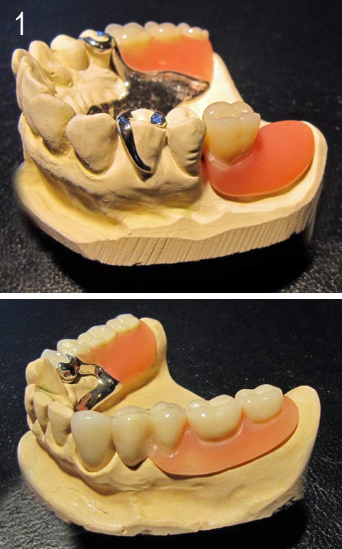
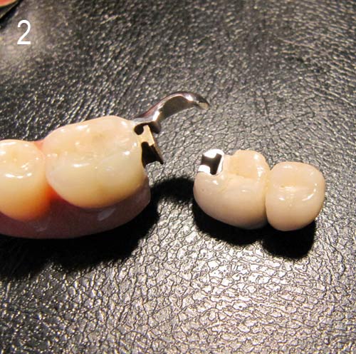
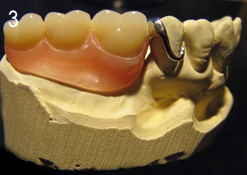
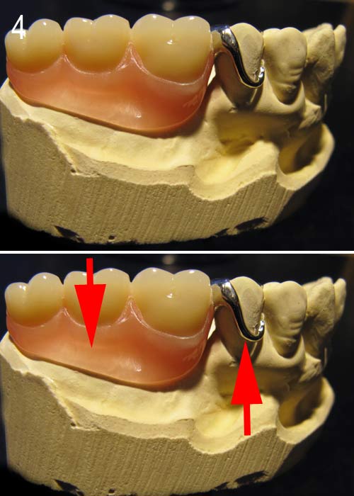