Diseases of the Lens
Glaucomas
ANATOMY AND PHYSIOLOGY
APPLIED ANATOMY
The lens is a transparent, biconvex, crystalline structure placed between iris tind the vitreous. Its diameter is 9-
Structure (Fig. 1)

Fig. 1. Structure of the crystalline lens.
1. Lens_capsule. It is a thin, transparent, hyaline membrane surrounding the lens which is thicker over the anterior than the posterior surface. The lens capsule is thickest at pre-equator regions (14 ) and thinnest at the posterior pole (3 ).
2. Anterior epithelium. It is a single layer of cuboidal cells which lies deep to the anterior capsule. In the equatorial region these cells become columnar, are actively dividing and elongating to form new lens fibres throughout the life. There is no posterior epithelium, as these cells are used up in filling the central cavity of lens vesicle during development of the lens.
3. Lens fibres. The epithelial cells elongate to form lens fibres which have a complicated structural form. Mature lens fibres are cells which have lost their nuclei. As the lens fibres are formed throughout the life, these are arranged compactly as nucleus and cortex of the lens.
(a) Nucleus. It is the central part containing the oldest fibres. It consists of different zones.
Embryonic nucleus is its innermost part (1 to 3 months of gestation). Its fibres meet around the Y-shaped sutures (Fig. 2). Outside the embryonic nucleus, successive nuclear zones are laid down as the development proceeds and, depending on the period of formation, are called as fetal nucleus (corresponding to lens from 3 months gestation till at birth), the infantile nucleus (corresponding to lens from birth to puberty) and the adult nucleus (corresponding to lens in early adult life).
Fig. 2. Y-shaped sutures of the embryonic nuclear fibres
(b) Cortex. It is the peripheral part which comprises the youngest lens fibres.
4.Suspensory ligaments of lens (Zonules of Zinn). Also called as ciliary zonules, these consist essentially of a series of fibres passing from ciliary body to the lens. These hold the lens in positio: enable the ciliary muscle to act on it.’ are arranged in three groups:
i. The fibres arising from pars plana and ante part of ora serrata pass anteriorly to get insertj anterior to the equator.
ii. The fibres originating from comparative^ anteriorly placed ciliary processes posteriorly to be inserted posterior t« equator.
iii. The third group of fibres passes from] summits of the ciliary processes almost direclB inward to be inserted at the equator.
APPLIED PHYSIOLOGY AND BIOCHEMISTRY
The crystalline lens, being an avascular structure, is dependent for its metabolism on chemical exchanges with the aqueous humour.
The chemical composition of crystalline lens vis-a-vis is aqueous humour and the chemical exch between the two is depicted in Fig. 3.
|
Metabolism
Metabolic activity of the lens is largely confined! the cortex and the nucleus is relatively inert. I uses the energy of metabolism for its growth! active transport process. In the lens eighty pert glucose is metabolised anaerobically by the glycolytic pathway, 15 percent by pentose or hexose monophosphate shunt and a small proportion via oxidative Krebs’ citric acid cycle. Sorbitol pathway is relatively inconsequential in the normal lens; however, it is extremely important in the production of cataract in diabetic and galactosemic patients.
CATARACT
The lens of the eye is normally clear. When we become older most of us get small opacities in the peripheral parts of the lens. As long as they do not disturb vision they are called opacities. If they start interfering with vision, the doctors talk about cataracts. Cataract means opacities, cloudiness of the lens. It is not a layer that grows inside the eye or on the eye preventing clear vision.
Peripheral opacities of the lens do not usually disturb vision during the day when the pupil is small. They may disturb night driving. The lights of the advancing cars become large halos and it is difficult to see in twilight and dark. Photophobia, increased light sensitivity is perhaps the most common symptom of small opacities that do not yet reduce visual acuity notably. Then it is important to use a hat or a visor so the light does not fall directly on the eyes. It is also much easier to see when walking on the shadow side of the street. Yellow tint in spectacles may decrease light sensitivity both outside and when reading.
Figure 14 shows different types of lens opacities. The opacities may look like thin lace, small bubbles, or as frosted glass. The center of the lens may become yellowish. Then the person often becomes nearsighted and may be able to read without reading glasses.
|
|
Fig.14. Lens opacities occur in different parts of the lens. |
If the lens opacities disturb too much, it is usually possible to remove the cloudy lens. In most cases it is possible to replace the remo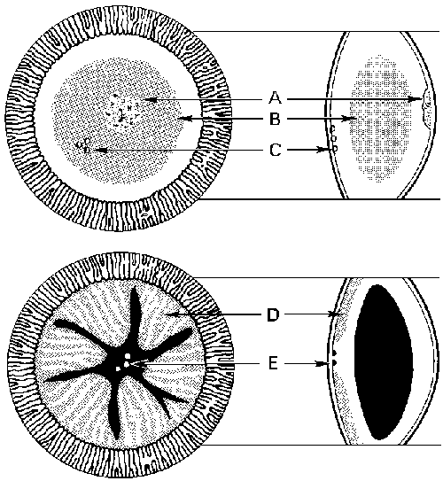 ved lens with a clear plastic lens. If it is not possible, the patient has to use catarcat spectacles or a contact lens after the operation. Cataract spectacles make everything look larger thaormal and straight lines seem to be curved. These disturbances disappear in a few weeks.
Fig.15. Cataract surgery.
After the cataract surgery many patients see nearly as well as they saw in their younger years, some other patients see only a little bit better than before the operation. If there are changes in the retina or the optic nerve the vision will not be clear even if the cloudy lens has been removed.
Cataract surgery is usually uneventful. Since the person with cataract is often elderly and may have several diseases, it is wise to ask the medical doctor whether an examination before the surgery is needed to make sure that there is nothing that might cause problems during the surgery. After the surgery the eye should not be touched or rubbed. Therefore it is good to cover it with a shield for the night.
CATARACT
Definition
The crystalline lens is a transparent structure. Its transparency may be disturbed due to degenerative process leading to opacification of lens fibres. Development of an opacity in the lens is known as cataract.
Classification
A. Etiological classification
I. Congenital and developmental cataract
II. Acquired cataract
1. Senile cataract
2. Traumatic cataract .
3. Complicated cataract
4. Metabolic cataract
5. Electric cataract
6. Radiational cataract
7. Toxic cataract e.g.
i. Corticosteroid-induced cataract
ii. Miotics-induced cataract
iii. Copper (in chalcosis) and iron (in sidero-sis) induced cataract.
8. Cataract associated- with skin diseases (Dermatogenic cataract).
9. Cataract associated with osseous diseases.
10.Cataract with miscellaneous syndromes e.g.
i. Dystrophica myotonica
ii. Down‘s syndrome.
B. Morphological classification (Fig. 4)
Fig. 4. Morphological shapes of cataract.
1. Capsular cataract. It involves the capsule and may be:
i. Anterior capsular cataract
ii. Posterior capsular cataract
2. Subcapsular cataract. It involves the Superficial part of the cortex (just below the capsule) and includes:
i. Anterior subcapsular cataract
ii. Posterior subcapsular cataract .
3. Cortical cataract. It involves the major part of the cortex.
4. Supranuclear cataract. It involves only the deeper parts of cortex (just outside the nucleus).
5. Nuclear cataract. It involves the nucleus of the crystalline lens.
6. Polar cataract. It involves the capsule and superficial part of the cortex in the polar region, only and may be:
i. Anterior polar cataract
ii. Posterior polar cataract
CONGENITAL AND DEVELOPMENTAL CATARACTS
These occur due to some disturbance in the normal growth of the lens. When the disturbance occurs before birth, the child is born with a congenital cataract. Therefore, in congenital cataract the opacity is limited to either embryonic or foetal nucleus. Development cataract may occur from infancy to adolescence. Therefore, such opacities infantile or adult nucleus, deeper parts of cortex or capsule. Developmental cataract typically affects the particular zone which is being formed when this process is disturbed. The fibres laid down previously and subsequently are ofteormally formed and remain clear. Congenital and developmental opacities assume most variegated appearance and minute opacities (without visual disturbance) are very common iormal population. These are detected with the beam of slit lamp under full mydriasis.
Etiology
It is not known exactly. Some factors which have been associated with certain types of cataracts are described below:
I.Heredity. Genetically determined cataract is due to an anomaly in the chromosomal pattern of the individual. About one-third of all congenital cataracts are hereditary. The mode of inheritance is usually dominant. Common familial cataracts include: cataracta pulverulenta, zonular cataract (also occurs as non-familial), coronary cataract and total soft cataract (may also occur due to rubella).
II. Maternal factors. These are as follows:
1. Malnutrition during pregnancy has been associated with non-familial zonular cataract.
2. Injections. Maternal infections like rubella are associated with cataract in 50 percent of cases. Other maternal infections associated with congenital cataract, include toxoplasmosis and cytomegalo-inclusion disease.
3. Drugs ingestion. Congenital cataracts have also been reported in the children of mothers who have taken certain drugs during pregnancy (e.g. thalidomide, corticosteroids).
4. Radiation. Maternal exposure to radiation during pregnancy may cause congenital cataracts.
III. Foetal or infantile factors. These include the following:
1. Deficient oxygenation (anoxia) owing to placental haemorrhage.
2. Metabolic disorders of the foetus or infant such as galactosemia, galactokinase deficiency and neonatal hypoglycemia.
3. Cataracts associated with other congenital anomalies e.g. as seen in Lowe’s syndrome, myotonia dystrophica and congenital icthyosis.
4. Birth trauma.
5. Malnutrition in early infancy may also cause developmental cataract.
IV. Idiopathic. About 50 percent cases are sporadic and of unknown etiology.
Clinical types
Congenital and developmental cataracts may be classified depending upon the stage of development at which cataract occurred and morphology (shape and pattern) of the opacity. There are numerous varieties of congenital and developmental cataracts. The common ones are described below:
1. Cataracta centralis pulverulenta (Embryonic nuclear cataract). It has dominant genetic trait and occurs due to inhibition of the lens development at a very early stage and thus, involves the embryonic nucleus. The condition is bilateral and is characterised by a small rounded opacity lying exactly in the centre of the lens. The opacity has a powdery appearance (pulverulenta) and usually does not affect the vision.
2. Lamellar (zonular) cataract. It is the most common type of congenital cataract, accounting for about 50 percent cases. Here, development of the lens is interfered at a later stage. Typically, this cataract occurs in a zone of foetal nucleus surrounding the embryonic nucleus (Fig. 5). Occasionally two such rings of opacity are seen. The ‘ main mass of the lens internal and external to the zone of cataract is clear, except for small linear opacities like spokes of a wheel (riders) which may be seen towards the equator.
Fig. 5. Lamellar (zonular) cataract as seen:
A, by oblique illumination;
B, in optical section with the beam of the slit-lamp.
It may be either genetic or environmental in origin. Genetic pattern is usually of dominant variety. Environmental form is associated with deficiency of vitamin D. Sometimes maternal rubella infection contracted between 7th and 8th week of gestation may also cause lamellar cataract.
It is usually bilateral and frequently causes severe visual defect.
3. Sutural cataract. It is a comparatively common variety and consists of a series of punctate opacities scattered around the anterior and posterior Y-sutures. Such cataracts are usually static, bilateral and do not have much effect on the vision. The individual opacities vary in size and shape and have different patterns and thus are named accordingly as under:
i. Floriform cataract. Here the opacities are arranged like the petals of a flower.
ii. Coralliform cataract. Here the opacities are arranged in the form of a coral.
iii. Spear-shaped cataract. The lenticular opacities are in the form of scattered heaps of shining crystalline needles.
iv. Anterior axial embryonic cataract occurs as fine dots near the anterior Y-suture.
4. Anterior polar cataract. It involves the central part of the anterior capsule and the adjoining superficial-most cortex. It may arise in the following ways:
i. Due to delayed development of anterior chamber. In this case the opacity is congenital.
ii. More commonly, such cataracts are acquired in infantile stage and follow contact of the lens capsule with the back of cornea, usually after perforation due to ophthalmia neonatorum of any other cause.
Morphological types: Anterior polar cataracts may occur as any of the following morphological patterns:
i. Thickened white plaque in the centre of capsule.
ii. Anterior pyramidal cataract. In it the thickened capsular opacity is cone-shaped with its apex towards cornea.
iii. Reduplicated cataract (double cataract): Sometimes along with thickening of central point of anterior capsule, lens fibres lying immediately beneath it also become opaque and are subsequently separated from the capsule by laying of transparent fibres in between. The buried opacity is called ‘imprint’ and the two together constitute reduplicated cataract
5. Posterior polar cataract. It is a very common lens anomaly and consists of a small circular circumscribed opacity involving the posterior pole. This is due to persistence of posterior vascular capsule of the lens.
6. Coronary cataract. It is an extremely common form of developmental cataract occurring about puberty; thus involving either the adolescent nucleus or deeper layer of the cortex. The opacities are often many hundreds iumber and have a regular radial-distribution in the periphery of lens (corona of club-shaped opacities) encircling the central axis. Since the opacities are situated peripherally, vision is usually unaffected. Sometimes the associated large punctate opacities may marginally reduce the vision.
7. Punctate cataract. It is also called blu-dot-cataract or cataracta-punctate-cerulea. It usually forms in the first two decades of life. The characteristic punctate opacities are in the form of rounded bluish dots situated in the peripheral part of adolescent nucleus and deeper layer of the cortex. Opacities are usually stationary and do not affect vision. However, large punctate opacities associated with coronary cataract may marginally reduce the vision.
8. Total congenital cataract. It is a common variety and may be unilateral or bilateral. In many cases there may be hereditary characler. Its other important cause is maternal rubella, occurring during the first trimester of pregnancy. Typically, the child is born with a dense white nuclear cataract. It is a progressive type of cataract. The lens matter may remain soft or may even liquefy (congenital Morgagriian cataract).
Congenital rubella cataract may occur alone or as part of the classical rubella syndrome which consists of:
i. Ocular defects (congenital cataract, salt and pepper retinopathy and microphthalmos).
ii. Ear defects (deafness due to destruction of organ of Corti).
iii. Heart defects (patent ductus arteriosus, pulmonary stenosis and ventricular septal defects).
9. Congenital membranous cataract. Sometimes there may occur total or partial absorption of congenital cataract, leaving behind thin membranous cataract. Rarely there is complete disappearance of all the lens fibres and only a fine transparent lens capsule remains behind. Such a patient may be misdiagnosed as having congenital aphakia.
Management of congenital and developmental
cataract
A. What to do and when to do?
1. Small stationary lens opacities, which do not interfere with vision, can safely be ignored.
2. Incomplete central stationary cataracts may be treated by optical iridectomy or use of mydriatics to improve vision considerably.
3.Complete bilateral cataracts should be removed as early as possible (within few weeks of birth) to prevent stimulus deprivation amblyopia.
4. Complete unilateral cataract should also be preferably removed within a few weeks of birth. However, it should be borne in mind that the chances of developing amblyopia with unilateral uncorrected aphakia and unilateral unoperated cataract are equal.
B. Surgical procedures. Childhood cataracts, (congenital, developmental as well as acquired) can be dealt with discission (needling) operation, anterior capsulotomy and aspiration or lensectomy. The needling operation (which was performed in the past) is now almost obsolete.
C. Correction of paediatric aphakia. It is still an unsolved query. The common views are as follows: (i) Children above the age of 5 years can be corrected by implantation of posterior chamber intraocular lens during surgery (ii) Children below the age of 5 years should preferably be treated by extended wear contact lens. Spectacles can be prescribed in bilateral cases. Later on secondary IOL implantation may be considered.
D. Correction of amblyopia. It is the central theme around which management of childhood cataract and aphakia revolves. In spite of best efforts, it continues to be the main cause of ultimate low vision in these children.
ACQUIRED CATARACT
We have studied that congenital and developmental cataracts occur due to disturbance in the formation of the lens fibres, i.e., instead of clear, opaque lens fibres are produced. While, in acquired cataract, opjlgification occurs due to degeneration of the already formed normal fibres. The exact mechanism and reasons for the degeneration of lens fibres are yet not clear. However, in general any factor, physical, chemical or biological, which disturbs the critical intra and extracellular equilibrium of water and electrolytes or deranges the colloid system within the lens fibres, tends to bring about opacification. The factors responsible for disturbing such an equilibrium of the lens fibres vary in different types of acquired cataracts and shall be discussed with the individual type. A few common varieties of acquired cataract are described here.
SENILE CATARACT
Also called as ‘age-related cararact, this is the commonest type of acquired cataract affecting equally persons of either sex usually above the age of 50 years. The condition is usually bilateral, but almost always one eye is affected earlier than the other. Classically, the senile cataract occurs in two forms, the cortical (soft cataract) and the nuclear (hard cataract). The corticalsenile cataract may start as cuneifojm (more commonly) or cupuliform cataract.
It is very common to find nuclear and cortical senile cataracts existing in the same eye; and for this reason it is difficult to give an accurate assessment of their relative frequency. In general, the predominant form can be given as cuneiform 70 percent, nuclear 25 percent and cupuliform 5 percent.
Etiology
Senile cataract is essentially an ageing process. Though its precise etiopathogenesis is not clear, the various factors implicated are as follows:
A. Factors affecting age of onset, type and maturation of senile cataract.
1. Heredity. It plays a considerable role in the incidence, age of onset and maturation of senile cataract in different families.
2. Ultraviolet irradiations. More exposure to UV irradiation from sunlight have been implicated for early onset and maturation of senile cataract in many epidemiological studies.
3. Dietary factors. Anomalous diet as regards certain proteins, amino acids, vitamins (riboflavin, vitamin E, vitamin C}, and essential elements have also been blamed for early onset and maturation of senile cataract.
4. Dehydrational crisis. An association with prior episode of severe dehydrational crisis (due to diarrhoea, cholera etc.) and age of onset and maturation of cataract is also suggested.
B. Mechanism of loss of transparency. It is basically different iuclear and cortical senile cataracts.
1. Cortical senile cataract. Its main biochemical features are decreased levels of total proteins, amino acids and potassium associated with increased concentration of sodium and marked hydration of the lens, followed by coagulation of proteins.
The probable course of events leading to senile opacification of cortex may be as shown below in the flowchart:
![]()
![]()
![]()
![]()
Opacification of cortical lens fibres
2. Nuclear senile cataract. In it the usual degenerative changes is intensification of the age-related nuclear sclerosis associated with dehydration and compaction of the nucleus resulting in a hard cataract. It is accompanied by a significant increase in water insoluble proteins. However, the total protein content and distribution of cations remaiormal. There may or may not be associated deposition of pigment urochrome and/or melanin derived from the amino acids in the lens.
Stages of maturation
A. Maturation of the cortical type of senile cataract
1 . Stage of lamellar separation. The earliest senile change is demarcation of cortical fibres owing to their separation by fluid. This phenomenon of lamellar separation can be demonstrated by slit-lamp examination only. These changes are reversible.
2. Stage of incipient cataract. In this stage early detectable opacities with clear areas between them are seen. Two distinct types of senile cortical cataracts can be recognised at this stage:
(a) Cuneiform senile cortical cataract. It is characterised by wedge-shaped opacities with clear areas in between. These extend from equator towards centre and in early stages can only be demonstrated after dilatation of the pupil. They are first seen in the lower nasal quadrant. These opacities are present both in anterior and posterior cortex and their apices slowly progress towards the pupil. On oblique illumination these present a typical radial spoke-like pattern of greyish white opacities (Fig.6). On distant direct ophthalmoscopy, these opacities appear as dark lines against the red fundal glow.
Since the cuneiform cataract starts at periphery and extends centrally, the visual disturbances are noted at a comparatively late stage.
Fig. 6. Immature senile cataract (cuneiform type): in optical section with the
beam of the slit-lamp.
(b) Cupuliform senile cortical cataract. Here part of posterior cortex (posterior subcapsular cataract), which gradually extends outwards. There is usually a definite demarcation between the cataract and the surrounding clear cortex. Cupuliform cataract lies right in the pathway of the axial rays and thus causes an early loss of visual acuity.
3. Immature senile cataract (ISC). In this stage opacification progresses further. The cuneiform (Fig 6.) or cupuliform patterns can be recognised till the advanced stage of ISC when opacification becomes more diffuse and irregular. The lens appears greyish white. But clear cortex is still present and so iris shadow is visible.
In some patients, at this stage, lens may become swollen due to continued hydration. This condition is called ‘intumescent cataract’. Intumescence may persist even in the next stage of maturation. Due to swollen lens anterior chamber becomes shallow.
4. Mature senile cataract (MSC). In this stage, opacification becomes complete, i.e. whole of the cortex is involved. Lens becomes pearly white in colour. Such a cataract is also labelled as ‘ripe cataract’ (Fig 7).
Fig. 7. Mature senile cataract.
5. Hypermature senile cataract (HMSC). When the mature cataract is left in situ, the stage of hypermaturity sets in. The hypermature cataract occur in any of the two forms:
(a) Morgagnian hypermature cataract: In some patients, after maturity the whole cortex lique-fies and the lens is converted into a bag of milky fluid. The small brownish nucleus settles al bottom, altering its position with change in position of the head. Such a cataract is called Morgagnian cataract (Fig.7). Sometime this stage, calcium deposits may also be seen on the lens capsule.
(b) Sclerotic type hypermature cataract: Sometimes after the stage of maturity, the cortex becomes disintegrated and the lens becomes shrunken due to leakage of water. The anterior capsule is wrinkled and thickened due to proliferation of anterior cells and a dense white capsular cataract may be formed in the papillary area. Due to shrinkage of lens, anterior chamber becomes deep and iris becomes tremulous (iridodonesis).
|
B. Maturation of nuclear senile cataract
In it the sclerotic process renders the lens inelastic and hard, decreases its ability to accommodate obstructs the light rays. These changes begin centrally and slowly spread peripherally al most up to the capsule when it becomes mature; however, a very thin layer of clear cortex may remain unaffected.
The nucleus may become diffusely cloudy (greyish) or tinted (yellow to black) due to deposition of pigments. In practice, the commonly observed pigmented nuclear cataracts are either amber, brown (cataracta brunescenm, Fig.9) or black (cataracta nigra) and rarely red (cataracta rubra) in colour.
Fig.9. Cataracta brunescenm
Clinical features
Symptoms. An opacity of the lens may be present without causing any symptoms; and may be discovered on routine ocular examination. Common symptoms of cataract are as follows:
1. Glare. One of the earliest visual disturbances with the cataract is glare or intolerance of bright light; such as direct sunlight or the headlights of an oncoming motor vehicle. The amount of glare or dazzle will vary with the location and size of the opacity.
2. Uniocular polyopia (i.e. doubling or trebling of objects): It is also one of the early symptoms. It occurs due to irregular refraction by the lens owing to variable refractive index as a result of cataractous process.
3. Coloured halos. These may be perceived by some patients owing to breaking of white light into coloured specturm due to presence of water droplets in the lens.
4. Black spots in front of eves. These may be perceived by some patients. These spots are stationary.
5. Image blur, distortion of images and misty vision may occur in early stages of cataract.
6. Loss of vision. Visual deterioration due to senile cataract has some typical features. It is painless and gradually progressive iature. Patients with central opacities (e.g. cupuliform cataract) have early loss of vision. These patients see better when pupil is dilated due to dim light in the evening (day blindness). In patients with peripheral opacities (e.g. cuneiform cataract) visual loss is delayed and the vision is improved in bright light when pupil is contracted. In patients with nuclear sclerosis, distant vision deteriorates due to progressive index myopia: Such patients may be able to read without presbyopic glasses. This improvement iear vision is referred to as ‘second sight’. As opacification progresses, vision steadily diminishes, until only perception of light and projection of rays remains in stage of mature cataract.
Signs. Following examination should be carried out to look for different signs of cataract:
1. Visual acuity testing.
2. Test for iris shadow. When an oblique beam of light is thrown on the pupil, a crescentric shadow of pupillary margin of the iris will be formed on the greyish opacity of the lens, as long as clear cortex is present between the opacity and the pupillary margin. When-lens is completely transparent completely opaque, no iris shadow is formed. Hence presence of iris shadow is a sign of immature cataract.
3. Oblique illumination examination. It reveals colour of the lens in pupillary area which varies in different types of cataracts.
4. Distant direct ophthalmoscopic examination. A reddish yellow fundal glow is observed in the absence of any opacity in the media. Partial cataractous lens shows black shadow against the red glow in the area of cataract. Complete cataractous lens does not even reveal red glow.
4. Slit-lamp examination should be performed with a fully dilated pupil. The examination reveals complete morphology of opacity (site, size, shape, colour and pattern).
Complications.
1.Phacoanaphylactic uveitis. A hypermature cataract may leak lens proteins into anterior chamber. These proteins may act as antigens and induce antigen-antibody reaction leading to uveitis.
2. Lens–induced glaucoma. It may occur by different mechanisms e.g. due to intumescent lens (phacomorphic glaucoma) and leakage of proteins into the anterior chamber from a hypermature cataract (phacolytic glaucoma).
3. Subluxatian or dislocation of Lens. It may occur due to degeneration of zonules in hypermature stage.
MANAGEMENT OF CATARACT IN ADULTS
Treatment of cataract essentially consists of its surgical removal. However, certain non-surgical measures may be of help, in peculiar circumstances, till surgery, is taken up.
A. Non-surgical measures
1. Treatment of cause of cataract. In acquired cataracts, thorough search should be made to find out the cause of cataract. Treatment of the causative disease, many a time, may stop progression and sometimes in early stages may cause even regression of cataractous changes and thus defer the surgical treatment. Some common examples include: (i) Adequate control of diabetes mellitus, when discovered. (ii) Removal of cataractogenic drugs such as corticosteroids, phenothiazenes and strong miotics, may delay or prevent cataractogenesis. (iii) Removal of irradiation (infrared or X-rays) may also delay or prevent cataract formation. (iv) Early and adequate treatment of ocular disease like uveitis may prevent complicated cataract.
2. Measures to delay progression. Many commercially available preparations containing iodide salts of calcium and potassium are being prescribed in abundance in early stages of cataract (especially in senile cataract) in a bid to delay its progression. However, till date no conclusive results about their role are available. Role of vitamin E and aspirin in delaying the process of cataractogenesis is also mentioned.
3. Measures to improve vision in the presence of incipient and immature cataract may be of great solace to the patient. These include:
i. Refraction, which often changes with considerable rapidity, should be corrected at frequent intervals.
ii. Arrangement of illumination. Patients with peripheral opacities (pupillary area still free), may be instructed to use brilliant illumination. Conversely, in the presence of central opacities, a dull light placed beside and slightly behind the patient’s head will give the best result.
iii. Use of dark goggles in patients with central opacities is of great value and comfort when worn outdoors.
iv. Mydriatics. The patients with a small axial cataract, frequently may benefit from pupillary dilatation. This allows the clear paraxial lens to participate in light transmission, image formation and focusing. Mydriatics such as 5 percent phenylephrine or 1 percent tropicamide; 1 drop b.i.d. in the affected eye may clarify vision.
B.Surgical management
Indications
1. Visual improvement. This is by far the most common indication. When surgery should be advised for visual improvement varies from person to person depending upon the individual visual needs. So, an individual should be operated for cataract, when the visual handicap becomes a significant deterrent to the maintenance of his or her usual life-style. In general, patients with a visual acuity of less than 6/36 (Snellen’s) may be advised surgical management.
2. Medical indications. Sometimes patients may be comfortable from the visual point (due to useful vision from the other eye or otherwise) but may be advised cataract surgery due to medical grounds such as lens induced glaucoma, phacoanaphylactic endophthalmitis and retinal diseases like diabetic retinopathy or retinal detachment, treatment of which is being hampered by the presence of lens opacities.
3. Cosmetic indicathion. Sometimes patient may insist for cataract extraction (even with no hope of getting useful vision), in order to obtain a black pupil.
Preoperative evaluation
Once it has been decided to operate for cataract, a thorough preoperative evaluation should be carried but before contemplating surgery. This should include:
II. Ocular examination. A thorough examination of eyes including slit-lamp biomicroscopy is desirable in all cases. The following useful information is essential before the patient is considered for surgery:
(a) Retinal function tests. The retinal function must be explored since, if it is defective, operation will be valueless, and patient must be warned of the prognosis, to avoid unnecessary disappointment and medicolegal problems. A few important retinal function tests are considered here.
1. Light perception (PL). Many sophisticated retinal function tests have been developed, but light perception must be present, if there is to be any potential for useful vision.
2. A test for Marcus-Gunn pupillary response (indicative of afferent pathway defect) should be made routinely. If present, it is a poor prognostic sign.
3. Projection of rays (PR). It is a crude but an important and easy test for function of the peripheral retina. It is tested in a semi-dark room with the opposite eye covered. A thin beam of light is thrown in the patient’s eye from four directions (up, down, medial and lateral) and the patient is asked to look straight ahead and point out the direction from which the light seems to come.
4. Two-light discrimination test. It gives information about macular function. The patient is asked to look through an opaque disc perforated with two pin-holes behind which a light is held. The holes are
5. Maddox rod test. The patient is asked to look at a distant bright light through a Maddox rod. An accurate perception of red line indicates normal function.
6. Colour perception. It indicates that some macular function is present and optic nerve is relatively normal.
7. Entoptic visualisation. It is evaluated by rubbing a point source of light (such as bare lighted bulb to torch) against the closed eyelids. If the patient perceives the retinal vascular pattern in black outline, it is favourable indication of retinal function. Being subjective iature, the importance of negative test can be considered if the patient can perceive the pattern with the opposite eye.
8. Laser interferometry. It is a very good test for measuring the macular potential for visual acuity in the presence of opaque media.
9. Objective tests for evaluating retina are required if some retinal pathology is suspected. These tests includes ultrasonic evaluation of posterior segment of the eye; electrophysiological studies such as ERG (electroretinogram), EOG (electrooculogram) and VER (visually-evoked response); and indirect ophthalmoscopy if possible.
(b) Search for local source of infection should be made by ruling out conjunctival infections, meibomitis, blepharitis and lacrimal sac infection. Lacrimal sac should receive special attention. Lacrimal syringing should be carried out in each patient. In cases where chronic dacryocystitis is discovered, either DCR (dacryocystorhinostomy) or DCT (dacryocystectomy) operation should be performed, before the cataract surgery.
(c) Anterior segment evaluation by slit-lamp examination. It is of utmost importance. Presence of keratic precipitates at the back of cornea, in a case of complicated cataract, suggests management for subtle uveitis before the cataract surgery. Similarly, information about corneal endothelial condition is also very important, especially if intraocular lens implantation is planned.
(d) lntraocular pressure (IOP) measurement. Preoperative evaluation is incomplete without the measurement of IOP. The presence of raised IOP needs a priority management.
Preoperative medication and preparation
1.Topical antibiotics such as tobramycin or gentamicin QID for 3 days just before surgery is advisable as prophylaxis against endophthalmitis.
2. Systemic antibiotics such as gentamicin 80 mg intramuscular at night and in the morning before surgery are preferred by a few surgeons.
3. Preparation of the eye to be operated. Eyelashes of upper lid should be trimmed at night and the eye to be operated should be marked.
4. An informed and detailed consent should be obtained.
5. Scrub bath and care of hair. Each patient should be instructed to have a scrub bath including face and hair wash with soap and water. Male patients must get their beard cleaned and hair trimmed. Female patients should comb their hair properly.
6. To lower IOP, acetazolamide 500 mg stat 2 hours before surgery and glycerol 60 ml mixed with equal amount of water or lemon juice, 1 hour before surgery, or intravenous mannitol 1 gm/kg body weight half an hour before surgery may be used.
7. To sustain dilated pupil (especially in extracapsular cataract extraction) the antiprostaglandin eyedrops such as indomethacin or flurbiprofen should be instilled three times one day before surgery and half hourly for two hours immediately before surgery. Adequate dilation of pupil can be achieved by instillation of 1 percent tropicamide and 5 percent or 10 percent phenylephrine eyedrops every ten minutes, one hour before surgery.
Anaesthesia
Cataract extraction can be performed under general or local anaesthesia. Local anaesthesia is preferred whenever possible (see page 408).
Types of surgical techniques
I. Intracapsular cataract extraction (ICCE)
In this technique, the entire cataractous lens along with the intact capsule is removed. Therefore, weak and degenerated zonules are a pre-requisite for this method. Because of this reason, this technique cannot be employed in younger patients where zonules are strong. ICCE can be performed between 40-50 chymotrypsin (which will dissolve the; Beyond 50 years of age usually there is no need of this enzyme.
Indications
ICCE has stood the test of time and has been widely employed for about 50 years over the world. Now (for the last 15 years) it has been almost entirely replaced by planned extracapsular technique. Indications of ICCE are as follows:
2. If facilities for microsurgery are not available then ICCE is performed in patients beyond the age of 40 years.
3. If surgeon is not trained in microsurgery then ICCE can be performed.
4. Absolute indications are markedly subluxated and dislocated lens.
II. Extracapsular cataract extraction (ECCE)
In this technique, major portion of anterior capsule with epithelium, nucleus and cortex are removet leaving behind intact posterior capsule.
Indications
1. Presently (in general) ECCE is being considered as the procedure of choice over ICCE.
2. The absolute indications of ECCE are:
• When posterior chamber intraocular lens implantation is to be performed.
• In patients having high myopia with degenerated fluid vitreous.
• Patients below the age of 40 years where ICCE is contraindicated due to strong zonules.
Advantages of ECCE over ICCE
1. ECCE is a universal operation and can be performed at all ages, except when zonules are not intact; whereas ICCE cannot be performed below 40 years of age.
2. Posterior chamber IOL can be implanted after ECCE, while it cannot be implanted after ICCE.
3. Postoperative vitreous related problems (such as herniation in anterior chamber, pupillary block and vitreous touch syndrome) associated with ICCE are not seen after ECCE.
METHODS OF EXTRACAPSULAR CATARACT EXTRACTION
1. Discission (Needling)
It is an obsolete operation and so not performed now-a-days. It is mentioned here only for historical interest. It used to be performed in congenital or acquired cataract during childhood. In this technique a cruciate incision was made in the anterior capsule with a Ziegler’s knife, the lens matter was stirred up and left as such for self-absorption. There used to be a high incidence of postoperative complications namely thick after cataract, uveitis and glaucoma.
2. Linear extraction (Curette evacuation)
It is also an obsolete procedure and thus abandoned and again mentioned only for historical interest. Before the modern ECCE, this procedure was performed in patients between the ages of 15 and 35 years of age after full mydriasis. In this technique after opening the anterior chamber a large piece of anterior capsule is removed with a large toothed capsule forceps. Lens matter including nucleus and cortex is expressed out using lens curette or lens spatula and lens expressor. Remaining cortical matter is aspirated with a cannula.
3. Conventional extracapsular cataract extraction
The ECCE is performed under an operating microscope, with full mydriasis. It is the surgery of choice for almost all types of childhood and adult cataracts unless contraindicated.
Absolute indications
1. All patients with cataract below the age of 40 years.
2. Cataract associated with high myopia and degenerated vitreous.
3. When posterior chamber intraocular lens is to be implanted.
4. Patients with history of vitreous prolapse and/or aphakic retinal detachment in previously operated eye.
Absolute contraindications. Markedly subluxated and dislocated lens.
Surgical steps
(a) Initial steps up to making a partial thickness groove at limbus are similar to ICCE except that the size of the limbal groove is slightly smaller (10 to 2 O’clock).
(b) Anterior capsulotomy. It can be performed by any of the following methods: 1. Can-opener’s technique. In it, an irrigating cystitome (or simply a 26 gauge needle, bent at its tip) is introduced into the anterior chamber and multiple small radial cuts are made in the anterior capsule for 360° (Fig. 10.A).
2. Linear capsulotomy (Envelop technique). Here a straight incision is made in the anterior capsule (in the upper part) from 2-10 O’clock position. The rest of the capsulotomy is completed in the end after removal of nucleus and cortex.
3. Continuous circular capsulorrhexis. (CCC) Recently this is the most commonly performed procedure. In this the anterior capsule is torn in a circular fashion either with the help of an irrigating bent-needle cystitome or with a capsulorrhexis forceps.
(c) Removal of anterior capsule. It is removed with the help of, a Kelman-McPherson forceps (Fig. 10.B).
(d) Corneoscleral section. It is completed from 2-10 O’ clock position as described in ICCE (Fig.
(e) Hydrodissection. After the anterior capsulotomy, the balanced salt solution (BSS) is injected under the peripheral part of the anterior capsule. This manoeuvre separates the corticonuclear mass from the capsule.
(f) Removal of nucleus. After hydrodissection the nucleus can be removed by any of the following techniques:
1. Pressure and counter-pressure method. In it the posterior pressure is applied at 12 O’clock position with corneal forceps or lens spatula and the nucleus is expressed out by counter-pressure exerted at 6 O’clock position with a lens hook (Fig. 10.D).
2. Irrigating wire vectis technique. In this method loop of an irrigating wire vectis is gently passed below the nucleus, which is then lifted out of the eye.
(g) Aspiration of the cortex. The remaining cortex is aspirated out using a two-way irrigating and aspiration cannula (Fig. 10Е).(h) Rest of the steps from closing the incision till the application of pad and bandage are the same as described in ICCE technique.
Fig. 10. rgical steps of extracapsular cataract extraction with posterior chamber intraocular lens implantation: A, anterior capsulotomy can-opener’s technique; B, removal of anterior capsule; C, completion of corneo-scleral section; D, removal ol nucleus (pressure and counter-pressure method); E, aspiration of cortex; F, insertion of inferior haptic of posterior chamber IOL; G, insertion of superior haptic of PC-IOL; H, dialing of the IOL; I, corneo-scleral suturing.
4. Phacoemulsification
It is presently the most popular method of extracapsular cataract extraction. It differs from the conventional ECCE as follows:
1. Corneoscleral incision required is very small (
2. Continuous curvilinear capsulorrhexis (CCC) of 4-
3. Hydrodissection i.e., separation of capsule from the cortex by injecting fluid exactly between tie two (Fig. 10.B) and hydrodelineation i.e., separating nucleus from epinucleus by injecting fluid between the two (Fig.
4. Nucleus is emulsified and aspirated by phacoemulsifier. Phacoemulsifier basically acts through a hollow
5. Remaining cortical lens matter is aspirated with the help of an irrigation-aspiration technique (Fig.10.F).
Laser phacoemulsification. This technique is under trial and perhaps soon may replace the conventional phacoemulsification. In it the lens nucleus is emulsified utilizing laser energy. The advantage of this technique is that the laser energy used to emulsify cataractous lens is not exposed to other intraocular structures (c.f. ultrasonic energy).
Advantages and disadvantages of phacoemulsi-lication vis-a-vis conventional ECCE, The potential advantages of phacoemulsification, compared with conventional ECCE, include more rapid wound healing, short convalescence and early stabilization of refractive error with less astigmatism. The main disadvantages are an expensive machine and a higher incidence of complications by beginners because the technique is relatively difficult to master.
Fig. 10. Surgical steps of pnacoemulsification : A, Continuous curvilinear capsulorrhexis; B, Hydrodissection; C, Hydrodelineation; D&E; Nucleus emulsification by divide and conquer technique (four quadrant cracking);
F, Aspiration of cortex.
CONGENITAL ANOMALIES OF THE LENS
1. Coloboma of the lens. It is seen as a notch in the lower quadrant the of the equator (Fig.11). It is usually unilateral and often hereditary.
2. Congenital ectopia lends.
3. Lenticonus. It is the cone-shaped elevation of the anterior pole (lenticonus anterior, Fig. 12) or posterior pole (lenticonus posterior). Lenticonus anterior may occur in Alport’s syndrome. On distant direct ophthalmoscopy, both present as an oil globule lying in the centre of the red reflex. Slit-lamp examination confirms the diagnosis.
4. Congenital cataract.
5. Microspherophakia. In this condition the lens is spherical in shape (instead of normal biconvex) and small in size. Microspherophakia may occur as an isolated familial condition or as a feature of other syndromesv e.g. Weil-Marchesani or Marfans syndrome.
Fig. 11. Coloboma of the lens.
Fig. 12. Lenticonus anterior.
ANATOMY AND PHYSIOLOGY
APPLIED AN ATOMY
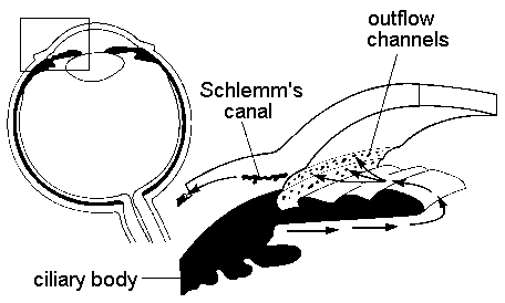
Pathophysiology of glaucoma revolves around the aqueous humour dynamics. The principal ocular structures concerned with it are ciliary body, angle of anterior chamber and the aqueous outflow system.
Ciliary body
It is the seat of aqueous production.
Angle of anterior chamber (Fig.1).
It plays an important role in the process of aqueous drainage. It is formed by root of iris, anterior-most part of ciliary body, scleral spur, trabecular meshwork and Schwalbe’s line (prominent end of Descemet’s membrane of cornea). The angle width varies in different individuals and plays a vital role in the pathomechanism of different types of glaucoma.
Glaucoma, macular degeneration, intraocular tumors, optic atrophy
Glaucoma is a group of diseases that cause loss of visual field because nerve fibers are damaged at the optic disk. In many but not all cases the pressure inside the eye, the intraocular pressure, is higher thaormal and causes changes in the nerve fibers of the optic nerve, over a very long period of time. Glaucoma is often diagnosed during health control visit, the person is unaware of the changes in his eyes. There is a more rare type of glaucoma in which the pressure rises acutely causing pain and redness of the eye.
Glaucoma is often difficult to diagnose because there are so many variants of normal structure of the optic disc and sometimes the noticeable changes in the optic disc appear late. An example of these problems is in the following Figure 11.
A B
Figure 11. This person was examined more than ten years ago and was found to have optic disc changes in the right eye (A) that were rather typical to glaucoma: enlargement of the optic cup and displacement of the vessels toward the nasal side. The optic disc of the left eye (B) was normal. However, no high pressures have been measured during the past years, the visual fields are normal and the structure of the optic disc has not changed. The question then is: is this a congenital difference or was there one time high pressure or circulatory changes that changed the structure of the optic disc. There is no sure answer and therefore the condition must be followed repeating visual fields and measurement of intraocular pressure.
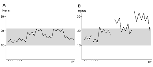
Even a normal eye has certain intraocular pressure. The pressure varies usually between 10 and 21 mmHg (quicksilver millimeter) and is often highest early in the morning. If the pressure rises or there are changes in the circulation of the nerve head, nerve fiber function becomes disturbed and some fibers die. In the corresponding areas of the visual field small blind patches, scotomas, are found. The areas of loss of vision are first very small and cannot be noticed by the person himself. Therefore it is important that the eyes are controlled by an ophthalmologist regularly when the person is past 50 years.
|
|
Fig.11c. Intraocular fluid is formed in the ciliary body behind the iris. It flows through the pupil into the anterior chamber. It drains through the small pores at the anterior chamber angle into the veins on the surface of the eye. |
Since the intraocular pressure (IOP) varies even in normal individuals, it is necessary to measure the pressure several times at different hours of the day if one suspects that the pressure might be too high at times.
|
Fig.12. Variations of the intraocular pressure. |
|
Mere measurement of the intraocular pressure is not sufficient for ruling out of glaucoma. The eyegrounds, especially the nerve heads, called the discs, have to be examined and the visual fields recorded.
|
|
Fig.13. Measurement of the intraocular pressure (IOP). |
|
|
B. Measurement with a Schiotz’ tonometer. |
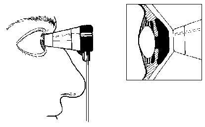
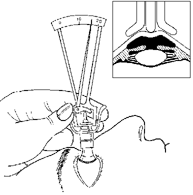
In hospitals and in doctors’ offices the IOP is measured with an applanation tonometer. It is a small device that gently touches the cornea. The patient may not notice the moment of measurement because the corneal surface is anesthetized with drops. (S)he sees the blue light approaching, then it stops to touch the cornea and then moves away.
An older instrument is Schiotz’ tonometer. The patient lies on his/her back looking at the ceiling, the instrument is placed on the cornea and the pressure is read on the scale. This instrument is sometimes loaned for measurements at home.
Measurement of the visual fields tries to reveal whether there are small poorly functioning areas, scotomas, in the field of vision. The instrument used is called perimeter and the examination is called perimetry. There are many different perimeters. In some perimeters tiny lights appear now and then in different parts of the visual field. When one or more lights appear the patient pressures the button as a sign that he has seen the light(s). In other perimeters the light moves from the edge of the perimeter inwards until it becomes visible. The patient always looks into the central spot of the perimeter. He/she does not move his/her eyes around to find the moving spot.
The anterior chamber angle is examined with a small contact lens that is placed on the cornea. In the contact lens there is a small mirror in which one can see the anterior chamber angle. This contact lens is different from the contact lens that are used instead of glasses (Fig.13c).
Figure 13c. The three mirror lens or the gonioscopy lens (gonio= angle, scopy= to look) can be used to examine the structures in the anterior chamber angle.
There are many kinds of medications used in the treatment of glaucoma, both eye drops and pills. Eye drops that lower the intraocular pressure are powerful medicines. If the drops get into the nose and from there into the stomach some of them may cause disturbance in the functions of the stomach and the gut. They may also cause allergic rhinitis and asthma. Therefore, the drops should be dropped on the conjunctiva of the lower lid. The eye is then closed and the inner corner of the eye pressed gently with paper handkerchief to prevent the drops from getting into the tear channels.
The eye drops should not get on the skin because they may cause excema. If it is difficult to drop one may ask the optician to drill small holes in old reading glasses. The holes are not drilled at the center of the lens, but slightly toward the side. The hole has to be large enough to let the tip of the dropper to go through. Otherwise the drops flow on the glasses. There are special supporting cups available for dropping the eye drops.
If medications do not control the intraocular pressure well enough, laser treatment and several different operations may be considered.
Glaucoma with closure of the anterior chamber angle develops if the iris is pushed forward and blocks the anterior chamber angle. The pressure rapidly rises very high, the eye becomes painful, reddish, vision becomes blurred and the patient often has nausea. The patient needs immediate treatment because the nerve fibers do not tolerate high pressure for an extented period of time.
GLAUCOMA
is a significant cause of blindness world-wide. It is the second leading cause of blindness in Caucasians and possibly the leading cause among African. In addition, glaucoma is prevalent iearly 3% to 4% of the population over age 70.
In ancient times, the term “glaucoma” referred to a broad group of blinding diseases. By the 1700s, elevation of intraocular pressure, detected by palpation of the globe, was known to be a distinct ocular disease. When the ophthalmoscope was introduced in 1854, glaucoma was redefined as diseases of the eye characterized by optic nerve damage and elevated intraocular pressure. The distinctly different mechanisms of open-angle glaucoma and angle-closure glaucoma were not revealed until the 1930s, when reliable gonioscopy allowed examination of the angle of the anterior chamber of the eye ).
Early detection and treatment of glaucoma are essential in preventing loss of vision and blindness. Definitive diagnosis and treatment are the domain of ophthalmologists, but primary care physicians play an essential role in screening patients through careful history taking and physical examination and in identifying those at high risk.
Glaucoma is a symptomatic condition and not a disease “sui generis”. It is a collection of physicals signs: raised intra-ocular pressure, visual field loss, enlargement of the blind spot and changes in the appearance of the optic nerve head.
Glaucoma may be defined as “those situations were IOP is too high for normal functioning of the optic nerve head (Shields, 1992.) IOP is closely linked to aqeous humor (clear, watery, fluid in the eye) dynamics. IOP is a function of aqeous humor outflow (AHO) and production (AHP) (IOP = AHO – AHP). Therefore pharmaocologica
treatment is aimed at either increasing outflow or decreasing production of aqeous humor. IOP is measured by many different types of machines by health care proffesionals. These exams are important because as with hypertension, when symptoms are noticed by the patient the damage has already been done.
Glaucoma is now defined as a group of ocular diseases characterized by elevated intraocular pressure that causes progressive damage to the optic nerve, resulting in optic nerve atrophy and blindness. The two primary types of disease, open-angle glaucoma and angle-closure glaucoma, are classified according to the anatomy of the anterior chamber angle. This is determined by visual inspection of the angle using a special lens (goniolens) at the slit-lamp biomicroscope. Both types of disease may be present in the same eye, a condition known as combined-mechanism glaucoma. Glaucoma is further differentiated as primary (not related to other conditions) or secondary (associated with an ocular condition, systemic condition, or both). Glaucoma ieonates and young children is referred to as congenital glaucoma and is not addressed here.
Glaucoma encompasses a number of conditions that are characterized by a particular pattern of blindness involving optic nerve damage and visual field loss. Most, but not all, of the conditions involve increased intraocular pressure (IOP) within the eye, which is by far, the most common risk factor for vision loss due to glaucoma. This increased pressure damages the optic nerve and can result in a progressive loss of peripheral vision leading to blindness if not properly diagnosed and treated.
It is a serious condition of the eye affecting approximately two percent of the population. It has robbed millions of people of their eyesight. If left untreated, it can cause total, irreversible blindness. Glaucoma can strike anyone, but it need not cause blindness. If glaucoma
is found early and treated properly, your eyesight can be preserved. Early diagnosis is the key to prevention of blindness from glaucoma.
Glaucoma is characterized by optic nerve damage and visual field loss. Typically, it involves increased pressure inside the eye that affects the delicate tissues of the optic nerve. Early detection and treatment are the keys to preventing unnecessary vision loss
Risk Factors For Glaucoma
• People over 35 years of age. The chances of developing
glaucoma increase with age.
• People with a family history of glaucoma. Since glaucoma
has genetic links, anyone with a blood relative who has glaucoma should
schedule an eye examination at minimum of once per year.
• People who are very nearsighted (Open Angle Glaucoma) or
farsighted (Angle Closure Glaucoma).
• People with diabetes and certain other chronic diseases.
• Race is a predisposing factor for glaucoma. Persons of
African ancestry are at an increased risk for developing Open Angle
Glaucoma. Conversely, persons of Chinese, Japanese, and Southeast
Asian descent are more prone to develop Angle Closure Glaucoma.
• Persons with cardiovascular disease or conditions resulting
in insufficient blood flow to the eye are at an increased risk for
developing glaucoma.
The following warning signs are indicative of the damage to vision caused by glaucoma. Their presence should send a strong signal that a thorough examination by an eye doctor is needed.
• Loss of peripheral (side) vision
• Need for frequent changes in glasses
• Difficulty in adjusting to a dark room
• Blurred vision
• Sore, reddened eyes
• Appearance of halos or rainbows around lights
• Severe headaches, nausea and eye pain in rare cases
Testing For Glaucoma
Persons diagnosed with glaucoma, or those persons exhibiting some of the symptoms of glaucoma (glaucoma suspects), are routinely followed with frequent tonometry tests to measure the pressure inside the eye. To monitor the effects glaucoma is having upon the optic nerve and the person’s overall vision, optic nerve photography and visual field testing are performed at regular intervals. The information from these tests gives an indication of the effectiveness of the treatment being used and whether stronger measures are in order.
It is important to remember that the vision lost due to damage to the optic cannot be restored. Medications, laser treatments, and surgery can only prevent further loss of sight. Glaucoma cannot be cured, but it can be controlled.
There are two main classifications of glaucoma: Open Angle Glaucoma and Closed /-ingle Glaucoma. The type of glaucoma relates to the cause of the increased pressure inside the eye.
Open Angle Glaucoma
Open angle glaucoma is the most common type of glaucoma. It is most often completely painless and causes a very gradual loss of peripheral vision, which may go unnoticed for many months or even years. Since it gives no obvious warning to its victim, glaucoma is often called “the sneak thief of sight.”
This form of glaucoma is characterized by an excessive production of fluid inside the eye. Although the drainage system of the eye, the trabecular meshwork of the angle of anterior chamber, remain open and
function properly, they are unable to remove the excess fluid at a pace sufficient to prevent a rise in pressure inside the eye.
Intraocular pressure may not reach extremely high levels, or it may rise very gradually, so patients may not experience discomfort or deterioration of vision for several years. Unfortunately, by the time vision is impaired, optic nerve damage is irreversible, except in unusual circumstances.
Generally, patients are asymptomatic until optic nerve damage is quite advanced.
Early detection and treatment are key to preventing optic nerve damage. Tonometry is the only method of accurately determining intraocular pressure; palpation is not reliable. However, even an accurate pressure reading may not detect open-angle glaucoma, because there is significant overlap in the intraocular pressures found in patients with and patients without open-angle glaucoma. A common misconception is that measurements within a distinct range of so-called normal intraocular pressure (ie, 10 to
The appearance of the optic nerve (ie, color and contour) and findings on visual field examination are the most important clues to diagnosis. Pathognomonic changes indicate glaucoma. Ophthalmoscopic examination of the optic nerve reveals changes in the cup and neuroretinal rim relatively early in disease, indicating the possibility of open-angle glaucoma. Particularly significant are the size of the cup
relative to the optic nerve, any thinning or notching of the disk rim, and the presence of disk hemorrhages. Visual field examination, which requires specialized equipment, detects defects in the field of vision that are characteristic for glaucomatous damage to the optic nerve relatively early in disease .
In contrast, in angle-closure glaucoma, the entrance to the drain is closed but the drain itself is perfectly functional. There is no reliable relationship between intraocular pressure and systemic blood pressure.
Treatment
Therapy for patients with glaucoma may include drugs, surgery, or both, depending on the type of disease.
Open angle glaucoma will usually respond well to medications when found in time. In most cases, the medication must be continued for life to keep this condition under control.
Since existing optic nerve damage in open-angle glaucoma is irreversible, the goal of treatment is to prevent progression of damage and protect the optic nerve from pressure elevations above the target therapeutic level. Initial treatment focuses on decreasing intraocular pressure. The level of reductioecessary to stabilize the optic nerve is debated, but a reduction of 20% to 50% from the initial pressure is most often sought. The need for further relief depends on whether the optic nerve and visual field remain stable or continue to deteriorate.
Medical treatment usually begins with a single topical agent, and additional topical and oral agents are added as necessary to stabilize the optic nerve and visual field. Currently, open-angle glaucoma is treated with topical beta adrenergics, such as noncardioselective betal and beta2 blockers (ie, timolol maleate [Timoptic], levobunolol hydrochloride [AKBeta, Betagan Liquifilm], metipranolol hydrochloride
[OptiPranolol]), and the relatively selective betal blocker, betaxolol hydrochloride (Betoptic). These agents reduce aqueous production.
When medical agents are unsuccessful, open-angle glaucoma is generally treated with argon laser trabeculoplasty (ALT). Low-powered, short-duration, 50-micrometer laser “burns” are applied to the trabecular meshwork, usually resulting in at least a 20% drop in intraocular pressure.
Glaucoma filtering surgery is performed if optic nerve damage progresses (evidenced by changes in the optic nerve cup, visual field, or both) despite drug therapy at maximum levels and ALT. A fistula is surgically created from the anterior chamber to the subconjunctival space. This allows aqueous humor to more readily exit the eye, resulting in lower intraocular pressure. Although effective, this procedure is associated with a low, but significant, complication rate, including loss of vision.
Closed Angle Glaucoma
The second type of glaucoma is known as Closed Angle Glaucoma. It is far more rare than open angle glaucoma. This condition is characterized by blockage of the drainage system of the eye, causing increased intraocular pressure.In open-angle glaucoma, elevated intraocular pressure is virtually always caused by obstruction of the outflow channels, especially the trabecular meshwork, rather than increased aqueous humor formation. To use an analogy, it is as if the entrance to the “drain” (the trabecular meshwork) is open, but the drain itself (the angle) is blocked.
Disease can be acute or chronic and is caused by anatomic narrowing of the anterior chamber angle, a factor that is fundamentally determined by genetics. This narrowing is primarily related to the size of the eyeball and the lens. Dim light or darkness, physical or emotional
stress, and mydriatic agents can lead to pupillary dilation, thereby precipitating angle-closure glaucoma.
Chronic angle-closure glaucoma is as insidious as open-angle glaucoma. Its fundamental mechanism relates to the anatomic narrowness of the anterior chamber angle. Often patients have a history of vague discomfort about the eyes or intermittent blurring of vision. Physical diagnosis relies on gonioscopic evaluation of the angle by an ophthalmologist. Chronic angle-closure glaucoma is treated with a laser, and any residual elevated intraocular pressure causing optic nerve damage is treated similarly to open-angle glaucoma.
Secondary glaucoma
Secondary glaucoma is usually treated according to the mechanism of the glaucoma. In addition, treatment is directed at the specific underlying cause of the associated ocular or systemic condition.
Ocular: Central retinal vein occlusion, High hyperopia High myopia, History of blunt trauma, Iris neovascularization, Lens-induced glaucoma, Uveitis
Systemic: Carotid vascular disease. Diabetes mellitus, Endocrine disorders, Herpes zoster, Long-term use of corticosteroids.
Lens induced glaucomas
The lens induced glaucoma patient is typically elderly, with a history of cataracts. Types of glaucomas associated with lens complications are: phacolytic glaucoma and phacomorphic glaucoma
In all of these cases, the glaucoma is typically very symptomatic with pain and redness in the involved eye, and cells and flare in the anterior chamber. Frequently, an advanced cataract in the involved eye severely reduces vision.
Phacolytic glaucoma This condition involves a hypermature cataract with severe visual reduction (typically light perception). It’s characterized
by acute onset of pain and redness and IOPs often of
Phacomorphic glaucoma In this case, an increase of lens thickness from an advancing cataract leads to a relative pupil block, posterior bomb and angle closure. The intumescence often develops quickly. Typically, the cataract reduces vision severely. The angle in this glaucoma is closed.
In cases where the lens precipitates a secondary glaucoma, the best management is surgical lens removal.
Neovascular Glaucoma
Ischemia to ocular tissues is theorized to be the genesis of NVG. The most common causes of NVG include ischemic central retinal vein occlusion (CRVO), diabetic retinopathy, and carotid artery disease and ocular ischemic syndrome (OIS). Neovascularization buds off of the capillaries of the posterior iris, grows along the posterior iris, through the pupil, along the anterior surface of the iris, and then into the angle. Once in the angle, the neovascularization, along with its attendant fibrovascular support membrane, acts to both physically block the angle as well as bridge the angle and physically pull the iris and cornea into apposition, thus blocking the trabecular meshwork.
If there is any degree of inflammation and ocular pain, prescribe a topical cycloplegic such as atropine 1% b.i.d. as well as a topical steroid such as Pred Forte, Vexol, or Flarex q.i.d. Aqueous suppressants may be used in order to temporarily reduce IOP. However, chronic medical therapy is not indicated for neovascular glaucoma. Aqueous suppressants
will temporize IOP and lead to a false sense of security as the neovascular process will continue with further angle closure. If a significant amount of the angle is in permanent synechial closure, filtering surgery must then be employed.
UVEITIC GLAUCOMA
In uveitic glaucoma the patient first develops uveitis, either due to trauma, systemic disease or idiopathically. The ensuing inflammation results in a rise in IOP through several mechanisms.
Patients with uveitic glaucoma will present with a unilateral red, painful, photophobic, lacrimating eye. Acuity will be moderately decreased. There will be corneal edema and folds in Descemet’s membrane, but the epithelium should be intact. There will be a profuse anterior chamber reaction, possibly with hypopyon. There are frequently posterior synechiae. There will be injection of the conjunctival and episcleral vessels. The angle may be closed with peripheral anterior synechiae (PAS). Intraocular pressure (IOP) may range from 30 to 80mm
Hg.
Often, the inflammatory cells physically block the trabecular meshwork, decreasing aqueous outflow, with the angle remaining open. Because the inflammatory cells and protein in the anterior chamber form adhesions between the posterior iris and anterior lens, posterior synechiae commonly form. This will lead to iris bombe’e, secondary angle closure and peripheral anterior synechiae formation. There may also be a combination of mechanisms that increases IOP.
In any inflammatory glaucoma, you must treat the inflammation first and the IOP secondarily. Strong cycloplegia in the form of atropine 1% BID immediately to put the uveal tissue at rest and begin healing, usage a steroid drops, if any posterior synechiae have formed, maximal pupil dilatation is necessary. Quelling the inflammation will temporize
the IOP rise. To further reduce the IOP, a topical beta-blocker BID or the topical carbonic anhydrase inhibitor are indicated.
OCULAR TRAUMA
Chemical burn: immediately irrigate the eye with normal saline (or equivalent isotonic solution) now. If nonsterile water is the only liquid available, it should be used.[1]
Refer the patient urgently while continuing irrigation.
How to irrigate:
· You will need a number of saline bags, a giving set and towels. Sit the patient by a sink. Instil anaesthetic drops and gently tilt the patient’s head back so that they are holding it over the rim of the sink, explaining what you are going to do (this is easy to forget in the rush and irrigation can be unpleasant in the first few moments, until a steady stream is achieved). Remove contact lenses if present.[2]
· Use a 500 mL bag of saline and empty it into the conjunctival sac, using a purpose-built irrigator if you have one – or through a standard giving set (cut the end of the tubing if necessary to deliver the fluid more quickly).
· Ensure that both upper and lower fornices are irrigated. As a rough guide, check the pH between bag change-overs. You will need several bags; the volume required to reach a neutral pH varies but may be up to
Once the eye has been irrigated, you can carry on reading this article. More information about chemical injuries is provided below.
The most urgent eye injuries are chemical burns, retrobulbar haemorrhage (RH) and open globe injuries including intraocular foreign bodies (FBs). This article covers these and also the assessment of eye injuries, blunt trauma, orbital fracture, lid laceration, glue in the eye, deterrent spray injuries and signs of nonaccidental injury (NAI). Specific practical techniques are explained in the last section.
See separate related articles Examination of the Eye, Corneal Foreign Bodies, Injuries and Abrasions (including arc eye), Corneal Problems Acute, Conjunctival Problems, Red Eye and Contact Lens Problems.
Background
Ophthalmic problems are a common cause of emergency department attendances. Trauma should be treated particularly seriously, as open wounds from penetrating injuries can rapidly lead to sight-threatening infections. A good basic assessment and documentation can minimise the medicolegal issues that may accompany these cases.
Terminology:[3] Injuries to the globe of the eye may be described as:
· Closed globe injuries – the eye wall is intact, eg corneal abrasion, contusion.
· Open globe injuries – the eye wall (cornea or sclera) has been breached. This can arise either from a penetrating object, or from a blunt injury severe enough to cause rupture of the globe. Open globe injuries may be termed penetrating injury, perforating injury or ruptured globe.
· An intraocular foreign body (FB) is a type of penetrating injury where a penetrating object remains in the globe.
Assessment
Your aim in assessing the patient is to:
· Determine what the injury is.
· Identify associated injuries.
· Identify factors that could potentially make it worse.
· Decide whether this can be managed by yourself or whether it needs referring after first treatment is administered
History
A detailed and accurate history is important:
· Time of injury.
· What was the patient doing at the time?
o Could this be a high-velocity injury (with risk of open globe injury or intraocular FB)? Think of this if the injury involved power tools, metal on metal work, hammer and chisel, grinding, lawn mowing, glass injuries, or an explosion.
o For young children or unconscious patients – try to get history from a witness and consider the possibility of serious or penetrating injury.
· Mode of injury:
o Physical, chemical, thermal.
o Sharp or blunt; speed of impact.
o Nature and size of object.
o Possible foreign body (on the surface or penetrating)?
· Were glasses or goggles worn, and what type – hugging the eye or with a space where an object could have entered?
· Other injuries sustained and treatment received so far.
· Previous acuity (even if just a rough estimate) and any eye problems.
· Current symptoms – pain, reduced vision, diplopia, flashes/floaters, foreign body sensation. If there is severe eye pain with progressive visual loss ± proptosis, think of retrobulbar haemorrhage (RH) – an emergency (see ‘Retrobulbar haemorrhage’, below).
· Past medical history, tetanus immunisation, medication and allergies.
Examination
If you suspect or find signs of an open globe (penetrating) injury, stop the examination and see ‘Open globe (penetrating) eye injuries’ section, below. DO NOT manipulate the eye, nor apply any pressure to the globe, nor patch the eye nor measure intraocular pressure.
Features suggesting a possible open globe injury are: history of sharp/high-velocity injury; deep eyelid laceration; distorted globe; subconjunctival haemorrhage; conjunctival laceration (may be subtle); black protruding uveal tissue; distorted iris or pupil, teardrop-shaped pupil; hyphaema, loss of intraocular pressure; shallow anterior chamber; positive Seidel’s test (see ‘Seidel’s test’, below).
Your examination will be dictated by the patient’s ability to co-operate (level of consciousness, pain, intoxication, age – although children as young as 3 or 4 can manage a slit lamp in the right conditions) and, to a certain extent, your confidence. Your examination must be complete – assume the worst until you have ruled it out. Note that the degree of pain or visual impairment in ocular trauma does not necessarily correlate with the seriousness of the of injury.
For an online Snellen chart, see ‘Internet and further reading’ section, below.
· Start with visual acuities of both eyes:
o The patient can often give an indication of whether the current acuity seems about right for them or not. Preferably use a Snellen chart; if this is not possible, document what the patient can see, eg signs in the waiting room, finger counting, and light perception (if the eye cannot be opened, check light perception through closed lids). Document what you find: this is invaluable when assessing how things are evolving.
· Examine the eye from front to back, doing as much as your equipment allows (you may need a drop of local anaesthetic if the patient cannot open their eyes due to pain):
o Orbits and lids: lacerations, subcutaneous emphysema, bruising, deformity of the orbital rim, oedema. If you think there may be a fracture, measure the medial intercanthal distance (this should be 35-
o Conjunctiva: look for haemorrhage and lacerations (small lacerations can be subtle – they may show up on staining with fluorescein) – these can indicate an open globe injury.
o Cornea: lacerations may be small and missed. Perform a Seidel’s test first (to assess for leakage from the cornea – see ‘Techniques’, below) and then assess for corneal abrasion with dilute fluorescein.
o Anterior chamber: look for hyphaema (seen as a fluid level of blood in an upright patient).
o Iris and pupils: shape, size, reactive and equal? Any pupil or iris damage is a serious sign.
o Fundus: a loss of red reflex could be due to opacification from blood in the vitreous or a large retinal detachment.
o Ideally, intraocular pressure should also be assessed unless you suspect an open globe injury.
· Do a functional examination: movement of the eyes (ask about diplopia before and during examination), pupil reactions and test visual fields. Test for relative afferent pupillary defect if possible.
Time may be of the essence where a peri-ocular haematoma develops: if this is severe, the window of opportunity to examine the eye may close quickly and not reopen for several days. If unable to examine fully, refer.
You may find the Examination of the Eye article useful. Techniques are outlined at the end of this article.
Worrying findings and reasons to refer
Serious symptoms:
· Reduced visual acuity, particularly if progressive.
· Pain unrelieved by local anaesthetic drops.
· Diplopia.
· Flashes and (new) floaters – can indicate retinal injury.
Serious signs:
· Deep lid laceration – there may be damage underneath it.
· Subconjunctival haemorrhage or conjunctival laceration:
o In context of eye trauma, can indicate open globe injury, especially if there is severe or diffuse haemorrhage. If it tracks posteriorly, it may indicate fracture.[2]
· Any pupil, iris or fundal abnormality:
o Hyphaema, irregular pupil, or decreased intraocular pressure suggest that an object has gone at least into the anterior chamber. Hyphaema indicates significant eye injury.
o A teardrop-shaped pupil indicates open globe injury.
o Vitreous haemorrhage suggests injury to the posterior segment of eye.
· A positive Seidel’s test (below) – indicates penetration of the cornea, ie open globe injury.
· Abnormalities of eye movements, proptosis or enophthalmos – indicate damage in the orbital area or to extraocular muscles.
The following injuries require urgent referral to an ophthalmologist:
· Chemical burn, open globe injury or retrobulbar haemorrhage (RH) (immediate referral).
· Difficulty in making a full assessment, eg unclear history, lid swelling, a young child, or reduced conscious level.
· Any of the ‘serious symptoms and signs’, above.
· Intraocular FB – known or suspected (if a high-velocity injury, this must be excluded).
· Corneal foreign body which cannot be removed.
· Corneal opacities, rust rings or large corneal abrasions.
Investigations
· CT scan is usually the first choice for evaluating orbital trauma, orbital fractures and for detecting intraorbital foreign bodies.
· Plain X-rays:
o Rarely used now for orbital injuries, as CT is more accurate.
o Plain X-rays of the orbit/face can be used to rule out known radiopaque foreign bodies, eg if there is a clear history of hammering metal and an apparently superficial wound of the periorbital area.[5]
· Ultrasound is useful for evaluating the globe and its contents, but is contra-indicated if open globe injury suspected.
· MRI is less used – may be difficult to perform emergently; contra-indicated if a metallic foreign body is suspected.
Chemical injuries
These range from trivial to potentially blinding. Alkali burns are more serious, as they may cause a penetrating eye injury. Common substances encountered include:[9]
· Acids – sulphuric, sulphurous, hydrofluoric, acetic, chromic and hydrochloric.
· Alkalis – ammonia, sodium hydroxide and lime. Car airbags contain alkali aerosol, which may be released even if the bag does not rupture.[10]
A chemical burn is the only eye injury that needs treatment before the history or examination. Copious irrigation is crucial (if you can, evert the lids to irrigate out any
Management:
· Refer immediately for surgery (requires a relaxing incision at the lateral canthus to relieve the high intraocular pressure).[14]
· Medical management can buy time, using intravenous (IV) mannitol, IV acetazolamide and IV dexamethasone.
Open globe (penetrating) eye injuries
· This is an injury which penetrates the cornea or sclera. An accurate history is important; the mechanism of injury and composition of the object will dictate the degree of damage. A penetrating injury may not be visible and is sometimes suspected on history alone.
Signs of open globe injury are listed above (see box, ‘Examination’ section).
penetrating injury, as it may occur with small or spontaneously sealing lacerations of the cornea.
Common Eye Trauma
|
|
When I picked ophthalmology as a career, never dreamed there would be so many midnight emergency room consults. Little did I know the number of people out there punching each other in the face and slamming their heads into airbags. Armies of welders, construction workers, and industrial cleaners ply their trade without proper eye-protection. Repeated exposure to this trauma can change your world outlook, such that I now dread baseball season, pellet guns, and fireworks. As odd and random as some of these injuries seem, it’s our duty to help these people and save those eyes!
Here are some of the common traumas you’ll likely see coming into an emergency room.
Corneal Abrasions:
The surface of the cornea is covered by a thin layer of epithelium. This “rug” of clear skin is only loosely adherent and is easily scraped off. These surface abrasions are common and we see them daily. The cornea contains more nerve innervation (per surface area) than any other place in the body so these abrasions “hurt like the dickens,” with patients complaining of excruciating pain and intense photophobia. Abrasions are easy to see, even without a microscope, as the raw surface will uptake fleurosceine and glow bright green under a blue light.
Fortunately, abrasions recover quickly and will often completely heal within 24 hours. Until complete epithelial healing you treat with aggressive lubrication and follow these eyes closely to insure the raw wound doesn’t become infected. Many physicians will treat an abrasion with empiric erythromycin ointment as well, reserving more aggressive antibiotics like ciprofloxacin for contact lens wearers and “dirty wounds” caused by tree branches, etc..
If an abrasion does become infected, you’ll see a white infiltrate at the wound. Any abrasion with an infectious infiltrate is officially called a “corneal ulcer.” Depending upon the size and location of an ulcer, you may need to culture the wound and tailor your antibiotic coverage accordingly.
Corneal Lacerations:
![]() Most corneal scratches only involve the surface epithelial layer. If the injury goes deeper into the stroma, then you have a laceration. With any laceration you want to insure that the cornea hasn’t perforated. You can check corneal integrity with the “Seidel test.” You wipe a strip of fluorescein paper over the wound and see if dye flows down the corneal surface, indicating leaking aqueous fluid.
Most corneal scratches only involve the surface epithelial layer. If the injury goes deeper into the stroma, then you have a laceration. With any laceration you want to insure that the cornea hasn’t perforated. You can check corneal integrity with the “Seidel test.” You wipe a strip of fluorescein paper over the wound and see if dye flows down the corneal surface, indicating leaking aqueous fluid.
If a patient is “Seidel positive” than you have an open-globe injury – time to call in your seniors for possible surgical repair!
Orbital Wall Fractures:
The bony orbital walls are thin and tend to break with blunt impact to the eye. This is especially true of the orbital floor and medial wall. These orbital fractures are common and you will see them weekly (usually at two in the morning).
Tim’s Bumper-Face Theory
![]() The face is designed like a modern crash-tested car, with many areas designed to crumple and diffuse energy upon violent impact. The sinuses are air-filled crumple zones that protect the brain and other vital structures. When the eye is hit, orbital contents (usually fat) break through and herniate into one of these sinuses. As bad as this sounds, this setup keeps the eye from exploding from high impact pressures.
The face is designed like a modern crash-tested car, with many areas designed to crumple and diffuse energy upon violent impact. The sinuses are air-filled crumple zones that protect the brain and other vital structures. When the eye is hit, orbital contents (usually fat) break through and herniate into one of these sinuses. As bad as this sounds, this setup keeps the eye from exploding from high impact pressures.
Most of the time these orbital bones heal fine with no long-term problems, with patients merely having a great deal of orbital and periorbital swelling that resolves over a few weeks. However, sometimes the broken bone creates a “hinge” or “trapdoor” that entraps fat or extraocular muscles. If there is significant entrapment or enophthalmos, we need to repair the break. During surgery we can release the muscle and bolster the floor to keep orbital contents from herniating back through the defect. This surgery is usually performed by an oculoplastics specialist.
When evaluating orbital fractures, focus on the following exam findings:
1. Vision, color: Make sure the optic nerve isn’t involved.
2. Extraocular movements: Usually decreased from swelling or muscle contusion, but make sure there isn’t any gross muscle entrapment. If concerned, you can perform forced ductions. This involves pulling on the eye with forceps to see if the eye is mobile.
3. Proptosis: Measure the degree of proptosis or enophthalmos using the Hertel exophthalmometer (a fancy ruler).
4. Palpate: Feel along the orbital rim for step-off fractures and subcutaneous emphysema (air crepitus).
5. Sensation: Check sensation of the V1 and V2 sensation on the forehead and cheek. V2 runs along the orbital floor and can be damaged with floor fractures.
Most of these patients do fine and we see them a week later with marked improvement in swelling and motility. In the meantime, you treat empirically with Keflex or Augmentin, advise Afriasal spray, and recommend “no nose blowing” (you don’t want to blow air from the sinuses into the orbit).
Lid Lacerations:
![]() When evaluating lid lacerations, you need to determine if the laceration involves the lid margin and how close the cut is to the canalicular (tear drainage) system. Most of these lid lacerations are straight-forward to repair, though special effort is made to align the lid margins to avoid lid notching and misdirected eyelashes.
When evaluating lid lacerations, you need to determine if the laceration involves the lid margin and how close the cut is to the canalicular (tear drainage) system. Most of these lid lacerations are straight-forward to repair, though special effort is made to align the lid margins to avoid lid notching and misdirected eyelashes.
If the laceration is medial (near the nose) you need to worry about the canalicular tear system – repair of this drain is much more involved and involves threading silicone tubes down into the nose to keep the canaliculus patent.
Metal on Metal:
![]() Small pieces of metal can fly into the eye – an unfortunate event occurring primarily in welders or construction workers. Particles of metal stick onto the cornea causing small abrasions and discomfort. Metal rusts quickly and will form a rust ring within a day. You can remove metal objects and rust rings at the slit-lamp using a needle. You can also use a small dremel-like drill to drill off the rust-ring. If the rust is deep, or aggressive pursuit seems to be making the situation worse, you can leave the residual rust in place as most of it will eventually migrate to the surface by itself.
Small pieces of metal can fly into the eye – an unfortunate event occurring primarily in welders or construction workers. Particles of metal stick onto the cornea causing small abrasions and discomfort. Metal rusts quickly and will form a rust ring within a day. You can remove metal objects and rust rings at the slit-lamp using a needle. You can also use a small dremel-like drill to drill off the rust-ring. If the rust is deep, or aggressive pursuit seems to be making the situation worse, you can leave the residual rust in place as most of it will eventually migrate to the surface by itself.
Anytime you have metal-striking-metal injuries, you must entertain the possibility of an intraocular foreign body. Small metal fragments can enter the eye at high speed and leave little or no signs of injury. Metal is very toxic to the retina and can kill the retinal cells if not detected. If you have any suspicion for penetrating injury, you should always order a thin-slice CT scan of the head to look for metal pieces not obvious on exam. You want to avoid MRI in this setting to avoid creating a moving projectile inside the eye.
Chemical Injuries:
Household cleaners contain abrasive solvents like bleach and ammonia that are extremely dangerous when splashed into the eye. The first thing you do with any chemical injury is:
Irrigate, Irrigate, Irrigate, Irrigate, Irrigate, Irrigate, Irrigate!
![]() The final visual outcome for a chemical burn is going to depend upon how quickly the chemical is washed out of the eye. If a patient calls you with a chemical conjunctivitis, tell them to immediately wash their eyes out! If the ER calls you with a chemical conjunctivitis, tell them to start irrigating immediately – several liters in each eye. Then grab your equipment and pH paper and head on down there!
The final visual outcome for a chemical burn is going to depend upon how quickly the chemical is washed out of the eye. If a patient calls you with a chemical conjunctivitis, tell them to immediately wash their eyes out! If the ER calls you with a chemical conjunctivitis, tell them to start irrigating immediately – several liters in each eye. Then grab your equipment and pH paper and head on down there!
Acids are less dangerous than bases as acids tend to precipitate denatured proteins and this limits tissue damage. Bases, on the other hand, just keep on going like the proverbial Energizer Bunny so you need to continually irrigate and check the pH until it normalizes.
On exam you want to carefully check the state of the cornea – hopefully, it is still clear. A red, inflammed conjunctiva is actually a good finding: if the conjunctiva is white, that means its blanched out from extreme damage. Be sure to flip the lids and irrigate/sweep the fornices to remove any material that may be retaining chemicals.
Chemical injuries can lead to significant scarring that may require corneal transplant if bad enough, so you want to be very aggressive with that irrigation!! The emergency room has access to a simple device called a Morgan lens to help irrigate via a suspended saline bag. Little kids hate this thing and have to be restrained when using it.
FUN FACT:
Speaking of abrasives: early Romans used human urine as a mouthwash to brush their teeth. The ammonia has strong cleaning powers. In fact, urine was an important component of toothpaste well into the 1700’s..
Traumatic Iritis:
![]() Blunt trauma to the eye can create swelling and inflammation in the front half of the eye. Because the cornea is clear, we have a direct window through which to view the inflammatory cascade. On exam you can actually see “cell and flare” in the anterior chamber. Cells are individual inflammatory cells floating within the aqueous fluid while “flare” is diffuse protein that has escaped through inflamed blood-vessel walls.
Blunt trauma to the eye can create swelling and inflammation in the front half of the eye. Because the cornea is clear, we have a direct window through which to view the inflammatory cascade. On exam you can actually see “cell and flare” in the anterior chamber. Cells are individual inflammatory cells floating within the aqueous fluid while “flare” is diffuse protein that has escaped through inflamed blood-vessel walls.
Patients will complain of painful sensitivity to light secondary to iris/ciliary spasm. Individual cells can be difficult to detect at the slit lamp … and it doesn’t help when the photophobic patient is squeezing their eyes shut and yelling at you. You’ll find it helpful to turn the lights completely off and to make your light beam narrow, bright, and at an angle (like in this drawing).
Fortunately, traumatic iritis generally runs a benign course with resolution of symptoms within a week of treatment. We give these patients topical steroids to decrease inflammation and a cycloplegic to dilate the eye. I generally use a medium-duration dilator like Cyclogyl several times a day – the induced paralysis of the ciliary muscle makes the patient less photophobic. Also, daily dilation forces the inflamed iris to move and keeps it from sticking to the underlying lens.
Hyphema:
![]() A hyphema describes blood floating in the anterior chamber, a common finding after blunt eye trauma. If the bleed is large, the blood will settle out in a layer at the bottom of the anterior chamber. If the entire AC is filled with blood, you’ll see an “8-ball hyphema.” Most of the time, however, the bleeding is microscopic and can only be seen as “red cells” floating in the aqueous fluid.
A hyphema describes blood floating in the anterior chamber, a common finding after blunt eye trauma. If the bleed is large, the blood will settle out in a layer at the bottom of the anterior chamber. If the entire AC is filled with blood, you’ll see an “8-ball hyphema.” Most of the time, however, the bleeding is microscopic and can only be seen as “red cells” floating in the aqueous fluid.
Blood typically clears well, though you can get staining of the cornea if the blood is persistant or coexists with high eye-pressure. Encourage your patient to sleep with their head elevated (to help the blood settle) and to avoid straining. You typically give steroids (to decrease the inflammatory response) and a cycloplegic dilating drop to help with photophobia. As with iritis, this dilation also keeps the iris from sticking to the underlying lens and forming synechia. With African Americans, consider checking for sickle cell disease. If they do have sickle cell, avoid carbonic anhydrase inhibitors as they cause a local acidosis that worsens sickling.
Follow these patients daily, as the bleeding can get worse. The main danger time is days 3 to 5 because this is when clots can contract and rebleed. You need to monitor their pressure as blood can clog the trabecular meshwork. After the blood has completely resolved and the eye is quiet, perform a thorough gonioscopy exam to access for “angle recession.” This is when the ciliary body splits from the blunt trauma — this is a sign (but not a causative factor) that the patient has also likely suffered trabecular meshwork damage and may eventually develop glaucoma in that eye sometime in the future.
FUN FACT:
![]() Speaking of fluid layering …
Speaking of fluid layering …
The “black and tan” tradition of beer mixing originated over a thousand years ago when Viking explorers raided the Celtic islands. The Vikings would mix their lighter northern beer with the local dark beers. Later, the term “black and tan” came in use to describe the uniforms worn by cruel British solders sent to Ireland in the early 1920’s to suppress uprisings.
A black and tan is most commonly constructed with Bass Ale (an English bitter) and Guinness (an Irish Dry Stout). The Guinness is pored over the lighter colored beer using an inverted spoon to disperse the Guinness and decrease mixing. The beers have different densities and so will remain “layered.”
The “black and tan” is enjoyed by beer enthusiasts who find a straight stout too harsh. However, you may want to avoid ordering one in Ireland because of its historical relevance.
Open Globe Injuries:
The eye can be perforated many ways … I’ve seen firecracker explosions, gunshot wounds, car wrecks, and domestic accidents that have perforated the eye. Visual outcome is usually terrible and a blind, painful eye may need later enucleation.
If you suspect an open globe injury you need to evaluate the eye in the operating room. One thing to remember – if you suspect an open globe injury, cover the eye with a shield and don’t push on it. You could extrude the eye contents (pop it like a grape) if you push on the eye.
Question: How are chemical injuries to the eye treated?
Answer: Irrigation, irrigation, irrigation. The quicker you can irrigate the offending agent, the better the visual prognosis.
With any chemical injury, you need to irrigate the eyes with saline copiously, as soon as possible. Have the patient irrigate at home, in route in the ambulance, and in the emergency before you even get to see them. In fact, a chemical injury is one of the few eye emergencies where you instruct the patient to begin treatment (irrigation) before you see them.
Findings on exam may be a hazy cornea, and inflammed conjunctiva. A white, bleached conjunctiva/sclera is a bad sign as it means that the chemical has cooked the conjunctival vessels. The eyelids need to be everted (even “double everted”) and swept of debris that may be harboring more chemical.
OCULAR ONCOLOGY.
The major objective of this lecture is to illustrate how eye cancer research has dramatically affected not only patients with these ocular tumors, but also our fundamental understanding of cancer biology and many other aspects of biomedical research. Retinoblastoma and uveal melanoma, the two most common eye cancers in children and adults, respectively, are very different clinically and biologically. Retinoblastoma is a rare childhood eye cancer that usually occurs before 5 years of age, is often hereditary and bilateral, is caused by the mutation of a single rate-limiting gene, and is rarely fatal if treatment of the eye cancer is successful. Uveal melanoma occurs mostly in older adults, is rarely hereditary or bilateral, is caused by an accumulation of multiple genetic lesions with no single disease-causing gene mutation, and is fatal in up to 50% of patients, even when the eye cancer is successfully treated. Despite these profound differences, retinoblastoma and uveal melanoma have provided unique and complementary insights into the cell cycle, differentiation, development, neoplasia, and other fundamental biological processes. Herein, we will consider some of the highlights in ocular oncology research over the past two decades, starting with the identification of the retinoblastoma gene in 1986. We will examine how the molecular genetics of retinoblastoma has fundamentally altered our view of development, cell cycle and cancer biology. We will then explore the role of the retinoblastoma tumor suppressor pathway in uveal melanoma. Finally, we will review recent exciting research in uveal melanoma that may lead to a significant change in our understanding of that cancer, and we will consider the far-reaching implications of this breakthrough in future research, clinical trials, and patient care.

