Clinical, instrumental and laboratory methods of examination in rheumatology. Rhrumatoid arthritis. Ostheoarthrosis. Gout. Classification. Clinical pattern. Treatment.
Approximately 1 of 7 patient visits to a primary care provider is prompted by musculoskeletal pain or dysfunction. While many or even most patients with these symptoms have benign, self-limited conditions, arthritis and chronic musculoskeletal disorders are leading causes of disability and work absenteeism, and are occasionally life-threatening. Determining whether the symptom is from local injury or inflammation, a mechanical problem, or a systemic illness will direct subsequent evaluation and management.
Introduction
Bones are strong, generally tubular structures composed of a dense collagen architecture infused with calcium phosphate crystals. The central component, or marrow, of bone is filled with tissues that produce the cellular elements of blood. Because they have to be rigid enough to support and protect the vital internal organs, bones are not intrinsically flexible enough to allow movement or locomotion. A joint is a skeletal articulation, where two separate bones are held together by strong yet flexible soft tissues that allow components of the skeleton to move when muscles on opposite sides of the joint contract or relax. These crucial soft tissues include the joint capsule and ligaments that actually connect the opposing bones, the tendons that attach muscles to the bones, the cartilage which cushions the surfaces of opposing bones, the synovial membrane which secretes the nutrients and lubricating substances that allow the joint to move smoothly and efficiently, and menisci, which are extra fibrous cushions found in various joints throughout the body.
Swelling in the joint, whether or not accompanied by pain and tenderness, is a sign of disruption in articular anatomy or function. The problem must be correctly defined before any appropriate corrective action can be taken. Therefore, the first step in the approach to joint swelling is diagnosis (a diagnosis is simply the definition of a medical problem). The two medical disciplines specifically directed toward the study and care of disruptions in skeletal function are orthopedics, a discipline which concentrates on the surgical treatment of these disorders, and rheumatology, a specialty directed towards treatment of the medical diseases that affect the joints.
The tools used by clinicians in arriving at a diagnosis include the history and physical examination, laboratory tests, imaging studies (x-ray, ultrasound, magnetic resonance imaging, etc), and biopsy. The sophisticated technologies now available to the clinician should not be used randomly. They are expensive, sometimes invasive, and should only be used in the proper clinical context. Therefore, the most important tool used by any physician is still the history and physical examination.
A systematic approach starts with a careful history and physical examination. Inadequate history and physical examination commonly lead to inappropriate diagnostic testing and treatment.
A history is simply the story of how the problem began and how it is affecting the patient. A physical examination is the visual observation, palpation, and manipulation of the affected area. The trained clinician gains valuable information from the physical examination by comparing his or her observations of the patient to normal anatomy. Deviations from the norm help the clinician define the physical problem. Every joint has a unique anatomy, and over the years clinicians have developed a myriad of physical maneuvers designed to distinguish specific disruptions iormal anatomy.
Diagnostic testing to reassure patients is generally unnecessary and test results may be abnormal in the absence of rheumatic disease. The following guidelines were prepared by an interdisciplinary group of primary care physicians who also practice rheumatology, rheumatologists, and allied health professionals.
These guidelines provide a framework for the initial evaluation of the adult patient with acute (i.e., less than 6 weeks’ duration) musculoskeletal pain who is seen in primary care settings and are recommended with the understanding that systematic followup will be undertaken.
History
The first question that has to be answered in the algorhythym used by clinicians in approaching a swollen joint is whether the problem is affecting the soft tissues surrounding the joint, the joint itself, or both articular and periarticular supporting structures. For instance, if the swelling of an ankle began during a game of basketball and the physical examination reveals pain with manipulation of the ankle but no significant bone tenderness, the clinician would probably suspect a sprained ankle. A sprain is the overstretching of a ligament, sometimes leading to a partial tear. If the forearm just above the wrist is askew after a fall, the patient probably has a fracture. The slow onset of painful swelling in the knee of an elderly patient may reflect the presence of osteoarthritis. If the swelling is accompanied by redness, heat, and tenderness, the clinician has to evaluate the patient for any of the many diseases that can cause an inflammatory arthritis. Inflammatory arthritides that typically affect just a single joint include the infectious or septic arthritides and the crystal-induced arthropathies, such as gout. Examples of inflammatory polyarthritides (arthritic conditions that affect 4 or more joints) include the autoimmune diseases, such as rheumatoid arthritis or systemic lupus Erythematosus, and the forms of arthritis associated with the human leukocyte antigen B27 (HLA-B27) gene, such as ankylosing spondylitis and the arthritis associated with the skin disease, psoriasis.

Diagram of a Normal Diarthrodial Joint (a joint with a synovial membrane).
Musculoskeletal emergencies may present with acute symptoms. These conditions include infection (for example, septic arthritis, subacute bacterial endocarditis and sepsis, osteomyelitis, necrotizing fasciitis), systemic vasculitis, acute myelopathy or spinal cord compression, fracture, deep vein thrombosis, and anterior compartment syndrome or tumor, and are suggested by the “red flag” signs or symptoms in Table . One should not miss these diagnoses because delayed recognition may lead to permanent disability or death. Once these have been excluded, an orderly evaluation will sort out the major diagnostic possibilities.
Details concerning the character of the pain, such as the location and the quality of the pain, its time of onset, and factors which make it worse or better, are important clues. Pain vaguely described as numbness, “falling asleep,” burning, shooting, or pins and needles is often due to a neurologic problem (neurogenic pain), especially when involving a dermatome, peripheral nerve, or stocking-glove distribution. Pain from arterial insufficiency is brought on by use and relieved promptly with rest (claudication pain pattern). In contradistinction, neurogenic claudication from lumbar spinal stenosis presents with leg pain when walking that improves slowly with sitting or spinal flexion.
Pain that originates from articular structures should be improved by resting the joint and made worse by moving it or by weight bearing.
Pain vaguely localized to a joint in which a careful examination cannot identify a specific structure of origin may be due to referred pain or a bone lesion. Bone lesions will often cause unrelenting pain at night.
Review of systems
Concomitant medical problems may have musculoskeletal manifestations or may affect treatment. Previous traumas, fractures, or surgical procedures of the symptomatic joint should be documented.
Symptoms involving multiple organ systems may suggest that the joint symptoms are part of a systemic illness. It is important to obtain a complete list of the patient’s medications; some may cause musculoskeletal symptoms, and previous experience with treatment may affect future treatment choices and compliance. The history should include visits to other physicians, adherence to previous treatments, and use of nontraditional remedies. Frequent use of medical services, a psychiatric disorder, or pending litigation about the condition should be noted.
Physical examination
Guided by the history, the physical examination helps to distinguish between mechanical problems, soft tissue disease, and noninflammatory and inflammatory joint disease. A major goal of the examination is to detect warmth over a joint, joint effusion, and pain on joint motion. These are the hallmarks of synovitis. Limitations in range of motion and instability are also important to assess.
The combination of point tenderness, reduced active range of motion, and preserved passive range of motion suggests soft tissue disorders, including bursitis, tendinitis, or muscle injury. Active range of motion is tested by having the patient imitate the examiner as she/he moves a joint through its key motions. If both active and passive range of motion are limited, soft tissue contracture, synovitis, or a structural abnormality of the joint are possibilities. Tendinitis may be suggested by tenderness to palpation along the course of the tendon, or pain or rub produced when the tendon is stretched or stressed during active range of motion against resistance. Inability to actively abduct the shoulder fully is strongly suggestive of a rotator cuff tear. Crepitus (joint noises or palpable grinding during joint motion) may be due to articular surface abnormalities or synovitis. Crepitus not associated with pain or limitation of motion is generally of no clinical significance.
Soft tissue swelling may be due to an effusion in the joint, synovial thickening, or edema in the surrounding soft tissues. A bulge sign and patellar ballottement are useful signs of small and moderate effusions in the knee, respectively.
The stability of a joint is of particular concern in knee and ankle pain. The medial and lateral collateral ligaments of the knee can be assessed by valgus and varus stress of the joint. Excess laxity of the knee on anterior drawer test may indicate an anterior cruciate ligament tear.
Extraarticular findings such as oral/nasal ulcers, iritis, rash, nodules, pericardial or pulmonary rub, enlargement of liver, spleen, or lymph nodes, and neurologic abnormalities suggest a systemic disease.
Sometimes the history and physical examination are so characteristic that no further diagnostic studies are needed, and appropriate therapy can be initiated. However, usually the history and physical examination just provide clues as to the nature of the underlying problem and further investigations are needed to adequately define the problem. The laboratory can provide the clinician with crucial diagnostic information through the examination of various body fluids, including blood, urine, sputum, or even feces. One of the most important laboratory tests available to the clinician evaluating a swollen joint is the analysis of synovial fluid. Synovial fluid is the lubricating and nutrient-filled fluid produced by the lining cells of the joint. Normally there is just enough fluid to coat the inner surfaces of the joint, so if joint fluid is detected on physical examination, there is something amiss. The physician can use a needle and syringe to withdraw synovial fluid from a swollen joint so that it can be analyzed. The number and type of inflammatory cells, the amount of protein, the presence of abnormal crystals, or the presence of invading microbes can all be determined by synovialysis (a term meaning analysis of synovial fluid) and can provide the clinician with invaluable diagnostic data. Using special stains of synovial fluid samples bacterial or mycobacterial invaders can be detected by normal light microscopy. Cultures of synovial fluid are also used to detect infections of the joint. Using polarized light with a red compensator, monosodium urate crystals (diagnostic of gout) or calcium pyrophosphate dihydrate crystals (typical of pseudogout) can be distinguished. The analysis of other bodily fluids, such as blood or urine, may be required to identify any of the many systemic medical problems that can cause arthritis. For instance, the presence of rheumatoid factors or elevated anti-cyclic citrullinated anti-body levels would be typical of a patient with rheumatoid arthritis. Patients with systemic lupus erythematosus have elevated antinuclear antibodies (a positive ANA) and depressed serum complement levels.
Clinical syndromes
Some distinct symptom patterns are useful in sorting out musculoskeletal symptoms and suggesting the diagnostic possibilities.
Monarthralgia or oligoarthralgia. Joint symptoms of one and up to a few joints may be due to trauma, infection, crystal-induced inflammation (gout, pseudogout), or primary inflammatory arthritis (including spondylarthropathies and atypical presentation of RA) (Figure 1). In acute monarthritis, it is essential that infection of a joint be diagnosed or excluded, and this can only be done by joint aspiration and synovial fluid culture. Chronic monarticular symptoms with little or no effusion are usually from OA. Tendinitis and bursitis generally involve one joint region, and the physical examination is usually diagnostic. Common syndromes include de Quervain’s tenosynovitis, olecranon bursitis, medial and lateral epicondylitis, bicipital and rotator cuff tendinitis, rotator cuff tear, trochanteric bursitis, patellar bursitis and prepatellar bursitis, anserine bursitis, plantar fasciitis, posterior tibial tendinitis, and Achilles tendinitis.
|
|
|
|
|
|
|||
|
|
|
|
|
|
|||
|
|
|
|
|
|
|||
|
|
|
|
|
||||
|
|
|
|
|
|
|||
Polyarthralgia or polyarthritis. A careful history and complete physical examination are essential to the evaluation of polyarthritis because the differential diagnosis is extensive. The presence of prolonged morning stiffness, systemic symptoms, Raynaud’s phenomenon, rash, or sicca symptoms, and manifestations of other organ involvement suggest a systemic rheumatic disease. The specific evaluation is guided by the clinical manifestations and should screen organ systems which can be involved without overt signs, such as the lung, heart, liver, kidney, and bowel, for potential involvement. Precise diagnosis and effective management require close followup as well as consultation and are beyond the scope of this guideline. An algorithm which outlines the minimum data that should be obtained is presented in Figure.
Figure. An initial approach to the patient with polyarticular joint symptoms. CBC = complete blood cell count; ESR = erythrocyte sedimentation rate; RF = rheumatoid factor; ANA = antinuclear antibodies.
Arthralgia and/or myalgia without physical findings has an extensive differential diagnosis. Often, no definitive diagnosis is possible at the initial presentation. Common causes include fibromyalgia, viral infection, an overuse syndrome (tendon strain associated with repetitive motion injuries or muscle fatigue), a neuropathy (e.g., carpal tunnel syndrome), or hypothyroidism. Rare causes include metabolic bone disease (e.g., osteomalacia, hyperparathyroidism). If the history and physical examination do not provide a diagnosis, symptomatic management and reassessment over several weeks is more productive initially than is laboratory testing or diagnostic imaging.
Myalgia is a common musculoskeletal symptom. This symptom may be secondary to a localized problem (trauma or overuse) or a systemic disorder (acute or chronic infection, toxic or metabolic disorders) or, less commonly, it may reflect a primary muscle disease. In otherwise healthy patients, the findings of normal strength and multiple tender points in characteristic locations should raise the possibility of fibromyalgia. Proximal weakness and elevated creatine phosphokinase enzyme levels suggest inflammatory myopathy. A patient 50 years or older with myalgias of the shoulder and hip girdle and normal strength should be evaluated for polymyalgia rheumatica, including measurement of the erythrocyte sedimentation rate (ESR).
Laboratory studies
In the initial evaluation of acute joint symptoms, diagnostic testing for rheumatic disease should be undertaken only after a careful history and physical examination, and is unnecessary when a mechanical problem or extraarticular source is diagnosed. Laboratory testing for monitoring an established disease or for obtaining prognostic information once a disease has been established is not addressed here. The frequency of abnormalities increases with age for the ESR, uric acid, antinuclear antibody (ANA), rheumatoid factor (RF), and imaging studies, even in the absence of disease.
The Westergren ESR is elevated in infection, inflammatory states, and malignancy and is not, by itself, diagnostic of a specific disease. Although the ESR is diagnostically nonspecific, in the setting of polyarthralgia and an equivocal joint examination, an elevated ESR suggests that an inflammatory arthritis is more likely. The ESR is almost always markedly elevated and, therefore, diagnostically useful in patients with giant cell arteritis and polymyalgia rheumatica.
Serum RF should be ordered when there is at least a moderate suspicion of RA: symmetric, small joint, polyarticular joint pain with inflammatory symptoms or signs. The utility of this test is limited when the likelihood of RF-associated disease (most notably, RA and Sjogren’s syndrome) is low. Patients with other inflammatory conditions (e.g., SLE, subacute bacterial endocarditis, vasculitis, viral infection) may also be RF-positive. Twenty-five percent or more of patients with RA never have a positive RF. Therefore, the diagnosis should never be based solely on the results of RF testing. The higher the RF titer, the more likely a positive RF is related to RA.
ANA tests should not be ordered in patients with focal problems (e.g., back pain, localized tendinitis) who do not have systemic symptoms. Nearly all patients with SLE show ANA positivity on human cell line substrates (HEp-2 cells), but positive test results without SLE are common when few manifestations of SLE are present. While a patient with a positive ANA with few or no compatible clinical features is unlikely to have SLE, the higher the titer, the more likely the result is related to SLE or other ANA-associated disease. A positive ANA can be further subclassified by the pattern and the specific autoantibody detected (anti-double-stranded DNA, anti-Ro, anti-La, anti-Scl-70, anti-RNP, anti-Sm, etc.) and can be useful in suggesting a specific rheumatic disease, but should not be ordered routinely.
A variety of serologic and biochemical tests have been bundled into “arthritis panels,” which increases the frequency of finding positive results unrelated to rheumatic disease. This may confuse the situation and lead to unnecessary or inappropriate further testing or treatment; therefore, panels are not recommended.
Definitive diagnosis of gout is based on the demonstration of monosodium urate crystals by polarized microscopy of synovial fluid. However, a compelling clinical presentation, such as recurrent, acute, self-limited podagra, may be sufficient. An elevated serum uric acid level without clinical evidence of gout adds no diagnostic information and requires no treatment. Measurement of uric acid may provide prognostic information but has limited diagnostic value in acute gout because high or normal levels may be found. Measurement of serum uric acid is most useful for monitoring the treatment of chronic hyperuricemia and gout.
Routine blood or urine tests such as a complete blood cell count with differential counts, urinalysis, and tests of renal and liver function should be performed if a multisystem disease is suspected. For patients with arthralgias and abnormal liver enzyme levels, hepatitis serologies should be ordered. When weakness or muscle pain is present, creatine phosphokinase should be measured to investigate for myositis.
Other tests such as HLA-B27, antineutrophil cytoplasmic antibody, Lyme or parvovirus serologies, myositis-specific antibodies (anti-Jo-1), and antiphospholipid antibodies are only useful when the clinical suspicion is high for a spondylarthropathy, Wegener’s granulomatosis, Lyme or parvovirus infection, inflammatory myositis, or the antiphospholipid antibody syndrome, respectively. They should not be routinely ordered.
Besides a complete blood count, including a white blood cell (WBC) and differential count, the routine evaluation should include determination of an acute-phase indicator, such as the erythrocyte sedimentation rate (ESR) or C-reactive protein (CRP), which can be useful in discriminating inflammatory from noninflammatory musculoskeletal disorders. Both tests are inexpensive and easily performed; the resulting values may be elevated with infections, inflammatory arthritis, autoimmune disorders, neoplasia, pregnancy, and advanced age. Serum uric acid determinations are only useful when gout has been diagnosed and therapy contemplated.
Serologic tests for rheumatoid factor, antinuclear antibodies (ANA), complement levels, Lyme disease antibodies, or antistreptolysin O (ASO) titer should be carried out only when there is substantive clinical evidence suggesting a relevant associated diagnosis, as these tests have poor predictive value when used in a screening fashion, especially when the pretest probability is low. They should not be performed arbitrarily in patients with minimal or nonspecific musculoskeletal complaints. For example, 4 to 5% of the general population will have positive tests for rheumatoid factor and ANAs, yet only 1% or 0.04% will have RA or SLE, respectively. IgM rheumatoid factor (autoantibodies against the Fc portion of IgG) is found in 80% of patients with RA and may also be seen in low titers in patients with chronic infections (tuberculosis, leprosy); other autoimmune diseases (SLE, Sjogren’s syndrome); or chronic pulmonary, hepatic, or renal diseases. ANAs are found iearly all patients with SLE and may also be seen in patients with other autoimmune diseases (polymyositis, scleroderma, antiphospholipid syndrome), drug-induced lupus (resulting from hydralazine, procainamide, or quinidine administration), or chronic hepatitic or renal disorders. The interpretation of a positive ANA determination may depend on the titer and on the pattern observed by immunofluorescence microscopy. Diffuse and speckled patterns are most common but least specific, whereas a peripheral, or rim, pattern is highly specific and is suggestive of autoantibodies against double-stranded (native) DNA. This pattern is seen only in patients with SLE.
Synovial fluid analysis is indicated in evaluating an acute monarthritis or in the febrile patient with established arthritis with an acute flare, to rule out septic arthritis. Inspection of fresh fluid, determination of the white cell and differential counts, culture with appropriate stains, and polarized light microscopy are the most useful tests. Noninflammatory fluids generally have fewer than 2,000 white blood cells/mm3, with less than 75% polymorphonuclear leukocytes. Analysis of synovial fluid by polarized light microscopy must be performed promptly by someone competent in the technique, since studies show considerable variation in laboratory accuracy (36). Any inflammatory fluid without an explanation, particularly when fever is present, should be assumed to be infected until proven otherwise by appropriate culture.
Imaging studies
Imaging studies are indicated when the examination cannot localize the anatomic structure that is causing symptoms, especially after significant trauma, when there is loss of joint function (e.g., unable to bear weight), when pain continues despite conservative management, when a fracture or bone infection is suspected, or when there is a history of malignancy. Plain radiographs will be unrevealing or unhelpful (and are therefore not indicated) for most patients with acute and new symptoms of RA, SLE, gout, mechanical back pain, or tendinitis/bursitis.
Radiographs may confirm the diagnosis of OA and assess its severity, but normal findings on radiographs do not rule out the presence of OA. The earliest radiographic changes in RA are nonspecific and include soft tissue swelling and periarticular osteoporosis, but these features are often absent at the initial presentation. In established RA or longstanding gout, erosions may be diagnostic: marginal erosions in the former, the “overhanging edge,” indicating reparative changes, in the latter. For patients with typical acute mechanical low back pain, a plain radiograph adds little to management decisions. An anteroposterior radiograph of the pelvis is a more specific test than an HLA-B27 but may be negative early in patients with sacroiliitis due to a seronegative spondylarthropathy. Calcification of fibrocartilage is often found in calcium pyrophosphate deposition disease, but is frequently an asymptomatic finding in elderly patients. Repeat radiographs after 7-10 days are appropriate when a fracture is suspected despite an unrevealing initial evaluation, because callus formation or abnormal alignment may be evident. Repeated imaging in patients with established rheumatic disease may be useful in assessing structural damage.
More specialized imaging such as MRI or radionuclide bone scanning is useful when specific disorders are suspected and the management would be altered according to the findings. An MRI may reveal the presence of a rotator cuff tear, spinal stenosis, avascular necrosis of bone, or mechanical derangement of the knee. A bone scan may be useful when osteomyelitis, stress fracture, or bony metastases are a concern. In general, MRI is better for assessing soft tissue and spinal cord elements, whereas nuclear medicine studies are best for assessing bone turnover. MRI and bone scanning are expensive, and the latter exposes the patient to significant radiation. In older patients, MRI of the shoulder and back commonly show rotator cuff degeneration and disc abnormalities, respectively; these may be incidental findings. These studies should be reserved for patients in whom specific disorders are suspected, when the diagnosis cannot be made in a less costly manner, and only after a thorough history and physical examination.
Referral criteria
To increase the likelihood of an optimal outcome, consultation is recommended in patients who have the following conditions:
· Suspected septic arthritis
· Undiagnosed multisystem or systemic rheumatic disease
· Acute myelopathy or mononeuritis multiplex
· Undiagnosed synovitis, in whom arthrocentesis or synovial biopsy may be needed
· Musculoskeletal pain undiagnosed after 6 weeks
· Unexplained immunochemical test abnormalities suggestive of an underlying rheumatic disease
· Musculoskeletal paiot adequately controlled with therapy
· Musculoskeletal pain associated with severe or progressive loss of function or work productivity
· Conditions for which treatment with steroids or immunosuppressive drugs is considered
· Systemic rheumatic disease in a pregnant or postpartum patient
· Dysfunction out of proportion to objective findings
· Suspected acute tendon/muscle rupture
· Acute internal derangement with severe pain, poor function, or instability
· End-stage joint disease
Conclusion
The most useful information in evaluating musculoskeletal pain comes from the history and physical examination, with reassessment as necessary. When the diagnosis and proper management are obscure, selective ordering of tests and/or consultation may be the most cost-effective approach.
Imaging
Generally, at some point in the evaluation of a swollen joint an imaging study of some kind will be done. It is difficult to envision the practice of modern medicine in general, let alone the practice of orthopedics or rheumatology, without x-rays. X-rays can be used to demonstrate or rule out a fracture. They can be used to detect the damage done to joints by erosive forms of arthritis, such as gout or rheumatoid disease. They can demonstrate the deposition of soft tissue calcifications in patients with tendonitis of the shoulder or in systemic rheumatologic disorders like scleroderma. Although very effective in the evaluation of boney or calcified structures, x-rays do not image soft tissue abnormalities very well. A number of new and sophisticated technologies have been developed that allow us to assess soft tissues throughout the body. Ultrasound machines use high frequency sound waves that rebound off soft tissue interfaces to outline soft tissue structures around joints. The images obtained by ultrasound are becoming better and better as our technology evolves. Computer assisted tomography (CAT) scanning, as the name implies, uses the computer to analyze a series of x-rays taken at different levels of the target organ, providing dramatic pictures of the inside of the body. No less dramatic are the images obtained by magnetic resonance imaging (MRI). With the help of powerful computers, these machines can detect differences in the magnetic resonances produced by the spin of nuclei in molecules found in different tissues throughout the body. These computers then produce detailed images that allow clinicians to visualize normal and abnormal soft tissue structures as never before. These tools can be used to identify torn ligaments, tendons, or menisci. They are used by clinicians to detect or quantify the amount of synovial proliferation or fluid in an inflamed joint or to detect boney fractures or erosions that are too subtle to be seen on x-ray. Other newer techniques, such as proton emission tomography (PET scanning) or single photon emission computed tomography (SPEC scanning) have not yet found a place in the routine evaluation of joint problems.
|
|
|
|
|
|
|
|
|
|
|
||||||||||||||||||||||||||||||||||||||||||||||||||||||||||||||||||||||||||||||||||||||||||||||||||||||||||||||||||||||||||||||||||||||||||||||
|
|
|
|
|
|
|||||||||||||||||||||||||||||||||||||||||||||||||||||||||||||||||||||||||||||||||||||||||||||||||||||||||||||||||||||||||||||||||||||||||||||||||||
|
|
|
|
|||||||||||||||||||||||||||||||||||||||||||||||||||||||||||||||||||||||||||||||||||||||||||||||||||||||||||||||||||||||||||||||||||||||||||||||||||||
|
|
|
|||||||||||||||||||||||||||||||||||||||||||||||||||||||||||||||||||||||||||||||||||||||||||||||||||||||||||||||||||||||||||||||||||||||||||||||||||||
Biopsy
Some disorders that affect the joint and periarticular supporting structures are difficult to diagnose without microscopic examination of histologic material obtained by biopsy (8). Some chronic infectious processes, such as tuberculosus of the joint, can often only be diagnosed by biopsy. A few chronic inflammatory arthropathies, such as sarcoidosis affecting the joint, some benigeoplastic processes, such as pigmented villonodular synovitis, and even some malignancies that affect the joints, such as sarcoma, lymphoma, or metastatic disease, can only be definitively diagnosed by biopsy. Advances in the technology of arthroscopy have led to the development of small flexible instruments that can be introduced into a joint space and have made biopsies much easier to obtain. Fortunately, the diagnosis is usually made using much less invasive techniques.
Musculoskeletal complaints account for more than 315,000,000 outpatient visits per year. Many of the musculoskeletal complaints that cause patients to seek medical attention are related to self-limited conditions requiring minimal evaluation and only symptomatic therapy and reassurance. However, some patients with similar symptoms have a more serious condition that requires further evaluation or additional laboratory testing to confirm the suspected diagnosis or determine the extent and nature of the pathologic process. A primary objective is to determine if a “red flag” or urgent rheumatologic condition is present and, if not, to formulate a differential diagnosis that leads to accurate diagnosis and timely therapy while avoiding excessive diagnostic testing and unnecessary treatment. There are several urgent conditions that must be diagnosed promptly to avoid significant morbid or mortal sequelae. These red flag diagnoses include septic arthritis, acute crystal-induced arthritis (e.g., gout), and fracture. Each of these may be suspected by an acute onset with a monoarticular or focal presenting complaint.

Individuals with musculoskeletal complaints should be evaluated in a uniform, logical manner by means of a thorough history, a comprehensive physical examination, and, if appropriate, laboratory testing. The goals of the initial encounter are to determine whether the musculoskeletal complaint is articular or nonarticular in origin, inflammatory or noninflammatory iature, acute or chronic in duration, and localized or widespread (systemic) in distribution.
With such an approach and an understanding of the pathophysiologic processes that underlie musculoskeletal complaints, an adequate diagnosis can be made in the vast majority of individuals. However, some patients will not fit immediately into an established diagnostic category. Many musculoskeletal disorders resemble each other at the outset, and some take weeks or months to evolve into a readily recognizable diagnostic entity. This consideration should temper the desire always to establish a definitive diagnosis at the first encounter.
The Cardinal Sign of Inflammatory Arthritis is Synovitis:
Thickening of the synovial membrane and/or joint effusion can be palpated when there is sufficient involvement. This thickening has been variously described as feeling “rubbery,” “like a partially inflated balloon,” “boggy,” ” doughy.” It is only palpable in the areas where the joint capsule and synovial reflections are near the surface. In early disease there may be insufficient synovial thickening to palpate; however, palpation will elicit pain.
•Pain and tenderness tend to be diffuse throughout the joint rather than localized.
Pain is not synonymous with inflammation. Additional signs must be present. The three classic, cardinal signs of inflammation are: Redness, Heat, and Swelling. Swelling deserves special mention. The swelling referred to here is specifically soft-tissue swelling and not the bony enlargement seen in osteoarthritis. In addition, swelling alone does not indicate inflammation. This is particularly true in the case of traumatic arthritis. However, arthritis due to trauma is usually obvious from a clear connection in the patients’ history between trauma to a joint and the onset of symptoms. If all three signs are present, your patient has an inflammatory arthritis. Occasionally, a patient with advanced, severe osteoarthritis will have evidence of an inflammatory synovitis. In addition, there is an uncommon form of osteoarthritis of the hands known as erosive osteoarthritis, which has an inflammatory component to the disease. However, in most cases, obvious signs of inflammation rule out osteoarthritis—the one major form of arthritis which is not inflammatory iature. One of the most important steps in formulating a diagnosis is determining whether your patient has mono- or polyarticular disease. This is true for both inflammatory and noninflammatory arthritis. Although your patient may complain of pain in only one joint a careful examination may reveal problems in other joints as well. Attention to certain features of the history and physical examination are particularly helpful when evaluating patients with rheumatologic complaints.
Monarticular
• Osteoarthritis
• Fracture
• Osteonecrosis
• Gout or Pseudogout
• Septic arthritis
• Lyme disease
• Reactive arthrtis
• Tuberculous/Fungal arthritis
• Sarcoidosis
Polyarticular
• Osteoarthritis
• Rheumatoid arthritis
• Psoriatic arthritis
• Viral arthritis
• Serum Sickness
• Juvenile arthritis
• SLE/PSS/MCTD
Nonarticular Pain
• Fibromyalgia
• Fracture
• Bursitis, Tendinitis, Enthesitis, Periostitis
• Carpal tunnel syndrome
• Polymyalgia rheumatica
• Sickle Cell Crisis
• Raynaud’s phenomenon
• Reflex sympathetic dystrophy
• Myxedema
Onset & Chronology
• Acute: Fracture, septic arthritis, gout, rheumatic fever, Reiter’s syndrome
• Chronic: OA, RA, SLE, psoriatic arthritis, fibromyalgia
• Intermittent: gout, pseudogout, Lyme, palindromic rheumatism, Behcet’s, Familial Mediterranean Fever
• Additive: OA, RA, Reiter’s syndrome, psoriatic
• Migratory: Viral arthritis (hepatitis B), rheumatic fever, GC arthritis, SLE
Laboratory Tests and Special Procedures
The position of this step as the fifth in your search for a diagnosis is particularly revealing. There are few tests or procedures in rheumatology that are specific and reliably diagnostic. The major exception to this rule is synovial fluid analysis—e.g., the finding of mono-sodium urate or calcium pyrophosphate crystals in synovial fluid is diagnostic for gout or pseudogout, respectively Culture of organisms from synovial fluid is likewise diagnostic. Frank blood in synovial fluid certifies trauma or a bleeding disorder. Aside from these exceptions, immunologic tests are useful but are of limited value.
Special attention to history-taking and physical examination will yield far more diagnostic information than laboratory tests or x-rays. The decision as to which tests to order and how to interpret the laboratory’ values rests on your clinical impressions. Two subsequent chapters will be devoted to tests in rheumatology and synovial fluid analysis. How and when to use these tests and procedures will be discussed in detail. In most cases, once you have the results of these tests, you will return to a review of the information gleaned from the history and physical examination in order to interpret the laboratory data and ultimately arrive at a diagnosis. In particular, you may want to review some of the patient s extraarticular problems for additional clues which -may aid you in the process of formulating a differential diagnosis.
Besides a complete blood count, including a white blood cell (WBC) and differential count, the routine evaluation should include determination of an acute-phase indicator, such as the erythrocyte sedimentation rate (ESR) or C-reactive protein (CRP), which can be useful in discriminating inflammatory from noninflammatory musculoskeletal disorders. Both tests are inexpensive and easily performed; the resulting values may be elevated with infections, inflammatory arthritis, autoimmune disorders, neoplasia, pregnancy, and advanced age. Serum uric acid determinations are only useful when gout has been diagnosed and therapy contemplated.
Serologic tests for rheumatoid factor, antinuclear antibodies (ANA), complement levels, Lyme disease antibodies, or antistreptolysin O (ASO) titer should be carried out only when there is substantive clinical evidence suggesting a relevant associated diagnosis, as these tests have poor predictive value when used in a screening fashion, especially when the pretest probability is low. They should not be performed arbitrarily in patients with minimal or nonspecific musculoskeletal complaints. For example, 4 to 5% of the general population will have positive tests for rheumatoid factor and ANAs, yet only 1% or 0.04% will have RA or SLE, respectively. IgM rheumatoid factor (autoantibodies against the Fc portion of IgG) is found in 80% of patients with RA and may also be seen in low titers in patients with chronic infections (tuberculosis, leprosy); other autoimmune diseases (SLE, Sjogren’s syndrome); or chronic pulmonary, hepatic, or renal diseases. ANAs are found iearly all patients with SLE and may also be seen in patients with other autoimmune diseases (polymyositis, scleroderma, antiphospholipid syndrome), drug-induced lupus (resulting from hydralazine, procainamide, or quinidine administration), or chronic hepatitic or renal disorders. The interpretation of a positive ANA determination may depend on the titer and on the pattern observed by immunofluorescence microscopy. Diffuse and speckled patterns are most common but least specific, whereas a peripheral, or rim, pattern is highly specific and is suggestive of autoantibodies against double-stranded (native) DNA. This pattern is seen only in patients with SLE.
Rheumatic Review of Systems
• Constitutional: fever, wt loss, fatigue
• Ocular: blurred vision, diplopia, conjunctivitis, dry eyes
• Oral: dental caries, ulcers, dysphagia, dry mouth
• GI: hx ulcers, Abd pain, change in BM, melena, jaundice
• Pulm: SOB, DOE, hemoptysis, wheezing
• CVS: angina/CP, arrhythmia, HTN, Raynauds
• Skin: photosensitivity, alopecia, nails, rash
• CNS: HA, Sz, weakness, paraesthesias
• Reproductive: sexual dysfunction, promiscuity, genital lesions, miscarriages, impotence
• MS: joint pain/swelling, stiffness, ROM/function, nodules
Additional imaging techniques may possess greater diagnostic sensitivity and facilitate early diagnosis in a limited number of articular disorders and are indicated in selected circumstances when conventional radiography is not adequate. Ultrasonography is useful in the detection of soft tissue abnormalities that cannot be appreciated fully by clinical examination. Although ultrasonography is inexpensive and easily performed, only in a limited number of circumstances is it the preferred method of evaluation. The foremost application of ultrasound is in the diagnosis of synovial (Baker’s) cysts, although rotator cuff tears and various tendon injuries may be evaluated with ultrasound by an experienced operator. Radionuclide scintigraphy provides useful information regarding the metabolic status of bone and, along with radiography, is well suited for total-body assessment of the extent and distribution of musculoskeletal involvement. It is a very sensitive but poorly specific means of detecting inflammatory or metabolic alterations in bone or periarticular soft tissue structures.
Computed tomography (CT) provides rapid reconstruction of sagittal, coronal, and axial images and thus of the spatial relationships among anatomic structures. It has proved most useful in the assessment of the axial skeleton because of its ability to visualize in the axial plane. Articulations that are difficult to visualize by conventional radiography, such as the zygapophyseal, sacroiliac, sternoclavicular, and hip joints, can be evaluated effectively using CT. CT has been demonstrated to be useful in the diagnosis of low back pain syndromes, sacroiliitis, osteoid osteoma, tarsal coalition, osteomyelitis, intraarticular osteochondral fragments, and advanced osteonecrosis.
Magnetic resonance imaging (MRI) has significantly advanced the ability to image musculoskeletal structures. MRI can provide multiplanar images with fine anatomic detail and contrast resolution. Other advantages are the absence of ionizing radiation and adverse effects and the superior ability to visualize bone marrow and soft tissue periarticular structures. However, the high cost and long procedural time of MRI limit its use in the evaluation of musculoskeletal disorders. MRI should be used only when it will provide necessary information that cannot be obtained by less expensive and noninvasive means.
MRI can image fascia, vessels, nerve, muscle, cartilage, ligaments, tendons, pannus, synovial effusions, cortical bone, and bone marrow. Visualization of particular structures can be enhanced by altering the pulse sequence to produce either T1-weighted or T2-weighted spin echo, gradient echo, or inversion recovery [including short tau inversion recovery (STIR) images. Because of its sensitivity to changes in marrow fat, MRI is a sensitive although nonspecific means of detecting osteonecrosis and osteomyelitis. Because of its enhanced soft tissue resolution, MRI is more sensitive than arthrography or CT for the diagnosis of soft tissue injuries (e.g., meniscal and rotator cuff tears), intraarticular derangements, and spinal cord damage following injury, subluxation, or synovitis of the vertebral facet joints.
OSTEOARTHRITIS
Osteoarthritis (OA, osteoarthrosis or degenerative joint disease) is not a single disease. Rather it is the end result of a variety of patterns of joint failure. To a greater or lesser extent it is always characterised by both degeneration of articular cartilage and simultaneous proliferation of new bone, cartilage and connective tissue. The proliferative response results in some degree of remodelling of the joint contour. Inflammatory changes in the synovium are usually minor and secondary.
Essentials of Diagnosis
· A degenerative disorder; no systemic symptoms.
· Commonly secondary to other articular disease.
· Pain relieved by rest; morning stiffness brief; articular inflammation minimal.
· Radiographic findings: narrowed joint space, osteophytes, increased density of subchondral bone, bony cysts.
Subchondral bone cyst Eburnated bone with focal necrosis of osteocytes Crack with leakage of synovial fluid into bone
Clinical features
Clinical symptoms of OA may include joint pain, tenderness, stiffness, inflammation, and creaking of joints. In OA, a variety of potential forces – hereditary, developmental, metabolic, and mechanical – may initiate processes leading to loss of cartilage a strong protein matrix that lubricates and cushions the joints. As the body struggles to contain ongoing damage, immune and regrowth process can accelerate damage. When bone surfaces become less well protected by cartilage, subchondral bone may be exposed and damaged, with regrowth leading to a proliferation of ivory-like, dense, reactive bone in central areas of cartilage loss, a process called eburnation. The patient increasingly experiences pain upon weight bearing, including walking and standing. Due to decreased movement because of the pain, regional muscles may atrophy, and ligaments may become more lax. OA is the most common form of arthritis, and the leading cause of chronic disability in the United States.
“Osteoarthritis” is derived from the Greek word “osteo”, meaning “of the bone”, “arthro”, meaning “joint”, and “itis”, meaning inflammation, although many sufferers have little or no inflammation. A common misconception is that OA is due solely to wear and tear, since OA typically is not present in younger people. However, while age is correlated with OA incidence, this correlation merely illustrates that OA is a process that takes time to develop. There is usually an underlying cause for OA, in which case it is described as secondary OA. If no underlying cause can be identified it is described as primary OA. “Degenerative arthritis” is often used as a synonym for OA, but the latter involves both degenerative and regenerative changes.
Primary osteoarthritis is mostly related to aging. With aging, the water content of the cartilage increases, and the protein makeup of cartilage degenerates. Eventually, cartilage begins to degenerate by flaking or forming tiny crevasses. In advanced cases, there is a total loss of cartilage cushion between the bones of the joints. Repetitive use of the worn joints over the years can irritate and inflame the cartilage, causing joint pain and swelling. Loss of the cartilage cushion causes friction between the bones, leading to pain and limitation of joint mobility. Inflammation of the cartilage can also stimulate new bone outgrowths (spurs, also referred to as osteophytes) to form around the joints. Osteoarthritis occasionally can develop in multiple members of the same family, implying a hereditary (genetic) basis for this condition.
The joints most frequently involved are those of the spine, hips, knees and hands. The disease is confined to one or only a few joints in the majority of patients. Common patterns of joint involvement include the nodal and non-nodal types of primary generalised OA with prominent involvement of the knees and hands (distal interphalangeal joints, proximal interphalangeal joints, carpometacarpal joints of thumbs), as well as OA confined to the knees or hips. The symptoms are gradual in onset. Pain is at first intermittent and is provoked by the use of the joint and relieved by rest. As the disease progresses, movement in the affected joint becomes increasingly limited, initially as a result of pain and muscular spasm, but later because of capsular fibrosis, osteophyte formation and remodelling of bone. There may be repeated effusions into joints, especially after minor twists or injuries. Crepitus may be felt or even heard. Associated muscle wasting is an important factor in the progress of the disease, as in the absence of normal muscular control the joint becomes more prone to injury. Pain arises from trabecular micro-fractures, traumatic lesions in the capsule and periarticular tissues, and a low-grade synovitis. Nocturnal aching may be attributable to hyperaemia of the subchondral bone.
Causes of Joint Pain and Patients with OA
|
Source
|
Mechanism |
|
Synovium |
Inflammation |
|
Subchondral bone |
Medullary hypertension, microfractures |
|
Osteophyte |
Stretching of periosteal nerve endings |
|
Ligaments |
Stretch |
|
Capsule |
Inflammation, distention |
|
Muscle |
Spasm |
In some cases it may be due to stretching of nerve endings in the periosteum covering osteophytes; in others, to microfractures in subchondral bone or medullary hyper-
tension caused by distortion of blood flow by thickened subchondral trabeculae. Joint instability, leading to stretching of the joint capsule, and muscle spasm may also be sources of pain.
The main symptom is acute pain, causing loss of ability and often stiffness. “Pain” is generally described as a sharp ache, or a burning sensation in the associated muscles and tendons. OA can cause a crackling noise (called “crepitus“) when the affected joint is moved or touched, and patients may experience muscle spasm and contractions in the tendons. Occasionally, the joints may also be filled with fluid. Humid and cold weather increases the pain in many patients.
Joints Affected in Osteoarthritis
OA commonly affects the hands, feet, spine, and the large weight bearing joints, such as the hips and knees, although in theory, any joint in the body can be affected. As OA progresses, the affected joints appear larger, are stiff and painful, and usually feel worse, the more they are used throughout the day, thus distinguishing it from rheumatoid arthritis.

Heberden’s nodes may form in osteoarthritis
In smaller joints, such as at the fingers, hard bony enlargements, called Heberden’s nodes (on the distal interphalangeal joints) and/or Bouchard’s nodes (on the proximal interphalangeal joints), may form, and though they are not necessarily painful, they do limit the movement of the fingers significantly.
OA at the toes leads to the formation of bunions, rendering them red or swollen.
OA is the most common cause of water on the knee, an accumulation of excess fluid in or around the knee joint.
Although it commonly arises from trauma, osteoarthritis often affects multiple members of the same family, suggesting that there is hereditary susceptibility to this condition. A number of studies have shown that there is a greater prevalence of the disease between siblings and especially identical twins, indicating a hereditary basis [10]. Up to 60% of OA cases are thought to result from genetic factors. Researchers are also investigating the possibility of allergies, infections, or fungi as a cause.
OA affects nearly 21 million people in the United States, accounting for 25% of visits to primary care physicians, and half of all NSAID (Non-Steroidal Anti-Inflammatory Drugs) prescriptions. It is estimated that 80% of the population will have radiographic evidence of OA by age 65, although only 60% of those will be symptomatic.
OA is a chronic degenerative disorder related to but not caused by aging, as there are people well into their nineties who have no clinical or functional signs of the disease. As a person ages, the water content of the cartilage decreases due to a reduced proteoglycan content, thus causing the cartilage to be less resilient. Without the protective effects of the proteoglycans, the collagen fibers of the cartilage can become susceptible to degradation and thus exacerbate the degeneration. Inflammation of the surrounding joint capsule can also occur, though often mild (compared to that which occurs in rheumatoid arthritis). This can happen as breakdown products from the cartilage are released into the synovial space, and the cells lining the joint attempt to remove them. New bone outgrowths, called “spurs” or osteophytes, can form on the margins of the joints, possibly in an attempt to improve the congruence of the articular cartilage surfaces. These bone changes, together with the inflammation, can be both painful and debilitating.
LABORATORY AND RADIOGRAPHIC FINDINGS
The diagnosis of OA is usually based on clinical and radiographic features. In the early stages, the radiograph may be normal but joint space narrowing becomes evident as articular cartilage is lost. Other characteristic findings include subchondral bone sclerosis, subchondral cysts, and osteophytosis. A change in the contour of the joint, due to bony remo-deling, and subluxation may be seen. Although tibiofemoral joint space narrowing has been considered to be a radiographic surrogate for articular cartilage thinning, joint space narrowing alone does not accurately indicate the status of the articular cartilage in patients with early OA who do not have radiographic evidence of bony changes (e.g., subchondral sclerosis or cysts, osteophytes). Similarly, osteophytosis alone, in the absence of other radiographic features of OA, may be due to aging rather than OA. As indicated above, there is often great disparity between the severity of radiographic findings, severity of symptoms, and functional ability in OA; while >90% of persons over age 40 have some radiographic changes of OA in weight-bearing joints, only 30% are symptomatic.
No laboratory studies are diagnostic for OA, but laboratory testing may help identify an underlying causes of secondary OA. Because primary OA is not systemic, the erythrocyte sedimentation rate, serum chemistry determinations, blood counts, and urinalysis are normal. Synovial fluid analysis reveals mild leukocytosis (< 2000 white blood cells per microliter), with a predominance of mononuclear cells. Synovial fluid analysis is of particular value in excluding other conditions, such as calcium pyrophosphate dihydrate deposition disease, gout or septic arthritis. Prior to the appearance of radiographic changes, the ability to clinically diagnose OA without an invasive procedure (e.g., arthroscopy) is limited.
Osteoarthritis of the hip showing joint space narrowing at the weight bearing surface and osteophyte formation.
The left image is OA of the spine with resulting scoliosis. Note the asymmetric disk space as well as the large osteophytes which develop in attempt to bear some of the weight of the body (arrow). The right image is a photo of a gross spine from another patient with OA of the spine. Note the the large bulky osteophytes and subchondral sclerosis of the abnormal disk as compared to the normal disk above (arrow).
Magnetic resonance imaging and ultrasonography have not been sufficiently validated to justify their routine clinical use for diagnosis of OA or monitoring disease progression.
The diagnosis of OA is usually based on clinical and radiographic features. In the early stages, the radiograph may be normal, but joint space narrowing becomes evident as articular cartilage is lost. Other characteristic radiographic findings include subchondral bone sclerosis, subchondral cysts, and osteophytosis. A change in the contour of the joint, due to bony remodeling, and subluxation may be seen. Although tibiofemoral joint space narrowing has been considered to be a radiographic surrogate for articular cartilage thinning, in patients with early OA who do not have radiographic evidence of bony changes (e.g., subchondral sclerosis or cysts, osteophytes), joint space narrowing alone does not accurately indicate the status of the articular cartilage. Similarly, osteophytosis alone, in the absence of other radiographic features of OA, may be due to aging rather than to OA.
As indicated above, there is often great disparity between the severity of radiographic findings, the severity of symptoms, and functional ability in OA. Thus, while more than 90% of persons over the age of 40 have some radiographic changes of OA in weight-bearing joints, only 30% of these persons are symptomatic.
No laboratory studies are diagnostic for OA, but specific laboratory testing may help in identifying one of the underlying causes of secondary OA. Because primary OA is not systemic, the erythrocyte sedimentation rate, serum chemistry determinations, blood counts, and urinalysis are normal. Analysis of synovial fluid reveals mild leukocytosis (<2000 white blood cells per microliter), with a predominance of mononuclear cells. Synovial fluid analysis is of particular value in excluding other conditions, such as calcium pyrophosphate dihydrate deposition disease, gout, or septic arthritis.
Prior to the appearance of radiographic changes, the ability to diagnose OA clinically without an invasive procedure (e.g., arthroscopy) is limited. Approaches such as magnetic resonance imaging (MRI) and ultrasonography have not been sufficiently validated to justify their routine clinical use for diagnosis of OA or monitoring of disease progression.
Osteoarthritis of the Hands. There is asymmetric cartilage space narrowing at the proximal and distal interphalangeal joints, thumb carpometacarpal joints, and the scaphoid trapezium trapezoid articulations. Subluxation at the thumb carpometacarpal joints and secondary hyperextension at the metacarpophalangeal joints (MCP’s) are characteristic deformities of osteoarthritis. Some secondary signs of osteoarthritic changes are noted at the thumb MCP’s, but the other MCP’s show only minimal cartilage narrowing. The osteoarthritic changes of the right radioulnar joint are likely posttraumatic.
Hemochromatosis. Hook-like osteophyte (arrowhead) at the third metacarpal head with cartilage loss at metacarpophalangeal joints (stars) and chondrocalcinosis of triangular fibrocartilage (arrow) are typical features of hemochromatosis.
Hemophilia. An anteroposterior radiograph of the knee shows severe cartilage space loss and secondary osteoarthritis with marginal osteophytes. There is widening of the intercondylar notch of the femur.
Diagnosis
Diagnosis is normally done through x-rays. This is possible because loss of cartilage, subchondral (“below cartilage”) sclerosis, subchondral cysts from synovial fluid entering small microfractures under pressure, narrowing of the joint space between the articulating bones, and bone spur formation (osteophytes) – from increased bone turnover in this inflammatory condition, show up clearly on x-rays. Plain films, however, often do not correlate well with the findings of physical examination of the affected joints.
With or without other techniques, such as MRI (magnetic resonance imaging), arthrocentesis and arthroscopy, diagnosis can be made by a careful study of the duration, location, the character of the joint symptoms, and the appearance of the joints themselves. As yet, there are no methods available to detect OA in its early and potentially treatable stages.
In 1990, the College of Rheumatology, using data from a multi-center study, developed a set of criteria for the diagnosis of hand osteoarthritis based on hard tissue enlargement and swelling of certain joints. These criteria were found to be 92% sensitive and 98% specific for hand osteoarthritis versus other entities such as rheumatoid arthritis and spondyloarthropities.
Investigations
The blood count and ESR are characteristically normal. Synovial fluid is viscous and has a low cell count; apatite crystals can rarely be detected. Cartilage degradation products such as keratan sulphate and j pyridinoline cross-links derived from collagen are increased ; in the synovial fluid and urine but these are too non-specific I and variable to be useful for diagnosis or following progress. I Radiographs show loss of joint space and formation of I marginal osteophytes.
Treatment
Generally speaking, the process of clinically detectable osteoarthritis is irreversible, and typical treatment consists of medication or other interventions that can reduce the pain of OA and thereby improve the function of the joint.
Conservative care
No matter the severity or location of OA, conservative measures such as weight control, appropriate rest and exercise, and the use of mechanical support devices are usually beneficial. In OA of the knees, knee braces, a cane, or a walker can be helpful for walking and support. Regular exercise, if possible, in the form of walking or swimming, or other low impact activities are encouraged. Applying local heat before, and cold packs after exercise, can help relieve pain and inflammation, as can relaxation techniques. Heat — often moist heat — eases inflammation and swelling, and may improve circulation, which has a healing effect on the local area. Weight loss can relieve joint stress and may delay progression (Prevention suggestion cited her). Proper advice and guidance by a health care provider is important in OA management, enabling people with this condition to improve their quality of life.
In
Treatment is with NSAIDs, local injections of glucocorticoid or hyaluronan, and in severe cases, with joint replacement surgery. There has been no cure for OA, as natural cartilage regrowth is, at best, very slow. However, if OA is caused by cartilage damage (for example as a result of an injury) Autologous Chondrocyte Implantation may be a possible treatment. Clinical trials employing tissue-engineering methods have demonstrated regeneration of cartilage in damaged knees, including those that had progressed to osteoarthritis. Further, in January 2007, Johns Hopkins University was offering to license a technology of this kind, listing several clinical competitors in its market analysis. Key issues in cartilage tissue engineering are obtaining and multiplying sufficient numbers of cartilage-generating chondrocytes (or stem cells that can be induced to produce them) in order to regenerate cartilage that is strong and able to lubricate joints. The chondrocytes are implanted into an area of damaged cartilage, and must be protected as they integrate into the joint, typically in a strong, biocompatible, biodegradeable scaffolding that allows for growth factors to stimulate cartilage production. As new cartilage is produced, the scaffolding is resorbed.
Supplements which may be useful for treating OA include:
There is still controversy about glucosamine’s effectiveness for OA of the knee. A 2005 review concluded that glucosamine may improve symptoms of OA and delay its progression. However, a subsequent large study suggests that glucosamine is not effective in treating OA of the knee, and a 2007 meta-analysis that included this trial states that glucosamine hydrochloride is not effective.
Along with glucosamine, chondroitin sulfate has become a widely used dietary supplement for treatment of osteoarthritis. A meta-analysis of randomized controlled trials found no benefit from chondroitin. However, the Osteoarthritis Research Society International is in support of the use of chondroitin sulfate for OA.
· Omega-3 fatty acid,a vitamin supplement comprised of important oils derived from fish.
· Frankincense resin from trees in the genus Boswellia. In Ayurvedic medicine, Indian frankincense (Boswellia serrata) has been used for hundreds of years for treating arthritis.
· Bromelain, protease enzymes extracted from the plant family Bromeliaceae (pineapple), blocks some proinflammatory metabolites.
· Antioxidants, including vitamins C and E in both foods and supplements, provide pain relief from OA.
· Hydrolyzed collagen (hydrolysate) (a gelatin product) may also prove beneficial in the relief of OA symptoms, as substantiated in a German study by Beuker F. et al. and Seeligmuller et al. In their 6-month placebo-controlled study of 100 elderly patients, the verum group showed significant improvement in joint mobility
· Ginger (rhizome) extract – has improved knee symptoms moderately.
· Selenium deficiency has been correlated with a higher risk and severity of OA.
· Vitamin B9 (folate) and B12 (cobalamin) taken in large doses has been thought to reduce OA hand pain in one very small, non-quantitative study of 25 people, the results of which are extremely vague at best. The risk from large doses would suggest that this is not a safe treatment.
· Vitamin D deficiency has been reported in patients with OA, and supplementation with Vitamin D3 is recommended for pain relief.
· Bone Morphogenetic Protein 6 (BMP-6) has recently been shown to have a functional role in the maintenance of joint integrity and is now being produced in an orally ingested form.
Other nutritional changes shown to aid in the treatment of OA include decreasing saturated fat intake[30] and using a low energy diet to decrease body fat. Lifestyle change may be needed for effective symptomatic relief, especially for knee OA.
Dealing with chronic pain can be difficult and result in depression. Communicating with other patients and caregivers can be helpful, as can maintaining a positive attitude. People who take control of their treatment, communicate with their health care provider, and actively manage their arthritis experience can reduce pain and improve function.
A mild pain reliever may be sufficiently efficacious. Paracetamol (tylenol/acetaminophen), is commonly used to treat the pain from OA. A randomized controlled trial comparing paracetamol with ibuprofen in x-ray-proven mild to moderate osteoarthritis of the hip or knee found equal benefit. However, paracetamol at a dose of
Non-steroidal anti-inflammatory drugs
In more severe cases, non-steroidal anti-inflammatory drugs (NSAID) may reduce both the pain and inflammation; they all act by inhibiting the formation of prostaglandins, which play a central role in inflammation and pain. Most prominent drugs in the class include diclofenac, ibuprofen, naproxen and ketoprofen. High oral drug doses are often required. However, diclofenac has been found to cause damage to the articular cartilage. Even more importantly all systemic NSAIDs are rather taxing on the gastrointestinal tract, and may cause stomach upset, cramping, diarrhea, and peptic ulcer. Such systemic adverse side effects are normally not observed when using NSAIDs topically, that is, on the skin around the target area. The typically weak and/or short-lived therapeutic effect of such topical treatments may be improved by using the drug in more modern formulations, including or ketoprofen associated with the Transfersome carriers or diclofenac in DMSO solution.
Another type of NSAID, COX-2 selective inhibitors (such as celecoxib, and the withdrawn rofecoxib and valdecoxib) reduce this risk substantially. These latter NSAIDs carry an elevated risk for cardiovascular disease, and some have now been withdrawn from the market.
Most doctors nowadays avoid the use of steroids in the treatment of OA as their effect is modest and the adverse effects may outweigh the benefits.
GLUCOCORTICOID INJECTION Systemic glucocorticoids have no place in the treatment of OA. However, intra- or periarticular injection of a depot glucocorticoid preparation may provide marked symptomatic relief for weeks to months. Because studies in animal models have suggested that glucocorticoids may produce cartilage damage, and frequent injections of large amounts of steroids have been associated with joint breakdown in humans, the injection should generally not be repeated in a given joint more often than every 4 to 6 months.
For moderate to severe pain, narcotic pain relievers such as tramadol, and eventually opioids (hydrocodone, oxycodone or morphine) may be necessary.
“Topical treatments” are treatments designed for local application and action. There are several NSAIDs available for topical use (e.g. diclofenac, ibuprofen, and ketoprofen) with little, if any, systemic side-effects and at least some therapeutic effect. The more modern NSAID formulations for direct use, containing the drugs in an organic solution or the Transfersome carrier based gel, reportedly, are as effective as oral NSAIDs.
Creams and lotions, containing capsaicin, are effective in treating pain associated with OA if they are applied with sufficient frequency.
Severe pain in specific joints can be treated with local lidocaine injections or similar local anaesthetics, and glucocorticoids (such as hydrocortisone). Corticosteroids (cortisone and similar agents) may temporarily reduce the pain. Certain anti-inflammatory medications, such as dexamethasone, can also be used in a procedure called iontophoresis, which uses mild electrical current to transfer the medication through the skin.
Transdermal glucosamine cream is another type of topical treatment that can help with degenerated cartilage. Unlike pain relieving medications which help to alleviate pain only, transdermal glucosamine cream containing glucosamine sulphate salt can help to repair and regenerate cartilage. There are superior transdermal glucosamine cream available that can deliver glucosamine into the body for treatment of degenerated cartilage.
Surgery
If the above management is ineffective, joint replacement surgery may be required. Individuals with very painful OA joints may require surgery such as fragment removal, repositioning bones, or fusing bone to increase stability and reduce pain. Surgical intervention for osteoarthritis of the knee may be no better than placebo at relieving symptoms.
A meta-analysis of randomized controlled trials of acupuncture for knee osteoarthritis concluded “clinically relevant benefits, some of which may be due to placebo or expectation effects”.
Prognosis
The most common course of OA is an intermittent, progressive worsening of symptoms over time, although in some patients the disease stabilizes. Prognosis also varies depending on which joint is involved.
Factors associated with progression of OA:
· Knees: High body mass index, varus or valgus knee deformity.
· Hips: Night pain, presence of femoral osteophytes, and subchondral sclerosis in females.
· Hands: Older age.
· Feet/Ankles
GOUT
Gout describes a number of disorders in which crystals of monosodium urate monohydrate derived from hyperuricaemic body fluids give rise to inflammatory arthritis, tenosynovitis, bursitis or cellulids, tophaceous deposits, urolithiasis and renal disease. Prolonged hyperuricaemia is necessary but not sufficient for the development of gout.
This illustration is by the caricaturist, Jame Gillray (1757-1815). The image conveys a feeling of intense pain brought on by gout!
Gouty arthritis is predominantly a problem of post-pubertal males and is seldom seen in women before the menopause. It is the most common cause of inflammatory joint disease in men over 40 years old. In a typical UK general practice of 2000 patients, there may be 17 men and 3 women with gouty arthritis and 10 times that number with asymptomatic hyperuricaemia. Serum uric acid concentrations are distributed in the community as a continuous variable and are determined by a number of demographic factors, of which age, sex, body bulk and genetic constitution are the most important. Serum uric acid levels are higher in urban than in rural communities and are positively correlated with intelligence, social class, weight, haemoglobin, serum proteins and a high protein diet.
Gout is a syndrome caused by an inflammatory response to the formation of monosodium urate monohydrate crystals which develop secondary to hyperuricemia. Acute and chronic forms are recognized. Hyperuricemia may be due to environmental and/or genetic factors.
Hyperuricaemia is arbitrarily denned as a serum uric acid level greater than two standard deviations from the mean, i.e. above 7.0mg/dl (0.42mmol/l) in adult males and 6.0mg/dl (0.36 mmol/l) in adult females
Clinical features
Acute gout
The metatarsophalangeal joint of a great toe is the site of the first attack of acute gouty arthritis in 70% of patients; the ankle, the knee, the small joints of the feet and hands, and the wrist and elbow follow in decreasing order of frequency. The onset may be insidious or explosively sudden. often waking the patient from sleep. The affected joint is hot, red and swollen, with shiny overlying skin and dilated veins; it is excruciatingly painful and tender. Very acute attacks may be accompanied by fever, leucocytosis and a raised ESRI and are occasionally preceded by prodromal symptoms such as anorexia, nausea or a change in mood. If untreated, the attack lasts for days or weeks but it eventually subsides spontaneously. Resolution of the acute attack may be accompanied by local pruritus and desquamation of the overlying skin.
Some patients have only a single attack, or suffer another only after an interval of many months or years. More often there is a tendency to have recurrent attacks. These increase in frequency and duration so that eventually one attack may merge into another and the patient remains in a prolonged state of subacute gout. Acute attacks are occasionally polyarticular, and tenosynovitis, bursitis or cellulitis may be the presenting feature.
Acute attacks may be precipitated by sudden rises in serum urate following dietary excess, alcohol, severe dietary restriction or diuretic drugs, or by sudden falls following initiation of therapy with allopurinol or uricosuric drugs. Acute attacks may also be provoked by trauma, unusual physical exercise, surgery or severe systemic illness.
A patients right foot showing moderate swelling of the big right toe caused by gout
Toe with Acute Attack of Gout
Similar to the previous image, inflammation of the skin caused by gout is characterised by swelling and a smooth appearance to the skin.
The classic picture is:
►Excruciating and sudden pain
►Stiffness in the joint.
►Low-grade fever may also be present
►Warmness
►Redness
►Swelling
The patient usually suffers from two sources of pain:
1-The crystals inside the joint cause intense pain whenever the affected area is moved.
2-The inflammation of the tissues around the joint also causes the skin to be swollen, tender and sore if it is even slightly touched. For example, a blanket draping over the affected area could cause extreme pain.
Gout usually attacks one joint at a time, while other arthritic conditions, such as systemic lupus and rheumatoid arthritis, usually attack multiple joints simultaneously.
Uric acid crystals can deposit in tiny fluid-filled sacs (bursae) around the joints. These urate crystals can incite inflammation in the bursae leading to pain and swelling around the joints, a condition called bursitis. In rare instances, gout leads to a more chronic type of joint inflammation which mimics rheumatoid arthritis.
Gout usually attacks the big toe (approximately 75% of first attacks), however it can also affect other joints such as the ankle, heel, instep, knee, wrist, elbow, fingers, and spine. In some cases the condition may appear in the joints of the small toes which have become immobile due to impact injury earlier in life, causing poor blood circulation that leads to gout.

The symptoms of gout usually appear at night and come on like a freight train. The weight of the bed sheets is often intolerable. One joint or several may be involved. The most common site is the first metatarsal phalangeal joint (big toe joint). The pain 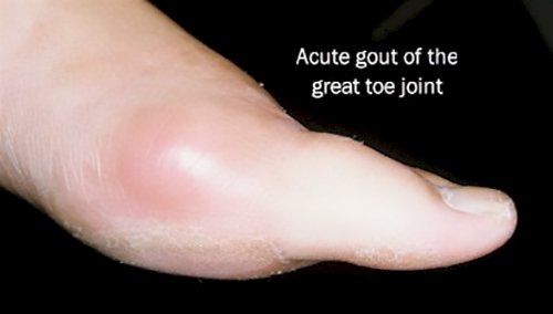
is described as crushing and excruciating. Attacks tend to last several days.
Acute gouty attacks occur in much the same manner. Most acute gouty attacks occur in the late hours of the night. As we sleep, our bodies tend to focus on the primary metabolic functions such as digestion, breathing, etc. The extremities, such as the feet tend to cool as a result of this ‘lack of attention’. As they cool, and if the dissolved amount of uric acid is high enough, the result is the change of uric acid from a liquid to a crystal. The hallmark symptoms of gout is the acute onset, usually at night with severe pain.
Chronic gout
First attacks of gouty arthritis are seldom associated with residual disability but recurrent acute attacks are followed by progressive cartilage and bone erosion in association with deposition of tophi and secondary degenerative changes. Severe functional impairment and gross joint deformities may occur in chronic tophaceous gout. Tophi are frequently found in the cartilage of the ear, bursae and tendon sheaths. Tophus formation is related to serum uric acid and to local factors. Tophi seldom develop in individuals with asymptomatic hyperuricaemia; however, they may develop rapidly in the feet or hands in post-menopausal women with heart failure and renal insufficiency who develop acute or subacute gouty arthritis following prolonged diuretic administration.
tophus over Achilles’ tendon
This patient has gout, and aspiration of this joint has confirmed the presence of uric acid crystals. Note the enlargement and deformity of the left second PIP joint.
Stages of gout
Investigations
The serum urate level is usually raised but it is important to appreciate that this does not prove the diagnosis because asymptomatic hyperuricaemia is very common. Whenever possible, synovial fluid should be aspirated and examined under polarising light. Acute attacks of gout can occur when the serum urate level is normal. This is frequently seen in patients who have received treatment with allopurinol or a uricosuric agent.
Joint radiographs may show characteristic punched-out erosions associated with the soft tissue swelling of urate tophi, occasionally flecked with calcium. The diagnosis will be clinically apparent in such patients. In others the erosions may be indistinguishable from those seen in various forms of inflammatory arthritis.
|
American College of Rheumatology Preliminary Criteria for Gout |
|
Gout may be diagnosed if one of the following criteria is present: Monosodium urate crystals in synovial fluid Tophi confirmed with crystal examination At least six of the following findings: Asymmetric swelling within a joint on a radiograph First metatarsophalangeal joint is tender or swollen (i.e., podagra) Hyperuricemia Maximal inflammation developed within one day Monoarthritis attack More than one acute arthritis attack Redness observed over joints Subcortical cysts without erosions on a radiograph Suspected tophi Synovial fluid culture negative for organisms during an acute attack Unilateral first metatarsophalangeal joint attack Unilateral tarsal joint attack |
|
|
RADIOGRAPHIC EVALUATION
Cystic changes, well-defined erosions described as punched-out lytic lesions with overhanging bony edges (Martel’s sign), associated with soft tissue calcified masses are characteristic radiographic features of chronic tophaceous gout. However, similar radiographic signs can also be observed in erosive osteoarthritis, destructive apatite arthropathies, and rheumatoid arthritis.
Proximal interphalangeal joint in gout demonstrates maintenance of normal mineralization and articular space, but well-defined erosion (closed arrow) and lobulated bone response (open arrows).
Shows classic location at first MTP and small erosion with “overhanging” edge. Notice that the joint space and bone density are preserved.
Another appearance showing multiple erosion locations including first MTP, base of third and fourth metacarpals, and possibly the head of the fifth metacarpal and second proximal phalanx.
Medications to Treat Acute Attacks of Gout
1. Non-steroidal anti-inflammatory drugs (NSAID’s) or COX-2 inhibitors. Examples of NSAID’s: Naproxen 500mg twice daily, indomethacin 25mg three times daily. Example of COX-2 inhibitor: celecoxib 200mg twice a day. Possible side-effects: Elevation of blood pressure, ankle swelling, upset stomach, ulcer (long-term use may have an increased risk of heart attack or stroke, but gout use is generally very short-term). Use with caution if kidney or liver problems.
2. Anti-Inflammatory corticosteroids. Examples: Prednisone 40mg first day, 30mg 2nd day, 20mg third day, 10mg fourth day. Possible side-effects: Elevation of blood pressure, elevation of blood sugar, mood changes. Short-term use, as in gout, generally much better tolerated than long-term use. Use with caution if diabetic.
3. Colchicine. Example: Oral dose of 0.6mg colchicine once an hour until improved or get diarrhea. Don’t exceed 4-5 doses. Possible side-effects: Other choices more commonly used due to frequent problems with diarrhea with colchicine (low doses often well-tolerated for prevention). Use with caution of kidney or liver problems.
4. Local steroid injections. Example: different doses used depending on the size of joint involved, and multiple preparations available. Possible side-effects: 1-2% of the time, a local reaction to the injection can occur, and the joint can can temporary worsening the next day, requiring ice application. In diabetics, a single local injection can temporarily raise blood sugar.
RHEUMATOID ARTHRITIS
Rheumatoid arthritis (RA) is a chronic, systemic inflammatory disorder that may affect many tissues and organs, but principally attacks the joints producing a inflammatory synovitis that often progresses to destruction of the articular cartilage and ankylosis of the joints. Rheumatoid arthritis (RA) is the most common form of chronic inflammatory joint disease. In its typical form RA is a symmetrical, destructive and deforming polyarthritis affecting small and large synovial joints, with associated systemic disturbance, a variety of extra-articular features and the presence of circulating antiglobulin antibodies (rheumatoid factors). Rheumatoid arthritis can also produce diffuse inflammation in the lungs, pericardium, pleura, and sclera, and also nodular lesions, most common in subcutaneous tissue under the skin. Although the cause of rheumatoid arthritis is unknown, autoimmunity plays a pivotal role in its chronicity and progression. Characteristically, the course of the disease is prolonged with exacerbations and remissions but atypical, asymmetrical and incomplete forms are not uncommon.
Etiology
The etiology of rheumatoid arthritis is not fully understood. Evidence points to a complex interplay between environmental and genetic factors. In monozygotic twins, there is a more than 30 percent concordance rate for rheumatoid arthritis development, and 80 percent of whites with rheumatoid arthritis express the HLA-DR1 or -DR4 subtypes. These and other regions of the Major Histocompatibility Complex may confer susceptibility to more severe disease by causing a specific arthrogenic peptide to be presented to CD4+ T cells.4
Joint damage in rheumatoid arthritis begins with the proliferation of synovial macrophages and fibroblasts after a triggering incident, possibly autoimmune or infectious. Lymphocytes infiltrate perivascular regions, and endothelial cells proliferate. Neovascularization then occurs. Blood vessels in the affected joint become occluded with small clots or inflammatory cells. Over time, inflamed synovial tissue begins to grow irregularly, forming invasive pannus tissue. Pannus invades and destroys cartilage and bone. Multiple cytokines, interleukins, proteinases, and growth factors are released, causing further joint destruction and the development of systemic complications.
Signs and symptoms
In the majority of patients the onset is insidious, with joint pain, stiffness and symmetrical swelling of a number of peripheral joints, but other disease patterns can occur. Initially, pain may be experienced only on movement of joints, but rest pain and prolonged early morning stiffness are characteristic features of all kinds of inflammatory arthritis.
While rheumatoid arthritis primarily affects joints, problems involving other organs of the body are known to occur. Extra-articular (“outside the joints”) manifestations other than anemia (which is very common) are clinically evident in about 15-25% of individuals with rheumatoid arthritis. It can be difficult to determine whether disease manifestations are directly caused by the rheumatoid process itself, or from side effects of the medications commonly used to treat it – for example, lung fibrosis from methotrexate, or osteoporosis from corticosteroids.
Joints
The arthritis of rheumatoid arthritis is due to synovitis, which is inflammation of the synovial membrane that lines joints and tendon sheaths. Joints become swollen, tender and warm, and stiffness limits their movement. With time, RA nearly always affects multiple joints (it is a polyarthritis). Most commonly, small joints of the hands, feet and cervical spine are affected, but larger joints like the shoulder and knee can also be involved, differing per individual. Synovitis can lead to tethering of tissue with loss of movement and erosion of the joint surface, causing deformity and loss of function.
Rheumatoid arthritis typically manifests with signs of inflammation and the affected joints are swollen, warm, painful and stiff early in the morning on waking or following prolonged inactivity. Increased stiffness early in the morning is often a prominent feature of the inflammatory disease which the person may experience and may last for more than an hour. Gentle movements may relieve symptoms in early stages of the disease.These signs help distinguish rheumatoid from non-inflammatory problems of the joints, often referred to as osteoarthritis or “wear-and-tear” arthritis.In arthritis of non-inflammatory causes signs of inflammation and early morning stiffness are absent and also movements aggravate pain due to the wear-and-tear. In RA, the joints are often affected in a fairly symmetrical fashion although this is not specific and the initial presentation may be asymmetrical.

Hands affected by RA
As the pathology progresses the inflammatory activity leads to tendon tethering and erosion and destruction of the joint surface, which impairs range of movement and leads to deformity. The fingers may suffer from almost any deformity, depending on which joints are most involved. Medical students are taught to learn names for specific deformities like ulnar deviation, boutonniere deformity, swaeck deformity and “Z-thumb” but these are of no more significance to diagnosis or disability than other variants.
In the typical case the small joints of the fingers and toes are the first to be affected. Swelling of the proximal, but not the distal, interphalangeal joints gives the fingers a ‘spindled’ appearance, and swelling of the metatarsophalangeal joints results in ‘broadening’ of the forefoot. As the disease progresses with or without intervening remissions, it tends to spread to involve the wrists, elbows, shoulders, knees, ankles, subtalar and midtarsal joints. The hips become involved only in the more severely affected, but neck pain and stiffness from cervical spine disease is common. The temporomandibular, acromio-clavicular, sternoclavicular and crico-arytenoid joints are sometimes affected.
In 10-15% of patients the disease starts as an acute polyarthritis with severe systemic symptoms. A systemic onset, with fever, weight loss, profound fatigue and malaise without joint symptoms, occurs less often, particularly in middle-aged men, and this can cause diagnostic confusion with malignant disease or chronic infections. The onset is palindromic in some patients, with recurrent acute episodes of joint pain and stiffness in individual joints lasting only a few hours or days. In about one-third of such cases the disease evolves into one more typical of arthritis. The onset of RA in the elderly can be indistinguishable from polymyalgia rheumatica with pain and stiffness in the region of the hip and shoulder girdles but no apparent synovitis. The presence of rheumatoid factor in such patients may be a clue to the true diagnosis before typical joint changes have developed.
Proximal Interphalangeal Joints.
Involvement of the proximal interphalangeal (PIP) joints usually is easily recognized from the fusiform soft tissue swelling, often accompanied by regional osteopenia. Uniform cartilage loss occurs early at this site and erosion appears somewhat later. Erosive disease parallels the synovial and capsular anatomy, being extensive over the proximal phalangeal condyles and more limited over the base of the distal phalanges. Flexion or extension deformities are frequent in advanced disease; bony ankylosis is rare. Erosive articular disease rarely is seen in the distal interphalangeal joints, although swelling and tenderness are common.
Metacarpophalangeal Joints.
Soft tissue swelling in the metacarpophalangeal (MCP) joints, although clinically prominent, is more difficult to evaluate than in the proximal interphalangeal joints, but in high-quality radiographs appears as discrete capsular distention. The cartilage at this site becomes narrow later, whereas erosion, particularly of the radial aspect of the metacarpal head, is an early and important sign of rheumatoid arthritis. Small, discrete, pocketed erosions develop at the proximal phalangeal base near the capsular insertion. As the erosive process evolves, narrowing ensues, and eventually complete destruction of the joint with “pencil in cup” deformity is seen, accompanied by palmar subluxation and ulnar deviation.
Ulnar Deviation
Wrist. In the wrist, soft tissue swelling is usually prominent and recognized relatively easily, particularly adjacent to the ulnar styloid, as a result of synovitis of the extensor carpi ulnaris tendon sheath. This involvement frequently leads to characteristic focal demin-eralization and surface erosion of the medial ulnar styloid. Additional erosion of the distal ulna results from synovitis of the prestyloid recess, eroding the tip of the styloid, and from synovitis in the inferior radio-ulnar compartment that erodes the foveal region. Other early and common sites of erosion include the waist of the navicular, the radial styloid, the pisiform and tri-quetrum, and the palmar aspect of the distal radial articular surface. Cartilaginous loss occurs relatively early at the wrist and tends to cause a uniform loss throughout the various compartments because of the development of early, abnormal communications between these separate spaces. In rheumatoid arthritis, joints that communicate are involved simultaneously and uniformly.
Anatomic alignment at the wrist generally becomes altered with medial migration of the carpus at the radiocarpal joint, allowing the navicular to occupy the hollow of the radius. This rotational change may result in foreshortening of the navicular and lunate. Frequently, a widened space is seen between the navicular and capitate. Involvement of the inferior radioulnar compartment with destruction of the triangular cartilage results in diastasis and, occasionally, in complete dorsal dislocation of the distal ulna. In the lateral view, either dorsal or palmar subluxation and deviation of the carpus in regard to the distal radius may be seen. Bony ankylosis is more common at the carpus than at any other site in rheumatoid arthritis. Finally, a near-total resorption or fragmentation, or both, of the carpus, penciling of the distal ulna, and cup-shape resorption of the distal radius are seen in end-stage disease.
Subchondral cortical line, a result of focal absorption and erosion accompanied by reactive sclerosis in the adjacent cancellous bone. The iliac side of the articulation is affected first because of its thinner cartilage; the sacral side may appear normal initially. At its inception, the process may be unilateral or asymmetric, but soon becomes bilateral, and eventually remarkably symmetric. With progressive erosion, joint widening becomes apparent in association with pronounced patchy sclerosis that extends some distance into the subarticular bone. The process may arrest at any phase but characteristically progresses to narrowing of the joint and finally to intracapsular and intra-articular bony anky-losis. Occasionally part of the original joint line may persist. Once the joint has fused solidly, the sclerosis resolves gradually and a generalized osteopenia of the pelvis develops.
RADIOGRAPHIC EVALUATION
Early in the disease, roentgenograms of the affected joints are usually not helpful in establishing a diagnosis. They reveal only that which is apparent from physical examination, namely, evidence of soft tissue swelling and joint effusion. As the disease progresses, abnormalities become more pronounced, but none of the radiographic findings is diagnostic of RA. The diagnosis, however, is supported by a characteristic pattern of abnormalities, including the tendency toward symmetric involvement. Juxtaarticular osteopenia may become apparent within weeks of onset. Loss of articular cartilage and bone erosions develop after months of sustained activity. The primary value of radiography is to determine the extent of cartilage destruction and bone erosion produced by the disease, particularly when one is monitoring the impact of therapy with disease-modifying drugs or surgical intervention. Other means of imaging bones and joints, including 99mTc bisphosphonate bone scanning and magnetic resonance imaging, may be capable of detecting early inflammatory changes that are not apparent from standard radiography but are rarely necessary in the routine evaluation of patients with RA.
Severe, erosive rheumatoid arthritis

Signs of destruction and inflammation on ultrasonography and magnetic resonance imaging in the second metacarpophalangeal joint in established rheumatoid arthritis. Thin arrows indicate an erosive change; thick arrows indicate synovitis. Ultrasonography (left side of image) in the (a) longitudinal and (b) the transverse planes shows both signs of destruction and inflammation. Axial T1-weighted magnetic resonance images were obtained (c) before and (d) after contrast administration, also demonstrating synovitis. Additionally, a coronal T1-weighted magnetic resonance image (e) before contrast administration visualizes the same bone erosion as shown in panels c and d.
Blood tests
When RA is clinically suspected, immunological studies are required, such as testing for the presence of rheumatoid factor (RF, a specific antibody). A negative RF does not rule out RA; rather, the arthritis is called seronegative. This is the case in about 15% of patients. During the first year of illness, rheumatoid factor is more likely to be negative with some individuals converting to seropositive status over time. RF is also seen in other illnesses, for example Sjögren’s syndrome, and in approximately 10% of the healthy population, therefore the test is not very specific.
Because of this low specificity, a new serological test has been developed, which tests for the presence of so called anti-citrullinated protein antibodies (ACPAs). Like RF, this test is positive in only a proportion (67%) of all RA cases, but is rarely positive if RA is not present, giving it a specificity of around 95%. As with RF, there is evidence for ACPAs being present in many cases even before onset of clinical disease Currently, the most common test for ACPAs is the anti-CCP (cyclic citrullinated peptide) test.
Also, several other blood tests are usually done to allow for other causes of arthritis, such as lupus erythematosus. The erythrocyte sedimentation rate (ESR), C-reactive protein, full blood count, renal function, liver enzymes and other immunological tests (e.g. antinuclear antibody/ANA) are all performed at this stage. Elevated ferritin levels can reveal hemochromatosis, a mimic RA, or be a sign of Still’s disease, a seronegative, usually juvenile, variant of rheumatoid.
Differential diagnosis
Several other medical conditions can resemble RA, and usually need to be distinguished from it at the time of diagnosis:
· Crystal induced arthritis (gout, and pseudogout) – usually involves particular joints and can be distinguished with aspiration of joint fluid if in doubt
· Osteoarthritis – distinguished with X-rays of the affected joints and blood tests
· Systemic lupus erythematosus (SLE) – distinguished by specific clinical symptoms and blood tests (antibodies against double-stranded DNA)
· One of the several types of psoriatic arthritis resembles RA – nail changes and skin symptoms distinguish between them
· Lyme disease causes erosive arthritis and may closely resemble RA – it may be distinguished by blood test in endemic areas
· Reactive arthritis (previously Reiter’s disease) – asymmetrically involves heel, sacroiliac joints, and large joints of the leg. It is usually associated with urethritis, conjunctivitis, iritis, painless buccal ulcers, and keratoderma blennorrhagica.
· Ankylosing spondylitis – this involves the spine and is usually diagnosed in males, although a RA-like symmetrical small-joint polyarthritis may occur in the context of this condition.
Rarer causes that usually behave differently but may cause joint pains:
· Sarcoidosis, amyloidosis, and Whipple’s disease can also resemble RA.
· Hemochromatosis may cause hand joint arthritis.
· Acute rheumatic fever can be differentiated from RA by a migratory pattern of joint involvement and evidence of antecedent streptococcal infection. Bacterial arthritis (such as streptococcus) is usually asymmetric, while RA usually involves both sides of the body symmetrically.
· Gonococcal arthritis (another bacterial arthritis) is also initially migratory and can involve tendons around the wrists and ankles.
Management of Rheumatoid Arthritis
There is no known cure for rheumatoid arthritis, but many different types of treatment can alleviate symptoms and/or modify the disease process.
The goal of treatment is two-fold: alleviating the current symptoms, and preventing the future destruction of the joints with the resulting handicap if the disease is left unchecked. These two goals may not always coincide: while pain relievers may achieve the first goal, they do not have any impact on the long-term consequences. For these reasons, most authorities believe that most RA should be treated by at least one specific anti-rheumatic medication, also named DMARD (see below), to which other medications and non-medical interventions can be added as needed
In a chronic and frequently progressive disease characterised by exacerbations and remissions over many years, as well as by systemic, psychiatric and social complications, periodic assessment of radiological progression, disease activity and disability is required. General practitioners and hospital physicians have a special responsibility to coordinate a team of medical specialists, orthopaedic surgeons, occupational therapists, physiotherapists, nurses, social workers and other health professionals in an integrated programme of multidisciplinary care and rehabilitation.
Patient education, counselling and continuing medical support are usually required for successful management, while physical rehabilitation, reconstructive surgery and environmental adaptation assume increasing importance when advancing joint damage and deformity are associated with functional impairment.
Repeated medical, functional and social assessment are required if patients are to maintain their maximum physical, psychological, social and vocational potential.
The aetiology of rheumatoid arthritis is unknown, and so treatment is empirically directed towards:
• relief of symptoms
• suppression of active and progressive disease
• conservation and restoration of function in affected joints.
To a greater or lesser extent these are achieved by combining:
• treatment of the patient—drugs, rest, physiotherapy, surgery
• modification of the environment—aids, appliances, housing, occupation, statutory social benefits.
General treatment in the active phase
Physical rest, anti-inflammatory drug therapy and maintenance exercises are the cornerstones of treatment for exacerbations of rheumatoid arthritis. Admission to hospital is necessary in a minority of patients when widespread active polyarthritis is associated with signs of constitutional disturbance and there has beeo response to rest at home and optimal doses of NSAIDs and disease-modifying antirheumatic drugs (DMARDs).
The rest from physical and emotional stress provided by 1-2 weeks in a hospital ward or day-patient unit is usually sufficient to induce a marked remission of symptoms without recourse to strict bed rest. Hospitalisation allows for detailed assessment by all members of the arthritis team followed by a programme of medical and physical rehabilitation best suited to the individual’s needs.
In a few patients a period of complete bed rest may be required to induce a remission. In these circumstances it is essential to prevent the development of ‘bed deformities’ by ensuring appropriate positioning of joints. Foot and quadriceps exercises should be performed daily, along with maintenance exercises for muscle groups in unaffected limbs.
Anaemia of chronic disease responds best to induction of disease remission, and oral iron is only indicated in those patients with superadded true iron deficiency. Folic acid is occasionally required to treat an associated macrocytic anaemia.
Prognosis
The course of the disease varies greatly. Some people have mild short-term symptoms, but in most the disease is progressive for life. Around 20%-30% will have subcutaneous nodules (known as rheumatoid nodules); this is associated with a poor prognosis.
Disability
· Daily living activities are impaired in most individuals.
· After 5 years of disease, approximately 33% of sufferers will not be working.
· After 10 years, approximately half will have substantial functional disability.
Prognostic factors
Poor prognostic factors include persistent synovitis, early erosive disease, extra-articular findings (including subcutaneous rheumatoid nodules), positive serum RF findings, positive serum anti-CCP autoantibodies, carriership of HLA-DR4 “Shared Epitope” alleles, family history of RA, poor functional status, socioeconomic factors, elevated acute phase response (erythrocyte sedimentation rate [ESR], C-reactive protein [CRP]), and increased clinical severity.
Mortality
Estimates of the life-shortening effect of RA vary; most sources cite a lifespan reduction of 5 to 10 years. According to the UK‘s National Rheumatoid Arthritis Society, “Young age at onset, long disease duration, the concurrent presence of other health problems (called co-morbidity), and characteristics of severe RA – such as poor functional ability or overall health status, a lot of joint damage on x-rays, the need for hospitalisation or involvement of organs other than the joints – have been shown to associate with higher mortality”. Positive responses to treatment may indicate a better prognosis. A 2005 study by the Mayo Clinic noted that RA sufferers suffer a doubled risk of heart disease, independent of other risk factors such as diabetes, alcohol abuse, and elevated cholesterol, blood pressure and body mass index. The mechanism by which RA causes this increased risk remains unknown; the presence of chronic inflammation has been proposed as a contributing factor.

